Comparative Study
. 2008 Jul 16;28(29):7370-5.
doi: 10.1523/JNEUROSCI.1881-08.2008.

Step training reinforces specific spinal locomotor circuitry in adult spinal rats

Affiliations
Comparative Study

Step training reinforces specific spinal locomotor circuitry in adult spinal rats

Ronaldo M Ichiyama et al. J Neurosci. .

Abstract

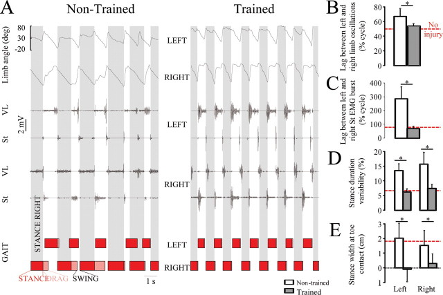
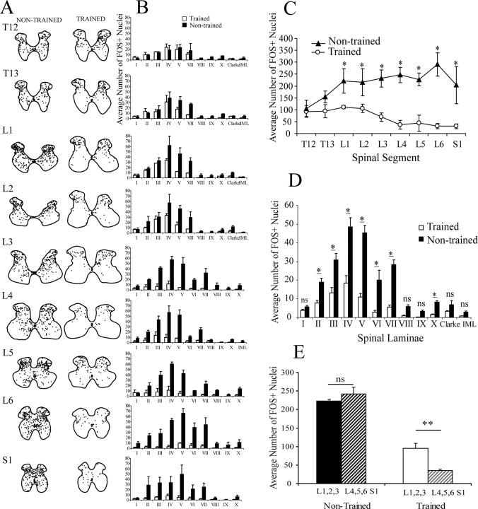
Locomotor training improves function after a spinal cord injury both in experimental and clinical settings. The activity-dependent mechanisms underlying such improvement, however, are sparsely understood. Adult rats received a complete spinal cord transection (T9), and epidural stimulation (ES) electrodes were secured to the dura matter at L2. EMG electrodes were implanted bilaterally in selected muscles. Using a servo-controlled body weight support system for bipedal stepping, five rats were trained 7 d/week for 6 weeks (30 min/d) under quipazine (0.3 mg/kg) and ES (L2; 40 Hz). Nontrained rats were handled as trained rats but did not receive quipazine or ES. At the end of the experiment, a subset of rats was used for c-fos immunohistochemistry. Three trained and three nontrained rats stepped for 1 h (ES; no quipazine) and were returned to their cages for 1 h before intracardiac perfusion. All rats could step with ES and quipazine administration. The trained rats had higher and longer steps, narrower base of support at stance, and lower variability in EMG parameters than nontrained rats, and these properties approached that of noninjured controls. After 1 h of stepping, the number of FOS+ neurons was significantly lower in trained than nontrained rats throughout the extent of the lumbosacral segments. These results suggest that training reinforces the efficacy of specific sensorimotor pathways, resulting in a more selective and stable network of neurons that controls locomotion.


Figures

Figure 1.
Figure 1.
A, Stick diagram decomposition, joint angular displacement, and EMG activity of selected hindlimb muscles of a step cycle during testing in a representative noninjured, spinal rat without ES or quipazine (Q), and nontrained and step-trained rat with ES (40 Hz at L2) and Q (0.3 mg/kg). B, Gait timing variability, an indicator of interim coordination variability, was higher in nontrained than noninjured or trained rats. Step height (C) and length (D), total quantity of joint movement (E), and TA EMG amplitude (F) were greater in trained than nontrained rats. The horizontal red dotted line in B–F indicates values for noninjured rats. Error bars indicate SEM. *p < 0.05, significant difference between trained and nontrained groups. MTP, Metatarsophalangeal joint; VL, vastus lateralis; St, semitendinosus; MG, medial gastrocnemius; TA, tibialis anterior.
Figure 2.
Figure 2.
A, Representative EMG recordings from the left and right VL and St, with corresponding gait cycle diagrams for a nontrained and a trained rat treated with ES and Q. B, C, The left and right limbs moved out of phase in both noninjured and trained rats (B), whereas nontrained rats showed impaired interlimb coordination and a significant lag between the left and right St EMG bursts over the duration of the stepping sequence (C). D, E, Variability of stance duration (computed as coefficient of variability) (D) and stance width at toe contact (E) were greater in nontrained than trained rats. The horizontal red dotted line in B–E indicates control values for noninjured rats. Error bars indicate SEM. *p < 0.01, significant difference between trained and nontrained groups. Abbreviations are the same as in Figure 1.
Figure 3.
Figure 3.
FOS immunohistochemistry after 1 h bipedal stepping under ES (L2; 40 Hz). A, Camera lucida drawing of the location of FOS+ nuclei for a representative nontrained and trained spinal rat. Note that FOS+ nuclei were distributed in all segments, and all laminas were analyzed in both groups. B, Mean (±SEM) number of FOS+ for each lamina at each spinal segment is shown. C, The average number of FOS+ nuclei per spinal segment was greater in nontrained than trained rats in spinal segments L1 to S1. D, Differences in the mean (±SEM) number of FOS+ nuclei per lamina for all spinal segments combined between spinal trained and nontrained rats are shown. *p < 0.05, significant difference between trained and nontrained groups. E, Comparison of the average number of FOS+ nuclei in the rostral (L1, L2, L3) and caudal (L4, L5, L6; S1) lumbosacral segments. Trained rats had significantly (p < 0.01) fewer FOS+ nuclei in the caudal segments, whereas nontrained rats did not show such a difference. ns, Nonsignificant.

Similar articles

Cited by

References

    1. Ahn SN, Guu JJ, Tobin AJ, Edgerton VR, Tillakaratne NJ. Use of c-fos to identify activity-dependent spinal neurons after stepping in intact adult rats. Spinal Cord. 2006;44:547–559. - PMC - PubMed
    1. Bareyre FM, Kerschensteiner M, Raineteau O, Mettenleiter TC, Weinmann O, Schwab ME. The injured spinal cord spontaneously forms a new intraspinal circuit in adult rats. Nat Neurosci. 2004;7:269–277. - PubMed
    1. Bonnot A, Morin D. Hemisegmental localisation of rhythmic networks in the lumbosacral spinal cord of neonate mouse. Brain Res. 1998;793:136–148. - PubMed
    1. Carr PA, Huang A, Noga BR, Jordan LM. Cytochemical characteristics of cat spinal neurons activated during fictive locomotion. Brain Res Bull. 1995;37:213–218. - PubMed
    1. Côté MP, Gossard JP. Step training-dependent plasticity in spinal cutaneous pathways. J Neurosci. 2004;24:11317–11327. - PMC - PubMed

Publication types