Review
. 2008 Aug;89(Pt 8):1789-1804.
doi: 10.1099/vir.0.2008/002089-0.

Bluetongue virus: dissection of the polymerase complex

Affiliations
Review

Bluetongue virus: dissection of the polymerase complex

Polly Roy. J Gen Virol. 2008 Aug.

Abstract

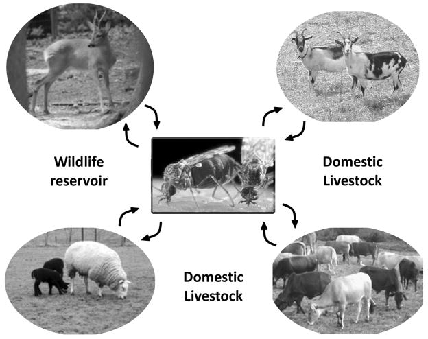
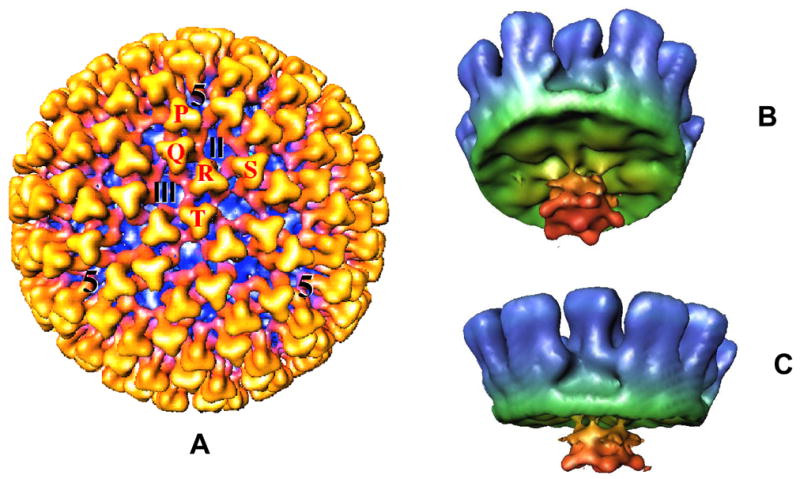
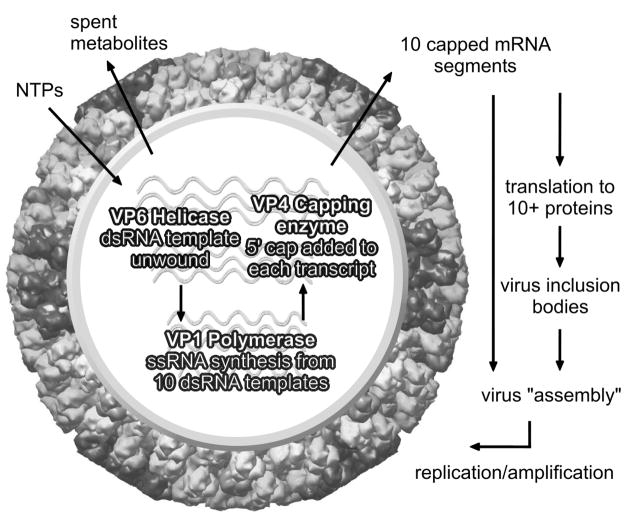
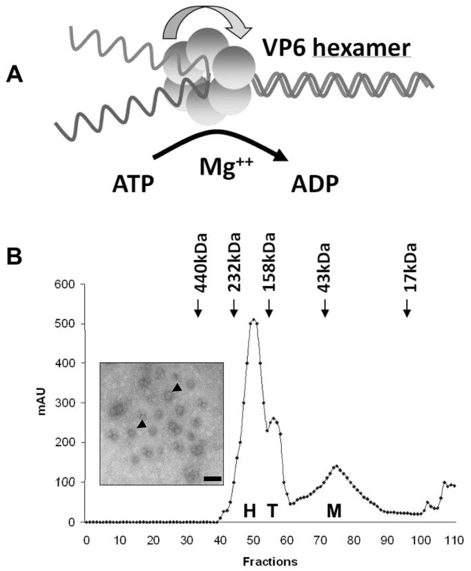
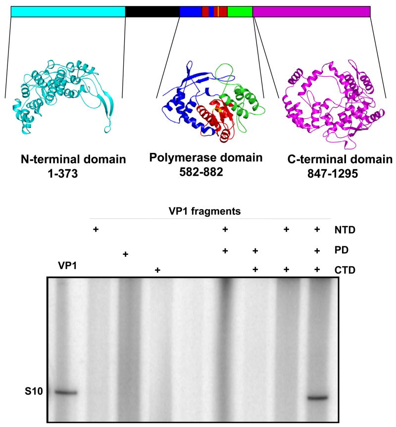
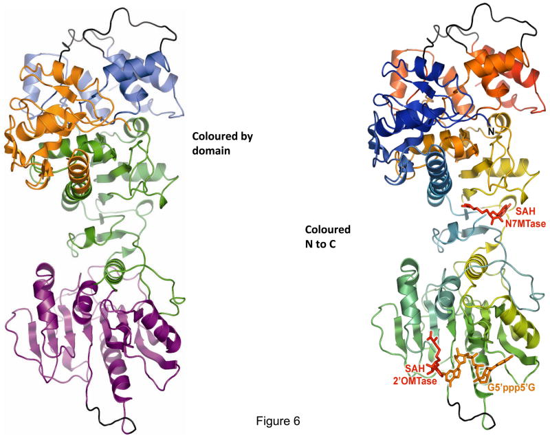
Bluetongue is a vector-borne viral disease of ruminants that is endemic in tropical and subtropical countries. Since 1998 the virus has also appeared in Europe. Partly due to the seriousness of the disease, bluetongue virus (BTV), a member of genus Orbivirus within the family Reoviridae, has been a subject of intense molecular study for the last three decades and is now one of the best understood viruses at the molecular and structural levels. BTV is a complex non-enveloped virus with seven structural proteins arranged in two capsids and a genome of ten double-stranded (ds) RNA segments. Shortly after cell entry, the outer capsid is lost to release an inner capsid (the core) which synthesizes capped mRNAs from each genomic segment, extruding them into the cytoplasm. This requires the efficient co-ordination of a number of enzymes, including helicase, polymerase and RNA capping activities. This review will focus on our current understanding of these catalytic proteins as derived from the use of recombinant proteins, combined with functional assays and the in vitro reconstitution of the transcription/replication complex. In some cases, 3D structures have complemented this analysis to reveal the fine structural detail of these proteins. The combined activities of the core enzymes produce infectious transcripts necessary and sufficient to initiate BTV infection. Such infectious transcripts can now be synthesized wholly in vitro and, when introduced into cells by transfection, lead to the recovery of infectious virus. Future studies thus hold the possibility of analysing the consequence of mutation in a replicating virus system.


Figures

Figure 1
Figure 1
A schematic showing BTV transmission by blood feeding Culicoides from infected to healthy animals that include both wildlife and domestic livestock.
Figure 2
Figure 2. Cryoelectron microscopy reconstruction of BTV core
(A): The whole core (700 Å in diameter) viewed along the icosahedral threefold axis showing the VP7 trimers (in yellow). The five quasi-equivalent trimers (P, Q, R, S and T) and the locations of channels are indicated. (B): Cryo-EM structure of a recombinant core-like particle showing the inside view of the CLP reconstruction with VP3 (green), VP7 (blue), VP1 and VP4. A flower-shaped density features (red) attached to the inside surface of VP3 at the 5 fold vertices is made up of VP1 and VP4 (Prasad et al., 1992; Grimes et al., 1997; Nason et al., 2004).
Figure 3
Figure 3. Cartoon of the transcriptionally active core
Diagram shows the activity of VP1, VP4 and VP6 which form the transcription complex within the core and are responsible for synthesis of ten ‘capped’ mRNA species that are extruded from the core, thereby initiating viral proteins synthesis and subsequent viral replication.
Figure 4
Figure 4. Helicase activity of VP6 and its oligomeric nature
(A). Model of helicase activity of VP6 showing that VP6 binds RNA and unwinds dsRNA to ssRNA product in presence of Mg++ and ATP. (B). Gel filtration analysis of VP6 monomer (M), tetramer (T) and hexamer (H). Inset shows the electron microscopy of ring-like structures formed by VP6 and ss- or dsRNA complexes, stained with 1% uranyl acetate. The scale bar represents 100 Å (Stauber et al., 1997; Kar & Roy, 2003).
Figure 5
Figure 5. 3D model structure of VP1 and reconstitution of polymerase activity from three VP1 fragments generated using the model
Upper panel: A structural model of BTV VP1 is generated based on other RNA dependent RNA polymerase structures, showing the PD domain as well as two other additional domains, NTD and CTD that were generated based on reovirus polymerase structure (Tao et al., 2002). The VP1 NTD modelled from amino acid residues 1–373 is shown in cyan. The PD model covers amino acid residues 581–880. Within the PD, the ‘fingers’ subdomain is blue, the ‘palm’ subdomain is red and the ‘thumb’ subdomain is green. The CTD modelled from amino acid residues 847 to 1295 is in magenta. The CTD model overlaps with the ‘thumb’ subdomain of the PD model. A region of VP1 that was not modelled is shown in black. Lower panel: Each of the three (NTD, PD and CTD) VP1 fragments was expressed, purified and tested for replicase activity. Positive controls for the replicase assay were purified VP1 (lane 1) which was at a similar concentration and purity to the fragments. The “+” indicates inclusion of each of the VP1 domains in the lane (Wehrfritz et al., 2007).
Figure 6
Figure 6. Cartoon representation of the X-ray structure of the VP4 molecule
Ribbon diagram of the VP4 structure showing the four discrete domains (domain nomenclature defined in text) that are arranged in a linear fashion (left). Each domain is coloured as follows: GTase domain (also possibly RTase active site), in red located in the C terminal 135 residues; N7MTase domain (underneath the GTase domain), in orange and 2′OMTase domain, in green are located at the centre of the polypeptide; KL domain (blue) is located in the first 108 residues of the molecule. On the right is the ribbon diagram of the VP4 structure coloured from blue at the N terminus to red at the C terminus, poorly ordered loops are represented in grey. The cartoon in left also shows the ligand (AdoHcy, SAH and G5′ppp5′G) binding sites (Sutton et al., 2007).
Figure 7
Figure 7. Comparisons of VP4 domains with available structures show conservative nature of functional domains
Upper panel: 2′OMTase VP4 (right) and superimposition (left) of VP4 (purple) and vaccinia virus VP39 (cyan). Lower panel: N7MTase domain of VP4 (right) and superimposition (left) of the N7MTase domain of VP4 with ecm1 (Sutton et al., 2007).
Figure 8
Figure 8. Recovery of infectious BTV virions from in vitro synthesized BTV RNA
10 transcripts were synthesized from BTV core in vitro or generated from cDNA clones. Both type of transcripts produced viral plaques upon transfection of BHK cells (Boyce & Roy, 2007; Boyce et al., 2008).

References

    1. Ahnert P, Patel SS. Asymmetric interactions of hexameric bacteriophage T7 DNA helicase with the 5′- and 3′-tails of the forked DNA substrate. J Biol Chem. 1997;272:32267–32273. - PubMed
    1. Appleby TC, Luecke H, Shim JH, Wu JZ, Cheney IW, Zhong W, Vogeley L, Hong Z, Yao N. Crystal structure of complete rhinovirus RNA polymerase suggests front loading of protein primer. J Virol. 2005;79:277–288. - PMC - PubMed
    1. Bannerjee AK, Shatkin AJ. Transcription in vitro by reovirus associated ribonucleic acid-dependent polymerase. J Virol. 1970;6:1–11. - PMC - PubMed
    1. Barbosa PR, Sousa MO, Barbosa EC, Bomfim Ade S, Ginefra P, Nadal J. Analysis of the prevalence of ventricular late potentials in the late phase of myocardial infarction based on the site of infarction. Arq Bras Cardiol. 2002;78:352–363. - PubMed
    1. Bartlett NM, Gillies SC, Bullivant S, Bellamy AR, Lawton JA, Zeng CQ, Mukherjee SK, Cohen J, Estes MK, Prasad BV, Smith RE, Furuichi Y, Gillies S, Bullivant S, Bellamy AR. Electron microscopy study of reovirus reaction cores. J Virol. 1974;14:315–326. - PMC - PubMed

Publication types

MeSH terms

LinkOut - more resources