. 2008 Oct;19(10):4141-53.
doi: 10.1091/mbc.e07-12-1217. Epub 2008 Jul 16.

YY1 protects cardiac myocytes from pathologic hypertrophy by interacting with HDAC5

Affiliations

YY1 protects cardiac myocytes from pathologic hypertrophy by interacting with HDAC5

Carmen C Sucharov et al. Mol Biol Cell. 2008 Oct.

Abstract

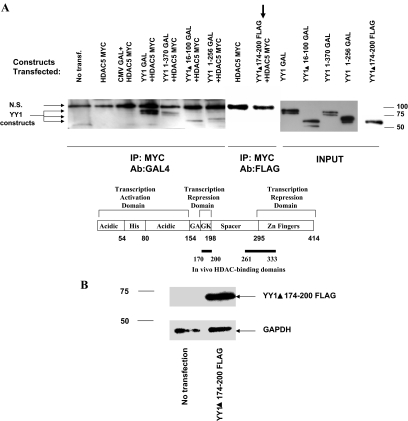
YY1 is a transcription factor that can repress or activate the transcription of a variety of genes. Here, we show that the function of YY1 as a repressor in cardiac myocytes is tightly dependent on its ability to interact with histone deacetylase 5 (HDAC5). YY1 interacts with HDAC5, and overexpression of YY1 prevents HDAC5 nuclear export in response to hypertrophic stimuli and the increase in cell size and re-expression of fetal genes that accompany pathological cardiac hypertrophy. Knockdown of YY1 results in up-regulation of all genes present during fetal development and increases the cell size of neonatal cardiac myocytes. Moreover, overexpression of a YY1 deletion construct that does not interact with HDAC5 results in transcription activation, suggesting that HDAC5 is necessary for YY1 function as a transcription repressor. In support of this relationship, we show that knockdown of HDAC5 results in transcription activation by YY1. Finally, we show that YY1 interaction with HDAC5 is dependent on the HDAC5 phosphorylation domain and that overexpression of YY1 reduces HDAC5 phosphorylation in response to hypertrophic stimuli. Our results strongly suggest that YY1 functions as an antihypertrophic factor by preventing HDAC5 nuclear export and that up-regulation of YY1 in human heart failure may be a protective mechanism against pathological hypertrophy.


Figures

Figure 1.
Figure 1.
YY1 represses fetal gene expression and activates adult gene expression in cardiac cells. Cells were infected with YY1-GFP and harvested 48 h after infection. Total RNA was analyzed by RT-PCR. Results were compared with cells infected with a control CMV-GFP adenovirus, defined as 100% (line at 100%). n = 8.
Figure 2.
Figure 2.
YY1 prevents up-regulation of the fetal isoforms and PE-induced cellular hypertrophy in response to PE. (A) NRVMs were treated with 10 μM PE for 48 h; white bars, no treatment; black bars, PE treatment. Total RNA was analyzed by RT-PCR. Results were compared with vehicle-treated cells infected with CMV-GFP, defined as 100%. n = 8. (B) Increase in protein synthesis was measured by leucine incorporation in untreated and PE-treated cells infected with a CMV control or YY1 adenovirus construct. (C) Cell size was measured using the ImageJ software. Fifty cells from four different experiments were measured. (D) Immunofluorescence with anti-actinin antibody of cells infected with a control or YY1-GFP virus in untreated and PE-treated cells.
Figure 3.
Figure 3.
Down-regulation of YY1 expression results in up-regulation of the fetal isoforms and in increase in cell size and protein synthesis. (A) YY1 expression is repressed by transfection with an YY1 siRNA oligonucleotide (lane 3) but not by a control or an unrelated siRNA oligonucleotide (lanes 2 and 4). Lane 1, no transfection. Total extract was used, and glyceraldehyde-3-phosphate dehydrogenase (GAPDH) was used as a loading control. (B) Cells were transfected with YY1 siRNA nucleotide and treated with vehicle (white bars) or PE (black bars) for 48 h. RNA expression was analyzed by RT-PCR and normalized to 18S. Results were compared with vehicle-treated cells transfected with a control siRNA, defined as 100%. n = 4. (C) Radiolabeled protein experiments show an increase in radiolabeled leucine incorporation in cells transfected with YY1 siRNA oligonucleotide. (D) Cell size measurements show an increase in cell size in cells that do not express YY1. Fifty-five cells from three different experiments were measured. (E) Immunofluorescence with anti-actinin antibody of cells transfected with a control or YY1 siRNA oligonucleotide.
Figure 3.
Figure 3.
Down-regulation of YY1 expression results in up-regulation of the fetal isoforms and in increase in cell size and protein synthesis. (A) YY1 expression is repressed by transfection with an YY1 siRNA oligonucleotide (lane 3) but not by a control or an unrelated siRNA oligonucleotide (lanes 2 and 4). Lane 1, no transfection. Total extract was used, and glyceraldehyde-3-phosphate dehydrogenase (GAPDH) was used as a loading control. (B) Cells were transfected with YY1 siRNA nucleotide and treated with vehicle (white bars) or PE (black bars) for 48 h. RNA expression was analyzed by RT-PCR and normalized to 18S. Results were compared with vehicle-treated cells transfected with a control siRNA, defined as 100%. n = 4. (C) Radiolabeled protein experiments show an increase in radiolabeled leucine incorporation in cells transfected with YY1 siRNA oligonucleotide. (D) Cell size measurements show an increase in cell size in cells that do not express YY1. Fifty-five cells from three different experiments were measured. (E) Immunofluorescence with anti-actinin antibody of cells transfected with a control or YY1 siRNA oligonucleotide.
Figure 4.
Figure 4.
YY1 binds to αMyHC and BNP promoters. ChIPs were done in noninfected cells, in cells overexpressing YY1 or in cells transfected with YY1 siRNA as described in the methods section. ChIPs from cells infected with YY1 or transfected with YY1 siRNA are a result of six independent experiments. YY1 or RNA polymerase antibodies, or IgG were used in the immunoprecipitation assay.
Figure 5.
Figure 5.
YY1 interacts with HDAC5 in NRVMs and in rat heart tissue. (A) Cells were infected with YY1-GFP adenovirus construct. Nuclear extracts were immunoprecipitated with anti-YY1 antibody and HDAC5 was detected by Western blot. (B) Rat heart nuclear fraction was immunoprecipitated with anti-YY1 antibody or IgG and interaction with HDAC5, HDAC4, or HDAC9 was detected by Western blot.
Figure 6.
Figure 6.
YY1 prevents nuclear export of HDAC5 in PE-treated cells, but down-regulation of YY1 does not result in cytoplasmic localization of HDAC5. (A) Western blot of nuclear and cytoplasmic fractions of cells infected with HDAC5-FLAG and GFP or HDAC5-FLAG and YY1 adenovirus constructs. Cells were treated with PE for 2 h before harvesting. GAPDH and Sp1 were used as loading control for cytoplasmic fraction and nuclear fraction respectively and to show that there was no cross-contamination between fractions. Calnexin was used as a control for both cytoplasmic and nuclear fractions. (B) The graph is representative of three individual Western Blot experiments shown in A. (C) YY1 prevents export of endogenous HDAC5 to the cytoplasm in response to PE treatment. Experiments were performed as described in A, but cells were infected only with YY1 or CMV adenovirus. Endogenous HDAC5 was detected using HDAC5 antibody. (D) Western blot of nuclear and cytoplasmic fractions of cells transfected with a control siRNA or YY1 siRNA and infected with HDAC5-FLAG. Calnexin was used as a loading control. (E) Immunostaining of NRVMs infected with HDAC5-FLAG–untreated cells (1), HDAC5-FLAG–PE-treated (2), HDAC5-FLAG and YY1–PE-treated (3), HDAC5-FLAG and transfected with YY1 siRNA (4). The panels show FLAG staining for HDAC5 localization, Hoechst staining for nuclear localization and merging of HDAC5 and nuclear staining.
Figure 7.
Figure 7.
The phosphorylation domain of HDAC5 is required for its interaction with YY1, and YY1 blunts HDAC5 phosphorylation. (A and B) Overexpression of YY1 blunts HDAC5 phosphorylation in response to PE. (A) NRVMs were infected with CMV or YY1-GFP. Cells were treated with PE for 1 h and total protein was extracted. Phosphorylated Ser 259 HDAC5 and total HDAC5 were detected by Western blot, and calnexin antibody was used as a loading control. (B) Quantification of Western in A. (C and D) COS cells were transfected with YY1-GAL and wild-type HDAC5-MYC (C) or YY1-GAL and HDAC5 del 260-615-FLAG (D), and total protein was extracted. Immunoprecipitation was done with a GFP antibody (control) or a GAL4 antibody, and HDAC5 was detected by Western blot using either MYC or FLAG antibodies. N.S., nonspecific.
Figure 8.
Figure 8.
YY1 is a transcription activator in the absence of or lack of interaction with HDAC5. (A) COS7 cells were transfected with various YY1 deletion constructs tagged with GAL4 or FLAG, and HDAC5-MYC. Total extract was immunoprecipitated using the MYC antibody, and YY1 interaction with HDAC5 was detected using antibodies to GAL4 or FLAG. The only YY1 construct that did not interact with HDAC5 lacked region 174–200 (arrow). Cells transfected with HDAC5-MYC or HDAC5-MYC and CMV-GAL4 constructs were used as controls for the immunoprecipitation experiments. (B) NRVMs were transfected using the Amaxa system. Overexpression of the YY1 del 174–200 FLAG construct results in expression of the protein in NRVMs as detected by Western blot using the FLAG antibody. (C) Gene expression was analyzed by RT-PCR. Results were compared with vehicle-treated cells transfected with pcDNA, defined as 100%. White bars, control; black bars, PE treatment. (D) NRVMs were transfected with siRNA for HDAC5 by using the Amaxa system. Down-regulation of HDAC5 was detected by Western blot using the HDAC5 antibody. HDAC4 expression was not changed. (E) Gene expression was analyzed by RT-PCR. Results of YY1 infection in siRNA control transfected cells were compared with control-infected cells transfected with control siRNA, defined as 100%. Results of YY1 infection in siRNA HDAC% transfected cells were compared with control-infected cells transfected with HDAC5 siRNA, defined as 100%.
Figure 8.
Figure 8.
YY1 is a transcription activator in the absence of or lack of interaction with HDAC5. (A) COS7 cells were transfected with various YY1 deletion constructs tagged with GAL4 or FLAG, and HDAC5-MYC. Total extract was immunoprecipitated using the MYC antibody, and YY1 interaction with HDAC5 was detected using antibodies to GAL4 or FLAG. The only YY1 construct that did not interact with HDAC5 lacked region 174–200 (arrow). Cells transfected with HDAC5-MYC or HDAC5-MYC and CMV-GAL4 constructs were used as controls for the immunoprecipitation experiments. (B) NRVMs were transfected using the Amaxa system. Overexpression of the YY1 del 174–200 FLAG construct results in expression of the protein in NRVMs as detected by Western blot using the FLAG antibody. (C) Gene expression was analyzed by RT-PCR. Results were compared with vehicle-treated cells transfected with pcDNA, defined as 100%. White bars, control; black bars, PE treatment. (D) NRVMs were transfected with siRNA for HDAC5 by using the Amaxa system. Down-regulation of HDAC5 was detected by Western blot using the HDAC5 antibody. HDAC4 expression was not changed. (E) Gene expression was analyzed by RT-PCR. Results of YY1 infection in siRNA control transfected cells were compared with control-infected cells transfected with control siRNA, defined as 100%. Results of YY1 infection in siRNA HDAC% transfected cells were compared with control-infected cells transfected with HDAC5 siRNA, defined as 100%.

References

    1. Abraham W. T., et al. Coordinate changes in myosin heavy chain isoform gene expression are selectively associated with alterations in dilated cardiomyopathy phenotype. Mol. Med. 2002;8:750–760. - PMC - PubMed
    1. Atchison L., Ghias A., Wilkinson F., Bonini N., Atchison M. L. Transcription factor YY1 functions as a PcG protein in vivo. EMBO J. 2003;22:1347–1358. - PMC - PubMed
    1. Bhalla S. S., Robitaille L., Nemer M. Cooperative activation by GATA-4 and YY1 of the cardiac B-type natriuretic peptide promoter. J. Biol. Chem. 2001;276:11439–11445. - PubMed
    1. Bushmeyer S., Park K., Atchison M. L. Characterization of functional domains within the multifunctional transcription factor, YY1. J. Biol. Chem. 1995;270:30213–30220. - PubMed
    1. Bushmeyer S. M., Atchison M. L. Identification of YY1 sequences necessary for association with the nuclear matrix and for transcriptional repression functions. J. Cell. Biochem. 1998;68:484–499. - PubMed

Publication types

LinkOut - more resources