. 2008 Oct;57(10):2637-43.
doi: 10.2337/db08-0326. Epub 2008 Jul 15.

Integrin-associated protein association with SRC homology 2 domain containing tyrosine phosphatase substrate 1 regulates igf-I signaling in vivo

Affiliations

Integrin-associated protein association with SRC homology 2 domain containing tyrosine phosphatase substrate 1 regulates igf-I signaling in vivo

Laura A Maile et al. Diabetes. 2008 Oct.

Abstract

Objective: Smooth muscle cell (SMC) maintained in medium containing normal levels of glucose do not proliferate in response to IGF-I, whereas cells maintained in medium containing 25 mmol/l glucose can respond. The aim of this study was to determine whether signaling events that have been shown to be required for stimulation of SMC growth were regulated by glucose concentrations in vivo.

Research design and methods: We compared IGF-I-stimulated signaling events and growth in the aortic smooth muscle cells from normal and hyperglycemic mice.

Results: We determined that, in mice, hyperglycemia was associated with an increase in formation of the integrin-associated protein (IAP)/Src homology 2 domaine containing tyrosine phosphatase substrate 1 (SHPS-1) complex. There was a corresponding increase in Shc recruitment to SHPS-1 and Shc phosphorylation in response to IGF-I. There was also an increase in mitogen-activated protein kinase activation and SMC proliferation. The increase in IAP association with SHPS-1 in hyperglycemia appeared to be due to the protection of IAP from cleavage that occurred during exposure to normal glucose. In addition, we demonstrated that the protease responsible for IAP cleavage was matrix metalloprotease-2. An anti-IAP antibody that disrupted the IAP-SHPS-1 association resulted in complete inhibition of IGF-I-stimulated proliferation.

Conclusions: Taken together, our results support a model in which hyperglycemia is associated with a reduction in IAP cleavage, thus allowing the formation of the IAP-SHPS-1 signaling complex that is required for IGF-I-stimulated proliferation of SMC.


Figures

FIG. 1.
FIG. 1.
A: Shc phosphorylation and Shc recruitment to SHPS-1 and SHPS-1 phosphorylation in aorta homogenates from hyperglycemic compared with control mice. A: Aortas from hyperglycemic (STZ) and control (CON) mice were homogenized. Shc phosphorylation was determined following immunoprecipitation (IP) with an anti-Shc antibody and immunoblotting (IB) with an anti-phosphotyrosine antibody (p-Tyr). Total Shc levels were determined by immunoblotting with an anti-Shc antibody after Shc immunoprecipitation. The graph shows the difference in Shc phosphorylation when the aortas from the STZ mice are compared with the CON mice expressed as arbitrary scanning units (mean ± SD, n = 12, ***P < 0.005). B: Aortas from hyperglycemic (STZ) and control (CON) mice were homogenized. Shc recruitment to SHPS-1 was determined after immunoprecipitation with an anti–SHPS-1 antibody and immunoblotting with an anti-Shc antibody (Shc) (top panel). SHPS-1 phosphorylation was determined from using the same homogenate by immunoprecipitation with the anti–SHPS-1 antibody and immunoblotting with an antiphosphotyrosine (p-Tyr) antibody (middle panel). Total SHPS-1 levels were determined after immunoprecipitation and immunoblotting with the anti–SHPS-1 antibody. The graph shows the difference in Shc recruitment to SHPS-1 between the aortas from the STZ compared with the CON mice expressed as arbitrary scanning units (mean ± SD, n = 12, ***P < 0.005).
FIG. 2.
FIG. 2.
Hyperglycemia regulates cleavage of IAP and TS-1 association with IAP. A: Homogenates from hyperglycemic (STZ) and control (CON) mice were separated by SDS-PAGE and IAP protein levels visualized by Western immunoblotting (IB) with the anti-IAP antibody that selectively recognizes intact IAP (R569) and the anti-IAP monoclonal antibody B6H12 that detects intact and fragmented IAP. MMP-2 activity in homogenates from both groups of mice was assessed by gelatin zymography. The graph shows the difference in the amount of intact IAP between the aortas from the STZ compared with the CON mice expressed as percent (mean ± SD, n = 10, ***P < 0.005). B: Homogenates from hyperglycemic (STZ) and control (CON) mice were either immunoblotted (IB) with an anti–TS-1 antibody (top panel), immunoprecipitated (IP) with the anti-IAP antibody B6H12 before immunoblotting with the anti–TS-1 antibody (middle panel), or immunoblotted with the anti–β-actin antibody. The graph shows the difference in the amount of TS-1 between the aortas from the STZ compared with the CON mice expressed as arbitrary scanning units (mean ± SD, n = 8, ***P < 0.005).
FIG. 3.
FIG. 3.
Disrupting the association between IAP and SHPS-1 inhibits SHPS-1 phosphorylation and downstream signaling in response to IGF-I. A: Homogenates from control (CON), hyperglycemic (STZ), and hyperglycemic mice that had been treated with the anti-IAP antibody (B6H12) in vivo were immunoprecipitated (IP) with the anti–SHPS-1 antibody, and IAP association with SHPS-1 was determined by immunoblotting for IAP (B6H12) (top panel). Equal quantities of homogenate from each sample were also immunoblotted with an anti–β-actin antibody (bottom panel). The graph shows the difference in IAP association with SHPS-1 between the aortas from the STZ (with or without injection with B6H12) compared with the CON mice, expressed as arbitrary scanning units (mean ± SD, n = 6, *P < 0.05). B: Homogenates from control, hyperglycemic (STZ), and hyperglycemic mice treated with the anti-IAP antibody B6H12 and/or IGF-I were immunoprecipitated with the anti–SHPS-1 antibody and immunoblotted with the anti-phosphotyrosine antibody (p-Tyr) (top panel). Homogenates were also immunoblotted directly with the anti-phospho ERK1/2 antibody. To demonstrate that there was no difference in the amount of protein in each sample, equal quantities of aortic homogenates were also immunoblotted with the anti–β-actin antibody (bottom panel). The graph shows the difference in SHPS-1 phosphorylation between the aortas from the different treatment groups expressed as arbitrary scanning units (mean ± SD, n = 6, **P < 0.01 when STZ-treated mice are compared with control, ##P < 0.01 when IGF-I treatment of STZ mice is compared with STZ-treated mice alone, and ++P < 0.05 when treatment with B6H12 is compared with STZ alone).
FIG. 4.
FIG. 4.
Hyperglycemia enhances the proliferative response of SMCs to IGF-I in vivo. Control (PBS) and hyperglycemic mice (STZ) were treated with IGF-I for 30 h (in the presence of the anti-IAP antibody B6H12 or control IgG). The aortas were removed and paraffin sections prepared. After staining with an anti-Ki67 antibody, the number of proliferating cells in the layer was counted and expressed as the percentage of Ki67 cells. The mean data from six mice per treatment group (with four sections counted/mouse) is shown graphically, and representative images are also shown. *P < 0.05 when the number of Ki67 cells are compared with control mice. **P < 0.01 when Ki67 staining in the presence of B6H12 is compared with STZ or STZ plus IGF-I.
FIG. 5.
FIG. 5.
Hyperglycemia-regulated activation of MMP-2 regulates IAP cleavage in murine SMCs. A: Murine SMCs were grown in medium containing either 25 or 5 mmol/l glucose before overnight incubation in serum-free medium. mSMCs grown in both 25 and 5 mmol/l glucose were then exposed to either an MMP-2 inhibitor (MMP-2i) or vehicle alone before lysis. Intact IAP was visualized after immunoblotting with the anti-IAP antibody that recognizes intact IAP (R569). Blots were then stripped and reprobed with the anti–SHPS-1 antibody. The graph shows the amount of IAP expressed as arbitrary scanning units (mean ± SD, n = 3, **P < 0.01 when IAP levels in lysate from SMCs grown in 5 mmol/l glucose are compared with lysate from SMCs grown in 25 mmol/l glucose and ##P < 0.01 when IAP levels in SMCs grown in 5 mmol/l glucose are compared with lysates grown in 5 mmol/l glucose and treated with the MMP-2 inhibitor). The scanning units were as follows: 25 mmol/l glucose 27,321 ± 3,862, 25 mmol/l glucose plus MMP-2 inhibitor 28,052 ± 3,641, 5 mmol/l glucose 9,697 ± 2,516, 5 mmol/l glucose plus MMP-2 inhibitor 30,637 ± 2,117 (mean ± SD, n = 3, P < 0.01). B: Conditioned medium collected from mSMCs grown in 25 and 5 mmol/l glucose and incubated overnight in serum-free medium was analyzed by gelatin zymography (top panel). The graph shows the difference in MMP-2 gelatinase activity expressed as arbitrary scanning units (mean ± SD, n = 3, *P < 0.05). The scanning units for medium collected from mSMCs in 25 mmol/l glucose were 2,173 ± 576 and for mSMCs in 5 mmol/l glucose they were 5,593 ± 1,671 (mean ± SD, n = 3, P < 0.05).
FIG. 6.
FIG. 6.
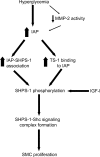
Proposed model by which hyperglycemia enhances IGF-I signaling in SMCs in vivo. Hyperglycemia protects IAP from cleavage by downregulating MMP-2 protease activity that enhances IAP binding to both TS-1 and SHPS-1. This in turn enhances SHPS-1 phosphorylation in response to IGF-I, generating a high-affinity binding site to recruit the SHP-2–c-Src–Shc. Phosphorylation of Shc then leads to activation of downstream signaling pathways including MAPK, which in turn lead to SMC proliferation.

References

    1. Kannel WB, McGee DL: Diabetes and cardiovascular risk factors: the Framingham study. Circulation 59:8–13, 1979 - PubMed
    1. Diabetes Control and Complications Trial Research Group: The effect of intensive treatment of diabetes on the development and progression of long-term complications in insulin-dependent diabetes mellitus. N Engl J Med 329:977–986, 1993 - PubMed
    1. Nathan DM, Lachin J, Cleary P, Orchard T, Brillon DJ, Backlund JY, O'Leary DH, Genuth S: Intensive diabetes therapy and carotid intima-media thickness in type 1 diabetes mellitus. N Engl J Med 348:2294–2303, 2003 - PMC - PubMed
    1. Nathan DM, Cleary PA, Backlund JY, Genuth SM, Lachin JM, Orchard TJ, Raskin P, Zinman B: Intensive diabetes treatment and cardiovascular disease in patients with type 1 diabetes. N Engl J Med 353:2643–2653, 2005 - PMC - PubMed
    1. Ross R: The pathogenesis of atherosclerosis: a perspective for the 1990s. Nature 362:801–809, 1993 - PubMed

Publication types

MeSH terms