. 2008 Oct;57(10):2728-36.
doi: 10.2337/db07-1252. Epub 2008 Jul 15.

Alterations in microRNA expression contribute to fatty acid-induced pancreatic beta-cell dysfunction

Affiliations

Alterations in microRNA expression contribute to fatty acid-induced pancreatic beta-cell dysfunction

Pascal Lovis et al. Diabetes. 2008 Oct.

Abstract

Objective: Visceral obesity and elevated plasma free fatty acids are predisposing factors for type 2 diabetes. Chronic exposure to these lipids is detrimental for pancreatic beta-cells, resulting in reduced insulin content, defective insulin secretion, and apoptosis. We investigated the involvement in this phenomenon of microRNAs (miRNAs), a class of noncoding RNAs regulating gene expression by sequence-specific inhibition of mRNA translation.

Research design and methods: We analyzed miRNA expression in insulin-secreting cell lines or pancreatic islets exposed to palmitate for 3 days and in islets from diabetic db/db mice. We studied the signaling pathways triggering the changes in miRNA expression and determined the impact of the miRNAs affected by palmitate on insulin secretion and apoptosis.

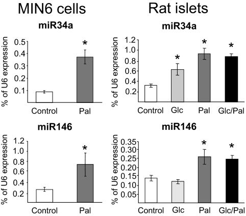
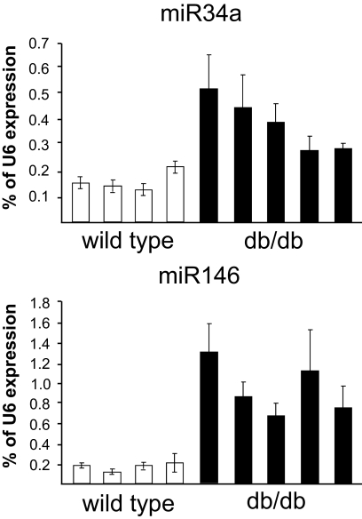
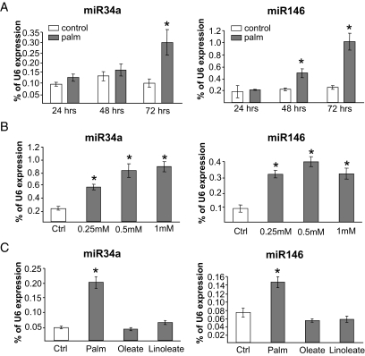
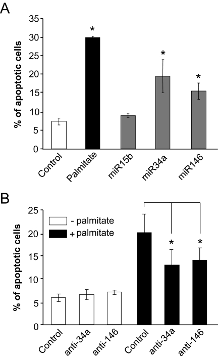
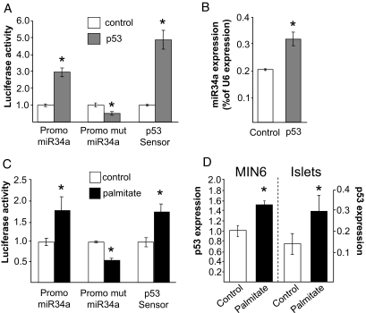
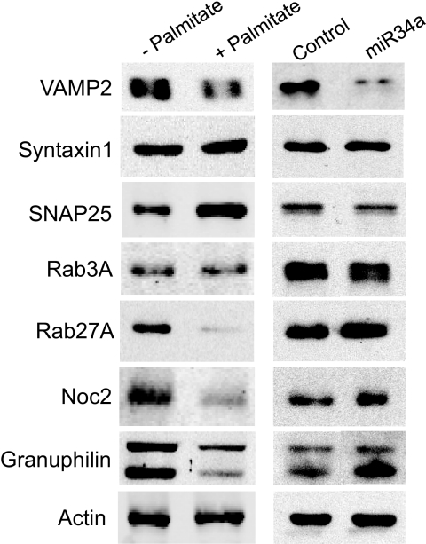
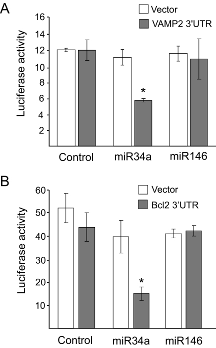
Results: Prolonged exposure of the beta-cell line MIN6B1 and pancreatic islets to palmitate causes a time- and dose-dependent increase of miR34a and miR146. Elevated levels of these miRNAs are also observed in islets of diabetic db/db mice. miR34a rise is linked to activation of p53 and results in sensitization to apoptosis and impaired nutrient-induced secretion. The latter effect is associated with inhibition of the expression of vesicle-associated membrane protein 2, a key player in beta-cell exocytosis. Higher miR146 levels do not affect the capacity to release insulin but contribute to increased apoptosis. Treatment with oligonucleotides that block miR34a or miR146 activity partially protects palmitate-treated cells from apoptosis but is insufficient to restore normal secretion.

Conclusions: Our findings suggest that at least part of the detrimental effects of palmitate on beta-cells is caused by alterations in the level of specific miRNAs.


Figures

FIG. 1.
FIG. 1.
Effect of palmitate on miR34a and miR146 expression. MIN6B1 cells (left) were cultured for 72 h in normal DMEM (25 mmol/l glucose concentration; Control) or in DMEM supplemented with 1 mmol/l palmitate (Pal). Freshly isolated rat pancreatic islets (right) were cultured for 72 h in normal RPMI 1640 (11 mmol/l glucose concentration; Control), in RPMI 1640 containing 25 mmol/l glucose (Glc), in RPMI 1640 containing 1 mmol/l palmitate (Pal), or in RPMI 1640 containing 25 mmol/l glucose and 1 mmol/l palmitate (Glc/Pal). The expression of miR34a and miR146 was assessed by quantitative RT-PCR. The level of U6 measured in parallel in the same samples was used to normalize the data. The asterisks indicate the conditions significantly different (P < 0.05) from control.
FIG. 2.
FIG. 2.
miR34a and miR146 expression is increased in pancreatic islets of db/db mice. Pancreatic islets were isolated from four wild-type mice and five diabetic db/db mice. Blood glucose level was 6.8 ± 0.4 mmol/l in control animals and 21.8 ± 1.4 mmol/l in db/db mice (P < 0.001). RNA extracts from each islet preparation were analyzed by quantitative RT-PCR for the expression of the indicated miRNAs. Data are expressed as percent U6 content measured in the same sample.
FIG. 3.
FIG. 3.
miR34a and miR146 expression is induced in a time- and dose-dependent manner by palmitate but not by unsaturated FFAs. A: MIN6B1 cells were cultured for the indicated time in normal medium (control) or in the presence of 1 mmol/l palmitate (palm). After RNA extraction, miR34a and miR146 levels were determined by quantitative RT-PCR. Data are expressed as percent U6 content measured in the same samples. *Conditions significantly different (P < 0.05, n = 3) from control. B: The cells were incubated for 72 h in normal DMEM (control) or in DMEM containing the indicated concentrations of palmitate. miR34a, miR146, and U6 levels were determined by quantitative RT-PCR. The results are expressed as percent U6 content in each sample. *Conditions significantly different (P < 0.05, n = 3) from control. C: The cells were incubated for 72 h in normal medium (control) or in the presence of 0.5 mmol/l palmitate (Palm), 0.5 mmol/l oleate, or 0.5 mmol/l linoleate. miR34a and miR146 levels were determined by quantitative RT-PCR and normalized to the U6 content. *Conditions significantly different (P < 0.05, n = 3) from control.
FIG. 4.
FIG. 4.
Effect of miR34a and miR146 on hormone secretion. The mouse insulin-secreting cell line MIN6B1 was transiently transfected with a plasmid encoding hGH and with control RNA duplexes (control, palmitate); with RNA duplexes with the mature sequence of miR15b, miR34a, or minR146; or with antisense miR34a (anti-miR34a). The cells were cultured for 3 days in normal DMEM (□ and □) or with DMEM containing 1 mmol/l palmitate (▪). hGH secretion under basal conditions (top) and in the presence of stimulatory concentrations of glucose and cAMP-raising agents (bottom) was measured by ELISA. The figure shows the results of three to five independent experiments performed in triplicate. *Conditions significantly different from controls (P < 0.05).
FIG. 5.
FIG. 5.
Effect of miR34a and miR146 on apoptosis. A: MIN6B1 cells were transfected with a control RNA duplex or RNA duplexes containing miR15b, miR34a, or miR146. The cells were cultured for 3 days in normal DMEM (□ and □) or with DMEM containing 1 mmol/l palmitate (▪). The number of cells displaying apoptotic nuclei was scored and divided by the total number of cells analyzed. Data are means ± SE of three independent experiments. *Conditions significantly different (P < 0.05, n = 3) from control. B: MIN6B1 cells were transfected with control oligonucleotides, anti-miR34a (anti-34a), or anti-miR146 (anti-146). The cells were then cultured for 3 days in normal DMEM (□) or in DMEM supplemented with 1 mmol/l palmitate (▪). The number of cells displaying apoptotic nuclei was scored and divided by the total number of cells analyzed. Data are means ± SE of four to five independent experiments. *Conditions that are significantly different (P < 0.05).
FIG. 6.
FIG. 6.
Identification of the signaling pathway leading to the induction of miR34a. A: MIN6B1 cells were transfected with luciferase reporter plasmids driven by the wild-type miR34a promoter (Promo miR34a), by a miR34a promoter lacking the p53 binding site (Promo mut miR34a), or by p53-responsive elements (p53 Sensor). Each of these luciferase reporters was cotransfected with an empty vector (control) or with a p53-overexpressing plasmid (p53). Luciferase activities were measured 3 days later and normalized to controls. *Conditions significantly different (P < 0.05, n = 3) from controls. B: MIN6B1 cells were transfected with an empty vector or with a plasmid encoding p53. The expression of miR34a was measured by quantitative RT-PCR 2 days later. Data represent the mean of three independent experiments. *P < 0.05 (n = 3). C: MIN6B1 cells were transfected with luciferase reporter plasmids as in A and were then cultured in the absence (control) or in the presence of 1 mmol/l palmitate. Luciferase activities were measured 3 days later and normalized to controls. The results are representative of three independent experiments. *Conditions significantly different (P < 0.05) from controls. D: MIN6B1 cells (left) and isolated rat pancreatic islets (right) were cultured for 3 days in the absence (Control) or in the presence of 1 mmol/l palmitate. p53 mRNA levels were assessed by quantitative RT-PCR and normalized to the level of housekeeping gene GAPDH. Data are means ± SE of three independent experiments. *P < 0.05.
FIG. 7.
FIG. 7.
Comparison of the effects of palmitate and miR34 on the expression of components controlling insulin exocytosis. Left: MIN6B1 cells were incubated for 72 h in the absence (−) or in the presence of 1 mmol/l palmitate (+). Right: MIN6B1 cells were transfected with a control RNA duplex or with an RNA oligonucleotide corresponding to the mature form of miR34a. The expression of the indicated components of the machinery controlling insulin exocytosis was assessed by Western blotting. The figure shows representative blots. Similar results were obtained in at least three independent experiments.
FIG. 8.
FIG. 8.
VAMP2 and BclII are direct targets of miR34a in insulin-secreting cells. A: MIN6B1 cells were cotransfected with a constitutively expressed Firefly luciferase construct; a Renilla luciferase reporter plasmid containing (▪) or lacking (Vector, □) the sequence of the 3′UTR of mouse VAMP2; and a control RNA duplex (control), miR34a, or miR146. Luciferase activities were measured 3 days later. Renilla luciferase activities were divided by the Firefly luciferase activities to correct for differences in transfection efficiencies. *Condition is significantly different (P < 0.05, n = 3) from control. B: MIN6B1 cells were cotransfected with a constitutively expressed Firefly luciferase construct; a Renilla luciferase reporter plasmid containing (▪) or lacking (Vector, □) the putative miR34 recognition sequence in the 3′UTR of BclII; and a control RNA duplex (control), miR34a, or miR146. Luciferase activities were measured 3 days later. Renilla luciferase activities were divided by the Firefly luciferase activities to correct for differences in transfection efficiencies. *Condition that is significantly different (P < 0.05, n = 3) from control.

References

    1. Weir GC, Bonner-Weir S: Five stages of evolving β-cell dysfunction during progression to diabetes. Diabetes 53 (Suppl. 3): S16–S21, 2004 - PubMed
    1. Prentki M, Nolan CJ: Islet beta cell failure in type 2 diabetes. J Clin Invest 116: 1802–1812, 2006 - PMC - PubMed
    1. Newsholme P, Keane D, Welters HJ, Morgan NG: Life and death decisions of the pancreatic beta-cell: the role of fatty acids. Clin Sci (Lond) 112: 27–42, 2007 - PubMed
    1. Bushati N, Cohen SM: microRNA Functions. Annu Rev Cell Dev Biol 23: 175–205, 2007 - PubMed
    1. Bartel DP: MicroRNAs: genomics, biogenesis, mechanism, and function. Cell 116: 281–297, 2004 - PubMed

Publication types

MeSH terms