

Muscle-specific IRS-1 Ser->Ala transgenic mice are protected from fat-induced insulin resistance in skeletal muscle
- PMID: 18633112
- PMCID: PMC2551673
- DOI: 10.2337/db06-0454
Muscle-specific IRS-1 Ser->Ala transgenic mice are protected from fat-induced insulin resistance in skeletal muscle
Abstract
Objective: Insulin resistance in skeletal muscle plays a critical role in the pathogenesis of type 2 diabetes, yet the cellular mechanisms responsible for insulin resistance are poorly understood. In this study, we examine the role of serine phosphorylation of insulin receptor substrate (IRS)-1 in mediating fat-induced insulin resistance in skeletal muscle in vivo.
Research design and methods: To directly assess the role of serine phosphorylation in mediating fat-induced insulin resistance in skeletal muscle, we generated muscle-specific IRS-1 Ser(302), Ser(307), and Ser(612) mutated to alanine (Tg IRS-1 Ser-->Ala) and IRS-1 wild-type (Tg IRS-1 WT) transgenic mice and examined insulin signaling and insulin action in skeletal muscle in vivo.
Results: Tg IRS-1 Ser-->Ala mice were protected from fat-induced insulin resistance, as reflected by lower plasma glucose concentrations during a glucose tolerance test and increased insulin-stimulated muscle glucose uptake during a hyperinsulinemic-euglycemic clamp. In contrast, Tg IRS-1 WT mice exhibited no improvement in glucose tolerance after high-fat feeding. Furthermore, Tg IRS-1 Ser-->Ala mice displayed a significant increase in insulin-stimulated IRS-1-associated phosphatidylinositol 3-kinase activity and Akt phosphorylation in skeletal muscle in vivo compared with WT control littermates.
Conclusions: These data demonstrate that serine phosphorylation of IRS-1 plays an important role in mediating fat-induced insulin resistance in skeletal muscle in vivo.
Figures

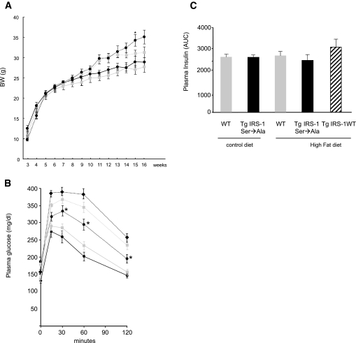
 with solid line, WT control diet; • with solid line, Tg IRS-1 Ser→Ala control diet; with dotted line, WT high-fat diet; • with dotted line, Tg IRS-1 Ser→Ala high-fat diet. B: IPGTT. A total of 1 g/kg of 10% glucose was injected in control diet–fed Tg IRS-1 Ser→Ala mice (• with solid line) and littermate control mice ( with solid line). A total of 1 g/kg of 10% glucose was also injected in high-fat–fed Tg IRS-1 Ser→Ala (• with dotted line), Tg IRS-1 WT (♦ with dotted line), and littermate control ( with dotted line) mice. C: Area under the curve of insulin concentration during IPGTTs. Results are expressed as means ± SE (WT, n = 11; Tg IRS-1 Ser→Ala, n = 16; Tg IRS-1 WT, n = 8). *P < 0.05 vs. WT high-fat diet.
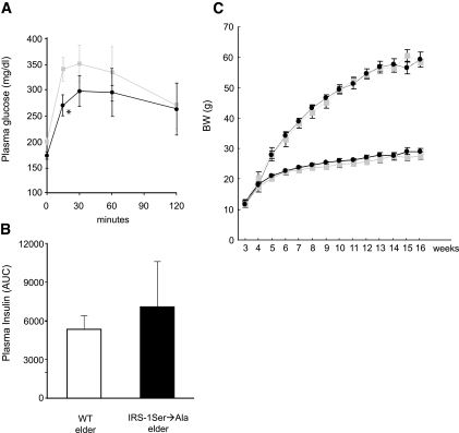 WT elder; ♦, Tg IRS-1 Ser→Ala elder. Results are expressed as means ± SE. C: Generation of Tg IRS-1 Ser→Ala/ob/ob. Growth curves: body weights were measured weekly under control diet until age 16 weeks (• with dotted line) and compared with WT/ob/ob ( with dotted line), Tg IRS-1 Ser→Ala/+/+ (• with solid line), and WT/+/+ ( with solid line); n = 7–10. *P < 0.05 vs. WT littermate mice.
References
-
- Shulman GI, Rothman DL, Jue T, Stein P, DeFronzo RA, Shulman RG: Quantitation of muscle glycogen-synthesis in normal subjects and subjects with non-insulin-dependent diabetes by C-13 nuclear magnetic-resonance spectroscopy. N Engl J Med 322:223–228, 1990 - PubMed
-
- Yu CL, Chen Y, Cline GW, Zhang DY, Zong HH, Wang YL, Bergeron R, Kim JK, Cushman SW, Cooney GJ, Atcheson B, White MF, Kraegen EW, Shulman GI: Mechanism by which fatty acids inhibit insulin activation of insulin receptor substrate-1 (IRS-1)-associated phosphatidylinositol 3-kinase activity in muscle. J Biol Chem 277:50230–50236, 2002 - PubMed
-
- Griffin ME, Marcucci MJ, Cline GW, Bell K, Barucci N, Lee D, Goodyear LJ, Kraegen EW, White MF, Shulman GI: Free fatty acid–induced insulin resistance is associated with activation of protein kinase C θ and alterations in the insulin signaling cascade. Diabetes 48:1270–1274, 1999 - PubMed
Publication types
MeSH terms
Substances
Grants and funding
LinkOut - more resources
Full Text Sources
Molecular Biology Databases
Miscellaneous
