. 2008 Jul 17;454(7202):291-6.
doi: 10.1038/nature07118.

Positive feedback of G1 cyclins ensures coherent cell cycle entry

Affiliations

Positive feedback of G1 cyclins ensures coherent cell cycle entry

Jan M Skotheim et al. Nature. .

Abstract

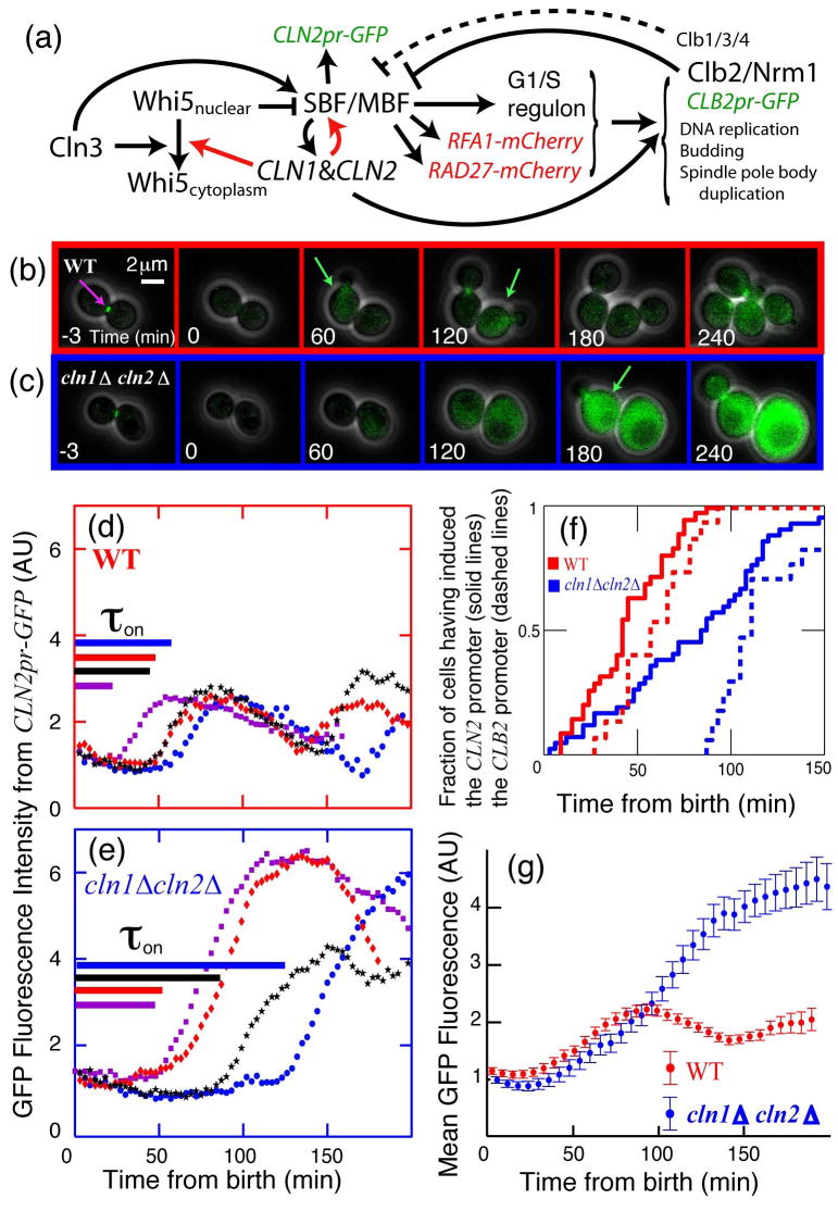
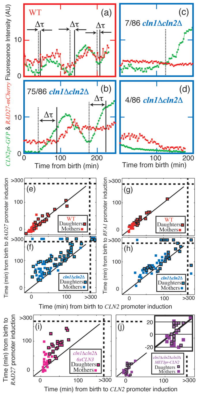
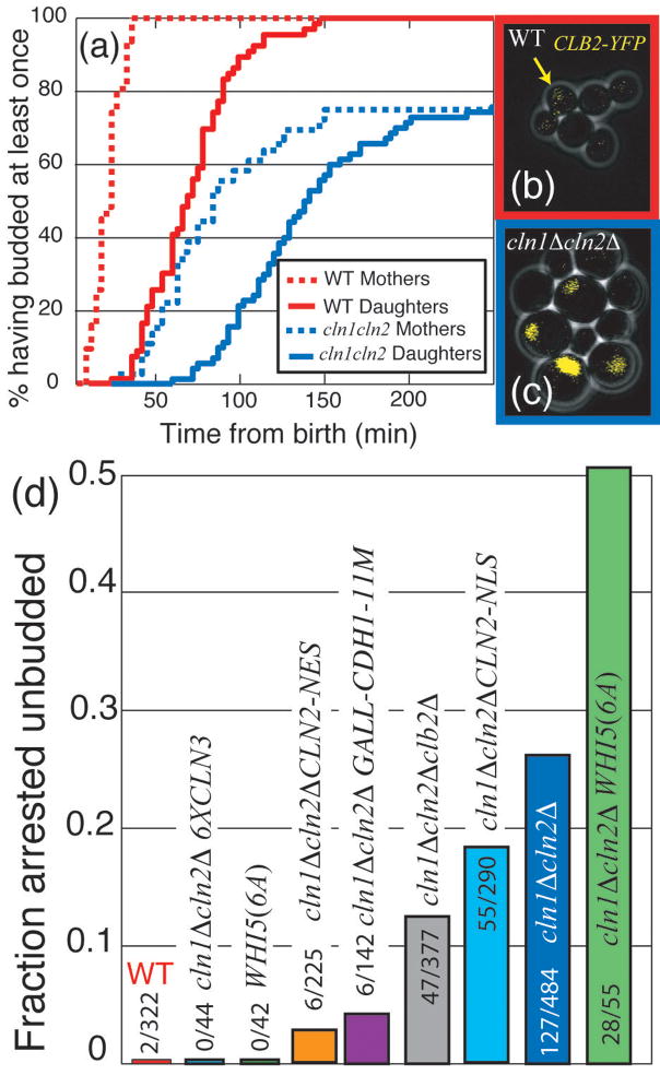
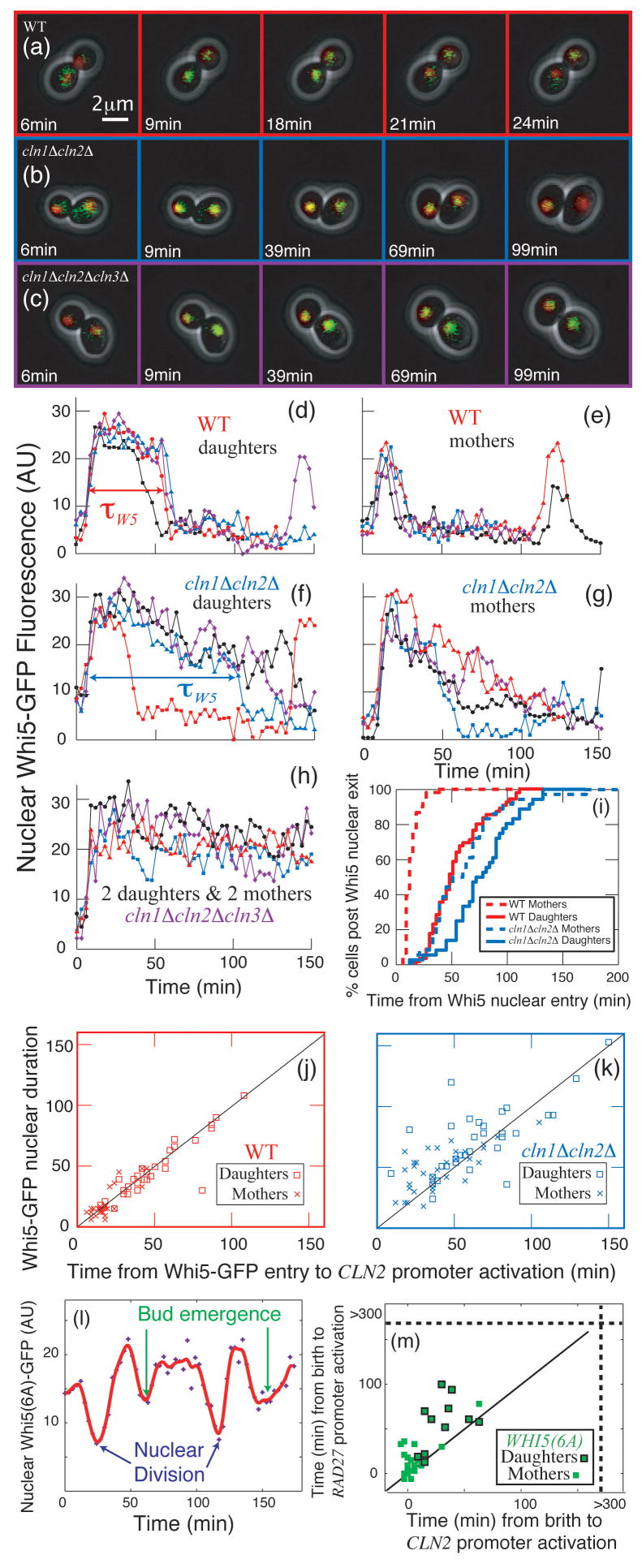
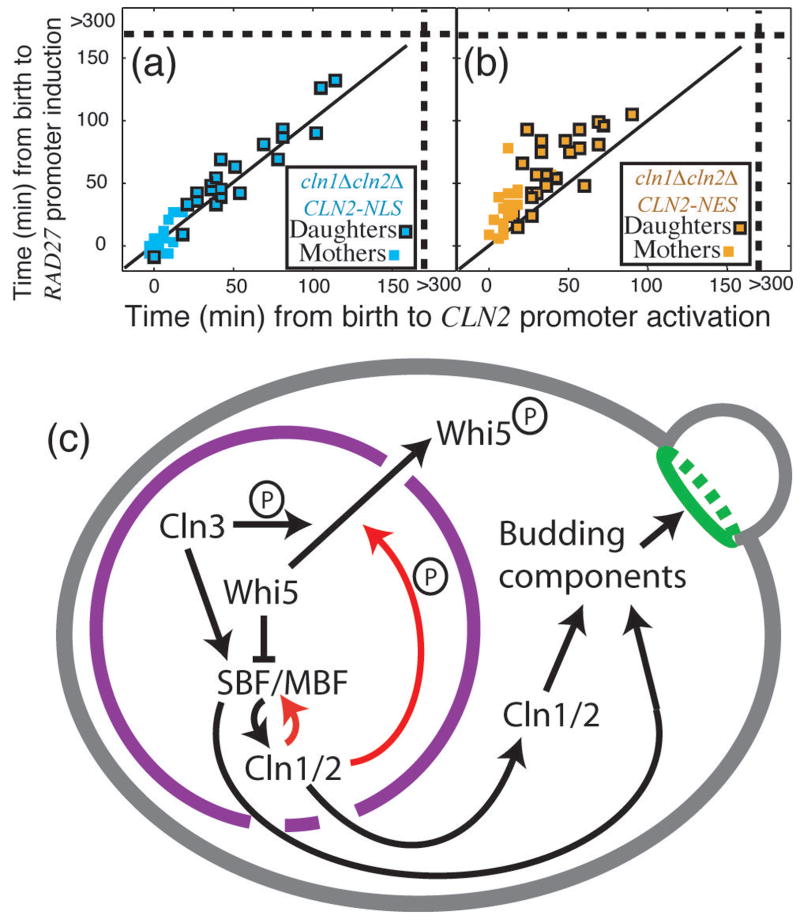
In budding yeast, Saccharomyces cerevisiae, the Start checkpoint integrates multiple internal and external signals into an all-or-none decision to enter the cell cycle. Here we show that Start behaves like a switch due to systems-level feedback in the regulatory network. In contrast to current models proposing a linear cascade of Start activation, transcriptional positive feedback of the G1 cyclins Cln1 and Cln2 induces the near-simultaneous expression of the approximately 200-gene G1/S regulon. Nuclear Cln2 drives coherent regulon expression, whereas cytoplasmic Cln2 drives efficient budding. Cells with the CLN1 and CLN2 genes deleted frequently arrest as unbudded cells, incurring a large fluctuation-induced fitness penalty due to both the lack of cytoplasmic Cln2 and insufficient G1/S regulon expression. Thus, positive-feedback-amplified expression of Cln1 and Cln2 simultaneously drives robust budding and rapid, coherent regulon expression. A similar G1/S regulatory network in mammalian cells, comprised of non-orthologous genes, suggests either conservation of regulatory architecture or convergent evolution.


Figures

Figure 1
Figure 1. Positive feedback drives the Start of the budding yeast cell cycle
(a) Schematic of the Start transition; novel interactions demonstrated in this paper are shown in red. (b,c): Combined phase and fluorescent images for CLN2pr-GFP MYO1-GFP MET3pr-CLN2 cells, either wild type (b) or cln1Δ cln2Δ (c), grown in -Met (inducing) and plated on +Met (repressing) to normalize initial conditions (Fig. S3). Green arrows indicate approximate peak GFP expression from CLN2pr-GFP. (d,e): Single-cell fluorescence intensity for 4 characteristic cells of each genotype; cells are synchronized at birth, marked by the disappearance of a Myo1-GFP ring at the bud-neck (purple arrow in a). Time from birth to CLN2 promoter activation (as defined in Methods),τon, for each individual cell is indicated by length of the corresponding line in (d,e). (f) Cumulative distribution of CLN2pr-GFP induction (solid lines) indicates that Cln1,2-dependent positive feedback contributes substantially to the early expression of CLN2; dashed lines indicate induction of CLB2pr-GFP marking the onset of negative regulation of CLN2. (g) Averaging fluorescence intensity for 87 WT and 83 cln1Δ cln2Δ daughter cells aligned at birth simulates a population study, which would obscure the effect of positive feedback. The results shown are for daughter cells in glucose; changes in cell type or nutrient conditions do not qualitatively influence the results (Table S3).
Figure 2
Figure 2. Cln1,2 drive coherent expression of the SBF/MBF regulon
(a–f): Strains containing both CLN2pr-GFP (green) and RAD27-mCherry (red) were examined (see Supplementary Information for detailed methods); τ marks the computed time between CLN2 and RAD27 inductions. WT (a, e): all cells transcribed both markers synchronously. cln1Δ cln2Δ, (b–d, f): 75 cells transcribed both markers, with variable intervening intervals; 7 cells transcribed CLN2pr-GFP, but not RAD27-mCherry; and 4 cells transcribed neither. Correlation of the initiation of RAD27 and CLN2 transcription in WT (e) and cln1Δ cln2Δ (f); points beyond the dotted lines in (f) represent no transcription within 300 min (movie limit; see also Table S3). (g–h): Substituting RFA1-mCherry for RAD27-mCherry yielded similar results. (i) cln1Δ cln2Δ 6xCLN3 cells and (j) cln1Δ cln2Δ cln3Δ cells expressing CLN2 from a MET3 promoter exhibited incoherent regulon expression compared to WT, although expression of both CLN2pr and RAD27pr were faster than in cln1Δ cln2Δ. P < 10−3 for all comparisons.
Figure 3
Figure 3. Stochastic unbudded arrest in cln1Δ cln2Δ cells, and its modulation by mitotic cyclins, Whi5, Cln3, and Cln2
(a) Cumulative plot of percentage of cells that budded at least once: 26% of cln1Δ cln2Δ cells arrest unbudded. (b,c): cells with integrated CLB2-YFP fusion protein (endogenous promoter); WT (b) and cln1Δ cln2Δ (c). Note high nuclear Clb2 specifically in large unbudded (arrested) cln1Δ cln2Δ cells. (d) Delaying (cln1Δ cln2Δ clb2Δ) or removing (cln1Δ cln2Δ GALL-CDH1-11M) mitotic cyclin accumulation reduced the fraction of arrested cells; addition of 5 copies of CLN3 eliminated this arrest, while the addition of WHI5(6A) exacerbated the arrest. Unbudded arrest was weakly rescued by nuclear Cln2 (CLN2-NLS), and strongly rescued by cytoplasmic Cln2 (CLN2-NES). Unless stated otherwise in text P < 10−3 for all comparisons.
Figure 4
Figure 4. Cln1,2 are required for rapid phosphorylation and inactivation of the rate- limiting repressor Whi5
(a–c): Combined phase and fluorescent images showing Whi5- GFP and Htb2-mCherry (to mark the nucleus) fusion proteins for (a) WT, (b) cln1Δ cln2Δ, and (c) cln1Δ cln2Δ cln3Δ cells. The difference between nuclear and non-nuclear fluorescence intensity was used to quantify nuclear Whi5 by automated image analysis. Compared to WT (d,e), in cln1Δ cln2Δ cells (f,g), Whi5 nuclear exit occurs later and is less sharp. In cln1Δ cln2Δ cln3Δ cells, Whi5 remains nuclear (h). (i) Percent of cells in which Whi5 has left the nucleus (defined as attaining half the maximum level) versus the time from Whi5 nuclear entry. (j,k) Whi5 nuclear exit is tightly correlated with CLN2 promoter activation in WT cells and less correlated in cln1Δ cln2Δ cells (See also Table S3). (l) WHI5(6A)-GFP, lacking 6 out of 12 Cln-dependent phosphorylation sites, reproducibly displayed significant, but slower and incomplete, shuttling out of the nucleus at Start and again at nuclear division. In WHI5(6A) strains containing CLN2pr-GFP and RAD27-mCherry (m), CLN2 and RAD27 induction were incoherent, correlating with the poor nuclear transport of Whi5(6A)-GFP.
Figure 5
Figure 5. Function of nuclear Cln2 and model for Start regulation by positive feedback
Comparison of cln1Δ cln2Δ cells with either a nuclear localized (a) or a nuclear excluded (b) CLN2 allele suggests that nuclear Cln2 is necessary and sufficient for regulon coherence. Strains contained CLN2pr-GFP and RAD27-mCherry. (c) Model for regulon activation and bud emergence: red lines indicate

Comment in

References

    1. Simchen G, Pinon R, Salts Y. Sporulation in Saccharomyces cerevisiae: premeiotic DNA synthesis, readiness and commitment. Exp Cell Res. 1972;75:207–18. - PubMed
    1. Nachman I, Regev A, Ramanathan S. Dissecting timing variability in yeast meiosis. Cell. 2007;131:544–56. - PubMed
    1. Shenhar G, Kassir Y. A positive regulator of mitosis, Sok2, functions as a negative regulator of meiosis in Saccharomyces cerevisiae. Mol Cell Biol. 2001;21:1603–12. - PMC - PubMed
    1. Ferrell JE, Jr, Machleder EM. The biochemical basis of an all-or-none cell fate switch in Xenopus oocytes. Science. 1998;280:895–8. - PubMed
    1. Xiong W, Ferrell JE., Jr A positive-feedback-based bistable ‘memory module’ that governs a cell fate decision. Nature. 2003;426:460–5. - PubMed

Publication types

MeSH terms