. 2009 Jan;129(1):79-88.
doi: 10.1038/jid.2008.194. Epub 2008 Jul 17.

Psoriasis is characterized by accumulation of immunostimulatory and Th1/Th17 cell-polarizing myeloid dendritic cells

Affiliations

Psoriasis is characterized by accumulation of immunostimulatory and Th1/Th17 cell-polarizing myeloid dendritic cells

Lisa C Zaba et al. J Invest Dermatol. 2009 Jan.

Abstract

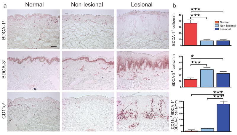
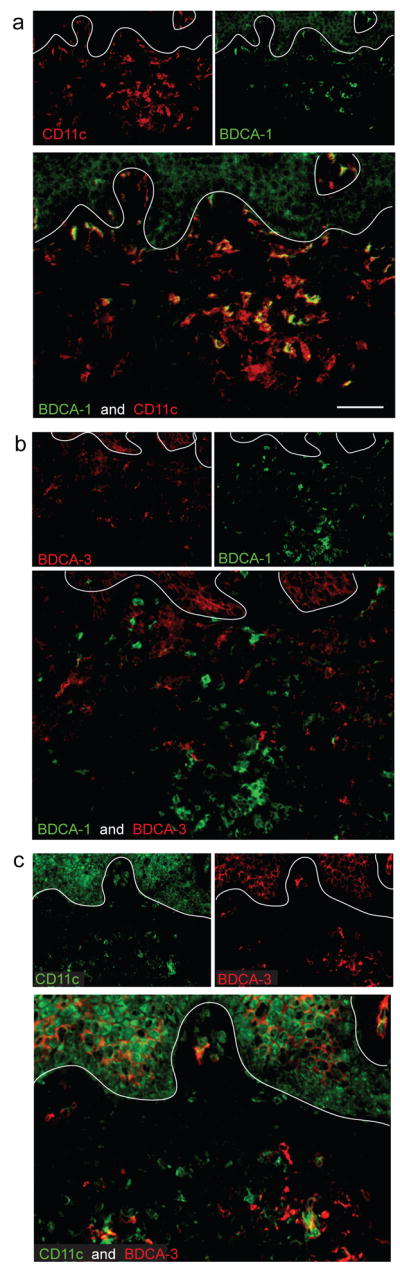
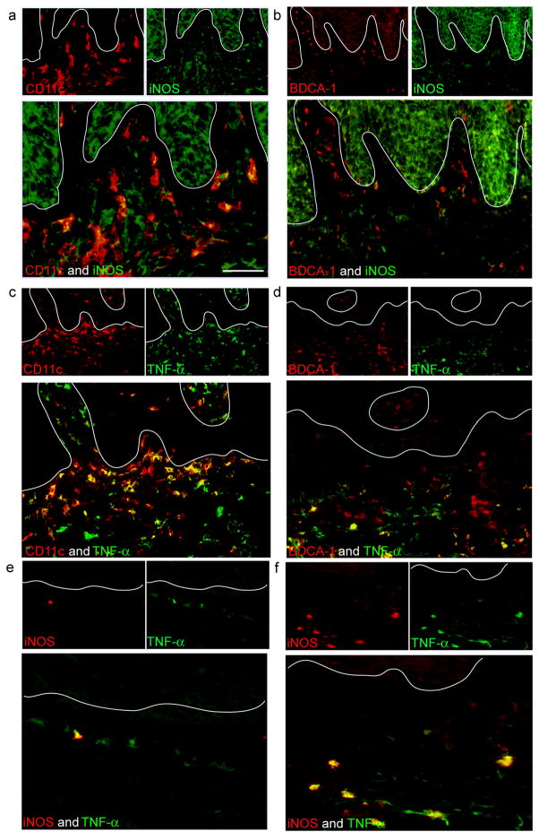
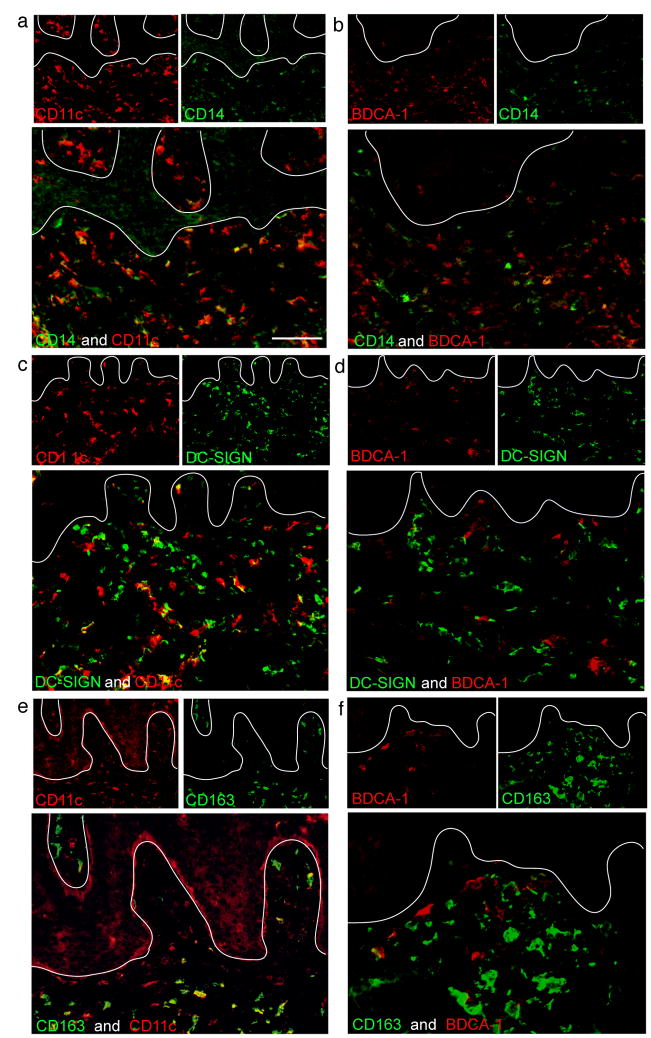
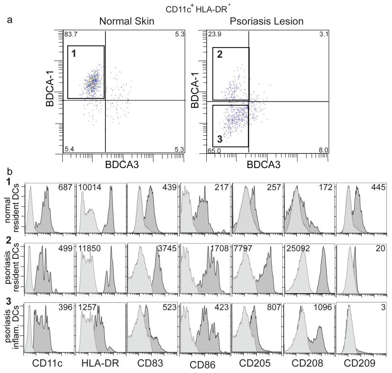
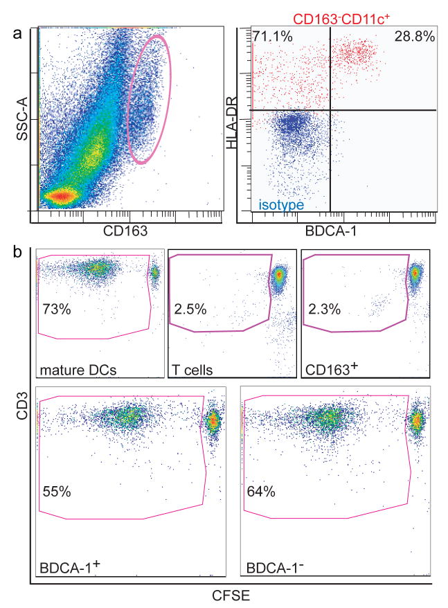
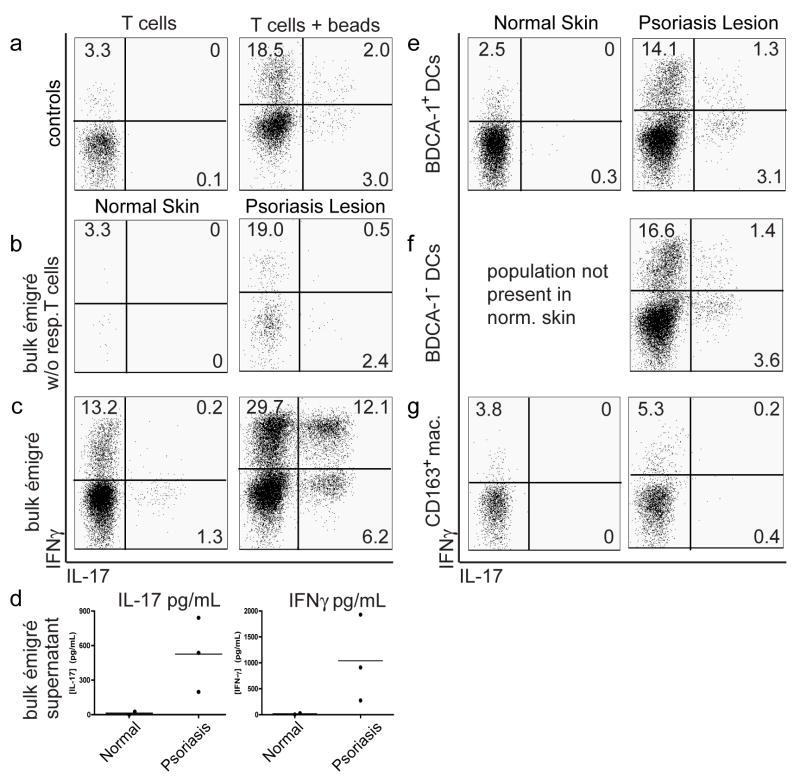
Myeloid dermal dendritic cells (DCs) accumulate in chronically inflamed tissues such as psoriasis. The importance of these cells for psoriasis pathogenesis is suggested by comparative T-cell and DC-cell counts, where DCs outnumber T cells. We have previously identified CD11c(+)-blood dendritic cell antigen (BDCA)-1(+) cells as the main resident dermal DC population found in normal skin. We now show that psoriatic lesional skin has two populations of dermal DCs: (1) CD11c(+)BDCA-1(+) cells, which are phenotypically similar to those contained in normal skin and (2) CD11c(+)BDCA-1(-) cells, which are phenotypically immature and produce inflammatory cytokines. Although BDCA-1(+) DCs are not increased in number in psoriatic lesional skin compared with normal skin, BDCA-1(-) DCs are increased 30-fold. For functional studies, we FACS-sorted psoriatic dermal single-cell suspensions to isolate these two cutaneous DC populations, and cultured them as stimulators in an allogeneic mixed leukocyte reaction. Both BDCA-1(+) and BDCA-1(-) myeloid dermal DC populations induced T-cell proliferation, and polarized T cells to become T helper 1 (Th1) and T helper 17 (Th17) cells. In addition, psoriatic dermal DCs induced a population of activated T cells that simultaneously produced IL-17 and IFN-gamma, which was not induced by normal skin dermal DCs. As psoriasis is believed to be a mixed Th17/Th1 disease, it is possible that induction of these IL-17(+)IFN-gamma(+) cells is pathogenic. These cytokines, the T cells that produce them, and the inducing inflammatory DCs may all be important new therapeutic targets in psoriasis.


Conflict of interest statement

Conflict of Interest

The authors do not have any financial interest related to this work.

Figures

Figure 1
Figure 1. CD11c+ dermal DCs are the major DC population accumulating in psoriasis lesional skin
(a) Representative immunohistochemistry of BDCA-1+ cells, BDCA-3+ cells, and CD11c+ cells in normal, non-lesional and lesional psoriatic skin. (b) Quantification of myeloid DCs per mm skin stained by immunohistochemistry of normal skin (red boxes; n=20), non-lesional skin (light blue boxes; n=20), and matched psoriatic lesional skin (dark blue boxes; n=20). CD11c+BDCA-1BDCA-3 cell numbers were calculated by subtracting BDCA-1 and BDCA-3 cell counts from CD11c cell counts. Error bars indicate SEM. (*) p<0.05, (***) p<0.001. Size bar = 100μm.
Figure 2
Figure 2. Most CD11c+ myeloid DCs are BDCA-1 in psoriasis lesional skin
(a) The majority of CD11c+ cells in psoriatic dermis were BDCA-1, while a small subset of CD11c+ cells co-expressed BDCA-1+. (b) BDCA-1 and BDCA-3 identified separate myeloid DC populations in the dermis. (c) Most BDCA-3+ cells co-expressed CD11c, and some BDCA-3 staining was observed on blood vessels. In all IF figures, single stained controls are above the merged image, white line denotes dermo-epidermal junction, dermal collagen fibers gave green autofluorescence, and antibodies conjugated with a fluorochrome often gave background epidermal fluorescence. Size bar = 100μm.
Figure 3
Figure 3. CD11c+BDCA-1 DCs contain the Tip-DC population
(a) Most CD11c+ dermal DCs in psoriatic lesional skin co-expressed iNOS compared to (b) BDCA-1+ cells. (c) Most CD11c+ dermal DCs in lesional psoriatic skin co-expressed TNF compared to (d) BDCA-1+ cells. Approximately 25% of BDCA-1+ cells co-expressed TNF. (e,f) iNOS and TNF were co-expressed in the same cell, identifying Tip-DCs in situ. Few Tip-DCs were observed in (e) psoriatic non-lesional skin compared to (f) lesional skin. Size bar = 100μm.
Figure 4
Figure 4. CD11c+BDCA-1 inflammatory dermal DCs express CD14 and DC-SIGN
(a) A subset of CD11c+ cells in the reticular dermis expressed low level CD14. (b) Few BDCA-1+ dermal DCs co-expressed CD14, and (c) 80% of CD11c+ cells were DC-SIGN+. This was not an exclusive myeloid DC marker as some DC-SIGN+ cells were not CD11c+. (d) BDCA-1+ cells did not co-express DC-SIGN. (e) CD163+ macrophages showed some CD11c co-expression. (f) BDCA-1+ cells did not express CD163. Size bar = 100μm.
Figure 5
Figure 5. CD11c+BDCA-1+ cells are phenotypically mature dermal DCs in psoriasis
DC-LAMP and DEC-205 identified mature DCs, often located in dermal aggregates. (a) DC-LAMP+ cells were CD11c+, and (b) BDCA-1+. (c) DEC-205+ cells were (c) CD11c+, and (d) BDCA-1+. There were many CD11c+ cells that were not positive for these two markers in the psoriatic dermis. Size bar = 100μm.
Figure 6
Figure 6. Psoriatic inflammatory dermal DCs (CD11c+ BDCA-1) are less mature than resident BDCA-1+ dermal DCs
Flow cytometric analysis of single cell suspensions of dermal émigrés from normal dermis or psoriatic dermis (n=3 for each). (a) Large cells gated on CD11c+ HLA-DRhi. In normal dermis, most myeloid DCs were BDCA-1+(box 1). In psoriatic dermis, myeloid DCs were either BDCA-1+ (box 2), or BDCA-1 (box 3). Percent of myeloid cells indicated for each quadrant. (b) Histograms in each row were gated on boxes (1–3) as identified above in Figure 6a. Dark grey histogram represents antigen expression, light grey is isotype. MFI is indicated in the upper right or upper left corner of each histogram. Resident BDCA-1+ myeloid DCs from normal and psoriatic dermis were phenotypically similar, while the additional population of BDCA-1 DCs in psoriasis showed lower HLA-DR, and were smaller cells. Psoriatic BDCA-1+ DCs demonstrated the highest expression of DC-LAMP/CD208, and DEC-205/CD205.
Figure 7
Figure 7. Both psoriatic CD11c+BDCA-1+ resident DCs and CD11c+BDCA-1 inflammatory DCs were immunostimulatory in an allo-MLR
Single-cell suspensions of psoriatic dermal émigrés were sorted into (a, left) CD163hi or (a, right), CD3CD45+HLA-DRhiCD11chiBDCA-1+ or CD3CD45+HLA-DRhiCD11chiBDCA-1 compared to isotype (red). (b) Gate contains CD3+ proliferating T cells with left-shifted CFSE as cells proliferated. Positive control (monocyte-derived mature DCs) on day 8-post sort at 1:50 stimulator to responder ratio. Background proliferation of T cells alone (2.5%), CD163hi cells did not stimulate above background. BDCA-1+ and BDCA-1 cells stimulated T cell proliferation similarly (both >55%). Representative graphs from 3 experiments.
Figure 8
Figure 8. Psoriatic dermal DCs induce IL-17/IFNγ producing T cells
Allogeneic T cell responders were mixed with FACS-sorted stimulator populations described in Figure 6, or with bulk émigrés from either normal or psoriatic dermal skin, at 1:10 ratio for 9 days. Intracellular cytokine expression of T cells (live CD3+CD4+CD8 cells) measured by flow cytometry (a,b,c,e,f,g) or protein supernatant (d). (b,c,e,f,g) Comparison of T cell polarization of normal dermal DCs (left panels) with psoriatic dermal DCs (right panels). (a) Controls: T cells alone (left panel) demonstrated baseline intracellular cytokine expression and T cells + beads (right panel) measured intracellular cytokine expressionfollowing CD3/CD28 bead stimulation. (b) Bulk émigrés w/o responder T cells measured baseline T cell cytokine production in single cell suspensions. Other stimulator populations mixed with responder T cells were (c) unsorted bulk émigrés, (e) sorted HLA-DR+CD11c+BDCA-1+ dermal DCs, (f) sorted HLA-DR+CD11c+BDCA-1 dermal DCs, or (g) sorted CD163+ macrophages. Psoriatic bulk dermal DCs induced a population of IL-17/IFNγ producing T cells, as did BDCA-1+ and BDCA-1 DCs (although to a lesser degree, possibly due to sorting). Quadrant percentages are given, representative graphs from 3 experiments. (d) Supernatant of the same cultures was collected for analysis of cytokine protein expression Bulk psoriatic dermal émigrés mixed with allogeneic T cells induced IL-17 and IFN-γ production, while normal dermal DCs did not.

References

    1. Abrams JR, Lebwohl MG, Guzzo CA, Jegasothy BV, Goldfarb MT, Goffe BS, et al. CTLA4Ig-mediated blockade of T-cell costimulation in patients with psoriasis vulgaris. J Clin Invest. 1999;103:1243–1252. - PMC - PubMed
    1. Barral DC, Brenner MB. CD1 antigen presentation: how it works. Nat Rev Immunol. 2007;7:929–941. - PubMed
    1. Blauvelt A. New concepts in the pathogenesis and treatment of psoriasis: key roles for IL-23, IL-17A and TGF-beta1. Expert Rev Dermatol. 2007;2:69–78.
    1. Bogunovic M, Ginhoux F, Wagers A, Loubeau M, Isola LM, Lubrano L, et al. Identification of a radio-resistant and cycling dermal dendritic cell population in mice and men. J Exp Med. 2006;203:2627–2638. - PMC - PubMed
    1. Dhodapkar KM, Banerjee D, Connolly J, Kukreja A, Matayeva E, Veri MC, et al. Selective blockade of the inhibitory Fcgamma receptor (FcgammaRIIB) in human dendritic cells and monocytes induces a type I interferon response program. J Exp Med. 2007;204:1359–1369. - PMC - PubMed

Publication types