. 2008 Sep 12;283(37):25660-25670.
doi: 10.1074/jbc.M804479200. Epub 2008 Jul 17.

Virus infection triggers SUMOylation of IRF3 and IRF7, leading to the negative regulation of type I interferon gene expression

Affiliations

Virus infection triggers SUMOylation of IRF3 and IRF7, leading to the negative regulation of type I interferon gene expression

Toru Kubota et al. J Biol Chem. .

Abstract

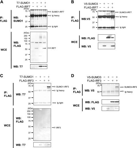
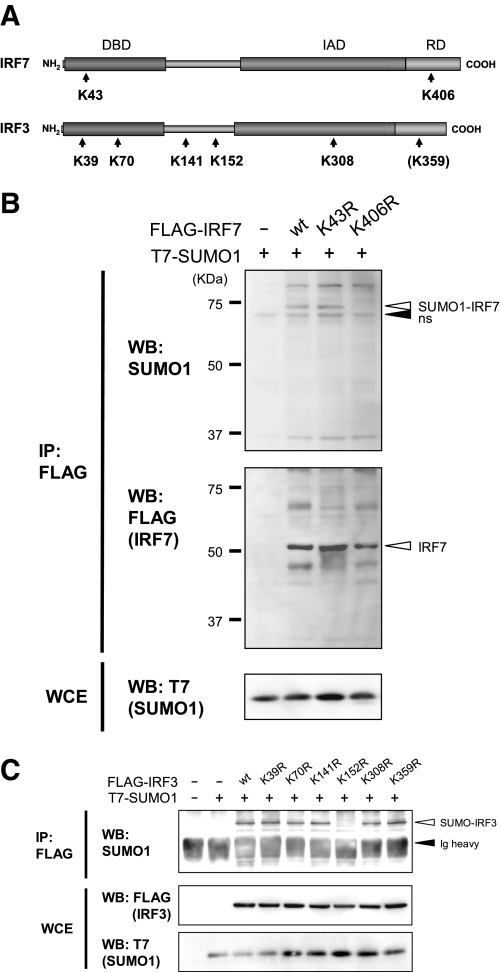
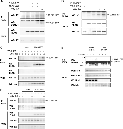
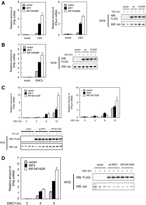
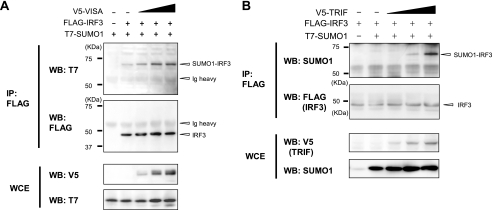
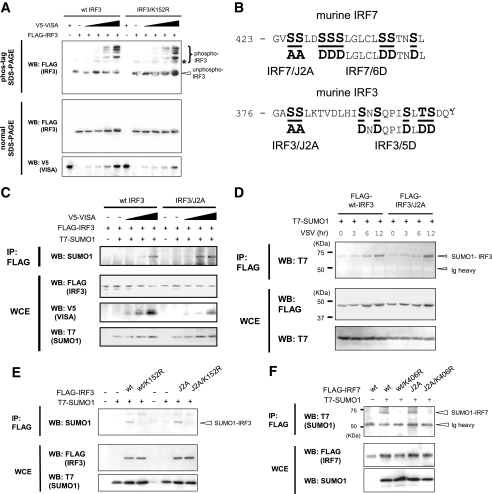
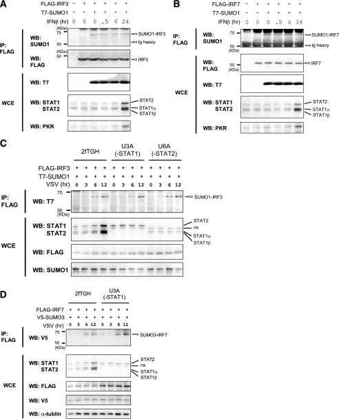
Viral infection activates Toll-like receptor and RIG-I (retinoic acid-inducible gene I) signaling pathways, leading to phosphorylation of IRF3 (interferon regulatory factor 3) and IRF7 and stimulation of type I interferon (IFN) transcription, a process important for innate immunity. We show that upon vesicular stomatitis virus infection, IRF3 and IRF7 are modified not only by phosphorylation but by the small ubiquitin-related modifiers SUMO1, SUMO2, and SUMO3. SUMOylation of IRF3 and IRF7 was dependent on the activation of Toll-like receptor and RIG-I pathways but not on the IFN-stimulated pathway. However, SUMOylation of IRF3 and IRF7 was not dependent on their phosphorylation, and vice versa. We identified Lys(152) of IRF3 and Lys(406) of IRF7 to be their sole small ubiquitin-related modifier (SUMO) conjugation site. IRF3 and IRF7 mutants defective in SUMOylation led to higher levels of IFN mRNA induction after viral infection, relative to the wild type IRFs, indicating a negative role for SUMOylation in IFN transcription. Together, SUMO modification is an integral part of IRF3 and IRF7 activity that contributes to postactivation attenuation of IFN production.


Figures

FIGURE 1.
FIGURE 1.
SUMO conjugation of IRF7 and IRF3. 293T cells were transfected with plasmids for FLAG-IRF7 (A and B) or FLAG-IRF3 (C and D) along with T7-SUMO1 (A and C) or V5-SUMO3 (B and D). Whole cell extracts (WCE) were immunoprecipitated (IP) with anti-FLAG antibody-agarose beads, and SUMO-conjugated proteins were detected with anti-T7, anti-V5, or anti-SUMO1 antibody in Western blot (WB) (top). Expression of transfected proteins was verified by Western blot analysis of whole cell extracts using the indicated antibody (bottom).
FIGURE 2.
FIGURE 2.
Identification of SUMO conjugation sites in IRF3 and IRF7. A, a schematic presentation of murine IRF7 and IRF3. The positions of lysine residues that conform to the putative SUMO conjugation sites are indicated by arrows. IRF3 and IRF7 mutants contained an Arg substitution at each of the Lys residues. Lys359 corresponds to Lys406 of IRF7 (in parenthesis). DBD, DNA binding domain; IAD, IRF association domain; RD, regulatory domain. B and C, 293T cells were transfected with mutant FLAG-IRF7 (B) or mutant FLAG-IRF3 (C) along with T7-SUMO1 for 48 h. Extracts were immunoprecipitated (IP) with anti-FLAG-agarose beads, and precipitates were tested by Western blotting (WB) with the indicated antibodies (top). ns, nonspecific band. Whole cell extracts were tested for expression of transfected proteins.
FIGURE 3.
FIGURE 3.
Increased SUMOylation of IRF3 and IRF7 following virus infection. A-D, 293T cells were transfected with FLAG-IRF7 (A and B) or FLAG-IRF3 (C and D) with or without T7-SUMO1 (A and C) or V5-SUMO3 (B and D) for 12 h. Cells were infected with VSV at an MOI of 1 for the indicated periods. Extracts were immunoprecipitated (IP) with anti-FLAG-agarose beads and tested for SUMO conjugation by Western blotting (WB) with the indicated antibodies. E, NIH3T3 cells expressing control shRNA or Ubc9 shRNA were infected with VSV at an MOI of 1, and cells were allowed to proceed for the indicated periods. Extracts were immunoprecipitated with anti-IRF3 antibody and tested in Western blotting with anti-SUMO1 antibody (top). Expression of endogenous IRF3, SUMO1, Ubc9, and tubulin α (tub) was tested by Western blot analysis of whole cell extracts (WCE).
FIGURE 4.
FIGURE 4.
Mutation of the SUMOylation site in IRF7 and IRF3 increases type I IFN production. NIH3T3 cells were transfected with WT IRF7 or IRF7/K406R (A and B) or with WT IRF3 or IRF3/K152R (C and D) for 12 h. For A and B, cells were infected with VSV or EMCV at an MOI of 1 for 9 h prior to harvest. For C and D, cells were harvested at varying periods after infection. The amounts of IFNβ or IFNα4 mRNAs were quantified by quantitative reverse transcription-PCR by normalizing with hypoxanthine guanine phosphoribosyltransferase mRNA. The values represent the average of three samples ± S.D. Comparable expression of WT IRF3/IRF7 and the mutants was verified by Western blotting (WB) of whole cell extracts (WCE) (right and bottom).
FIGURE 5.
FIGURE 5.
Induction of IRF3 and IRF7 SUMOylation by VISA and TRIF. 293T cells were transfected with FLAG-IRF3 along with increasing amounts of V5-VISA (A) or V5-TRIF (B) plus a constant amount of T7-SUMO1 for 12 h. SUMOylated IRF3 was detected by immunoprecipitation (IP) with anti-FLAG-agarose, followed by Western blot analysis (WB) with the indicated antibodies. WCE, whole cell extract.
FIGURE 6.
FIGURE 6.
Independence of SUMOylation of IRF3 and IRF7 from the phosphorylation. A, 293T cells were transfected with WT FLAG-IRF3 or FLAG-IRF3/K152R along with increasing doses of V5-VISA for 24 h. Whole cell extracts (WCE) were tested in the phos-tag SDS-PAGE (top panel) or normal SDS-PAGE (lower panels) by Western blot analysis (WB). B, the serine/threonine cluster phosphorylation sites involved in the activation of type I IFN genes. Phosphorylated Ser and Thr are in boldface type and underlined. Alanine or aspartic acid substitutions were placed in the indicated residues to create IRF7/J2A, IRF7/6D, IRF3/J2A, and IRF3/5D. C, cells were transfected with WT IRF3 or IRF3/J2A along with T7-SUMO1 and increasing amounts of V5-VISA. SUMOylated IRF3 was detected by immunoprecipitation (IP) and Western blotting as in Fig. 1. D, cells transfected with WT IRF3 or IRF3/J2A along with T7-SUMO1 for 12 h were infected with VSV at an MOI of 1. Cells were harvested at the indicated period, and SUMOylated IRF3 was detected by immunoprecipitation (IP) and Western blot analysis as above. E and F, cells were transfected with WT IRF3, WT IRF7, or mutants with indicated substitutions along with T7-SUMO1. SUMOylated IRF3 or IRF7 was detected by immunoprecipitation and Western blot analysis as above.
FIGURE 7.
FIGURE 7.
The lack of requirement of IFN signaling for SUMOylation of IRF7 and IRF3. A and B, 293T cells were transfected with FLAG-IRF3 (A) or FLAG-IRF7 (B) along with T7-SUMO1 for 12 h and treated with 1000 units/ml human IFNβ for the indicated periods. SUMOylated IRF3 and IRF7 were detected by immunoprecipitation (IP), followed by Western blot (WB) (top). Whole cell extracts (WCE) were analyzed for the IFN-stimulated expression of STAT1, STAT2, and PKR by Western blotting (bottom). C and D, the parental 2fTGH, STAT1-deficient U3A, and STAT2-deficient U6A cells were transfected with FLAG-IRF3 and FLAG-IRF7, along with T7-SUMO1 or V5-SUMO3, and infected with VSV at an MOI of 1 for the indicated periods. SUMOylated IRF3 and IRF7 were detected by immunoprecipitation, followed by Western blot analysis (top). Whole cell extracts (WCE) were analyzed for expression STAT1α, STAT1β, and STAT2 as above. ns, nonspecific band.

References

    1. Akira, S., Uematsu, S., and Takeuchi, O. (2006) Cell 124 783-801 - PubMed
    1. Andrejeva, J., Childs, K. S., Young, D. F., Carlos, T. S., Stock, N., Good-bourn, S., and Randall, R. E. (2004) Proc. Natl. Acad. Sci. U. S. A. 101 17264-17269 - PMC - PubMed
    1. Kawai, T., and Akira, S. (2006) Nat. Immunol. 7 131-137 - PubMed
    1. Takeda, K., and Akira, S. (2005) Int. Immunol. 17 1-14 - PubMed
    1. Yoneyama, M., Kikuchi, M., Natsukawa, T., Shinobu, N., Imaizumi, T., Miyagishi, M., Taira, K., Akira, S., and Fujita, T. (2004) Nat. Immunol. 5 730-737 - PubMed

Publication types

LinkOut - more resources