. 2008 Sep 19;283(38):26217-27.
doi: 10.1074/jbc.M803287200. Epub 2008 Jul 17.

Early mitochondrial dysfunction in long-lived Mclk1+/- mice

Affiliations

Early mitochondrial dysfunction in long-lived Mclk1+/- mice

Jérôme Lapointe et al. J Biol Chem. .

Abstract

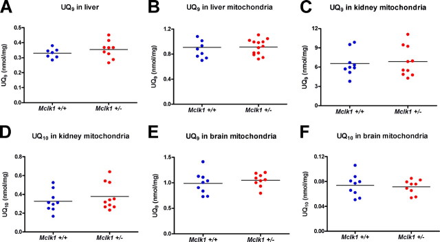
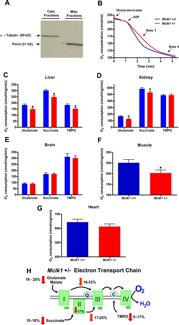
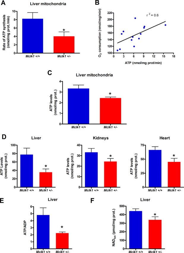
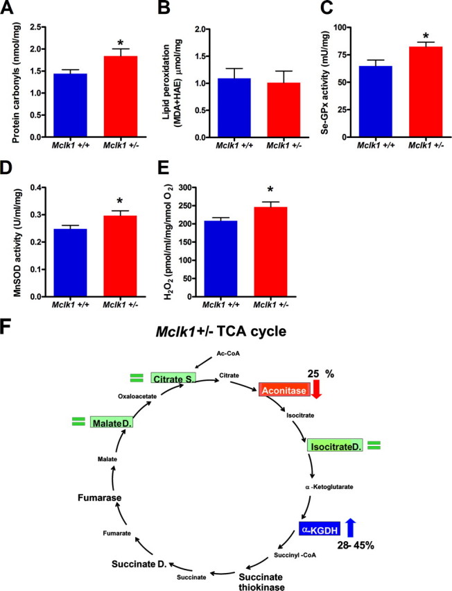
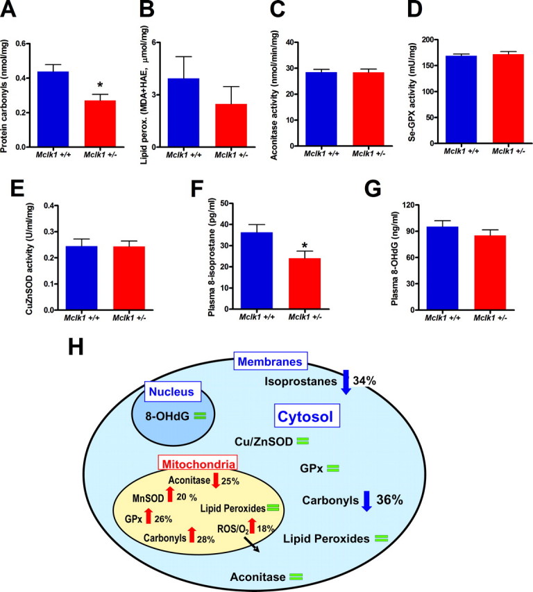
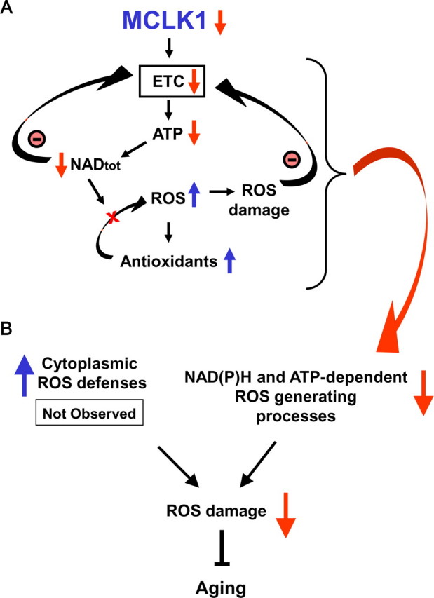
Reduced activity of CLK-1/MCLK1 (also known as COQ7), a mitochondrial enzyme that is necessary for ubiquinone biosynthesis, prolongs the lifespan of nematodes and mice by a mechanism that is distinct from that of the insulin signaling pathway. Here we show that 2-fold reduction of MCLK1 expression in mice reveals an additional function for the protein, as this level of reduction does not affect ubiquinone levels yet affects mitochondrial function substantially. Indeed, we observe that the phenotype of young Mclk1(+/-) mutants includes a severe reduction of mitochondrial electron transport, ATP synthesis, and total nicotinamide adenine dinucleotide (NAD(tot)) pool size as well as an alteration in the activity of key enzymes of the tricarboxylic acid cycle. Surprisingly, we also find that Mclk1 heterozygosity leads to a dramatic increase in mitochondrial oxidative stress by a variety of measures. Furthermore, we find that the mitochondrial dysfunction is accompanied by a decrease in oxidative damage to cytosolic proteins as well as by a decrease in plasma isoprostanes, a systemic biomarker of oxidative stress and aging. We propose a mechanism for the conjunction of low ATP levels, high mitochondrial oxidative stress, and low non-mitochondrial oxidative damage in a long-lived mutant. Our model helps to clarify the relationship between energy metabolism and the aging process and suggests the need for a reformulation of the mitochondrial oxidative stress theory of aging.


Figures

FIGURE 1.
FIGURE 1.
Ubiquinone levels are normal in Mclk1+/- mice. A, quantification of UQ9 levels in whole liver homogenates from 3-month-old Mclk1+/+ and Mclk1+/- Balb/c males was performed by HPLC analysis. B, UQ9 levels in isolated liver mitochondria from 3-month-old Mclk1+/+ and Mclk1+/- males. UQ10 is barely detectable in liver (not shown). C-F, UQ9 and UQ10 levels in kidney and brain mitochondria from 3-month-old Mclk1+/+ and Mclk1+/- Balb/c males. Each dot in the graphs represents an individual mouse.
FIGURE 2.
FIGURE 2.
Reduction of MCLK1 levels alters mitochondrial function in young Mclk1+/- mice. A, purity of cytosolic and mitochondrial fractions was assessed by Western blotting with antisera against cytosolic (Cyto.) α-tubulin and mitochondrial (Mito.) porin. B, typical trace graph representing oxygen consumption by Mclk1+/+ and Mclk1+/- liver mitochondria after glutamate/malate addition in respiratory reaction medium. Oxygen consumption levels of isolated liver (C), kidney (D), and brain (E) mitochondria from young (3 months) males were measured with glutamate/malate, succinate, and TMPD as specific respiratory substrates. Results with Mclk1+/+ are represented with blue bars, and those with Mclk1+/- are represented with red bars. For muscle (F) and heart (G) mitochondria, respiration was measured with glutamate/malate only. Each bar in the graphs represents the mean ± S.E. of 12 Mclk1+/+ and 10 Mclk1+/- animals. The asterisk denotes statistical significance of the difference between Mclk1+/+ and Mclk1+/- animals, p < 0.05. H, schematic representation of the electron transport chain in affected tissues of Mclk1+/- mice. The rates of oxygen consumption by isolated mitochondria are decreased with complex I-, complex II-, and cytochrome c/complex IV-specific substrates (red arrows). The rates of electron transport between complexes I and III as well as between complexes II and III are reduced, as shown in orange arrows. Complex II activity is down-regulated (red arrow), whereas the activities of the other complexes are unaffected (green bars). See Table 1 for complete numerical results.
FIGURE 3.
FIGURE 3.
Mitochondrial ATP synthesis and cellular ATP and NAD levels are decreased in Mclk1+/- mutants. A, rates of ATP production by isolated mitochondria from liver of young (3 months) Balb/c males of both genotypes. B, correlation between the rates of ATP production and oxygen consumption by mitochondria isolated from individual animals. C, ATP levels in freshly isolated liver mitochondria from Mclk1+/+ and Mclk1+/- animals. D, cellular ATP levels measured in liver (*, p < 0.03), kidney and heart (*, p < 0.03) samples of both genotypes. E, ATP/ADP ratio measured in liver. F, total cellular NAD (NAD+, NADH) levels measured in liver. Each bar in the graphs represents the mean ± S.E. of 6 Mclk1+/+ and 7 Mclk1+/- animals. The asterisk denotes statistical significance of the difference between Mclk1+/+ and Mclk1+/- animals unless stated otherwise p < 0.05.
FIGURE 4.
FIGURE 4.
Young Mclk1+/- mice have higher mitochondrial oxidative stress and altered TCA cycle. Mitochondria of young Mclk1+/- mice display high oxidative stress as revealed by accumulation of protein carbonyls (A), higher levels, but not significantly, of lipid peroxidation (B), up-regulation of the major enzymatic antioxidant defenses, such as Se-GPx (C) and manganese-dependent superoxide dismutase (MnSOD; D), and increased ROS production per molecule of reduced O2 from isolated mitochondria (E). Each bar in the graphs represents the mean ± S.E. of 12 Mclk1+/+ and 10 Mclk1+/- animals. The asterisk denotes statistical significance of the difference between Mclk1+/+ and Mclk1+/- animals, p < 0.05. F, schematic representation of the effects of Mclk1 heterozygosity on key TCA cycle enzymes. Aconitase (in red) is partially inactivated, and α-ketoglutarate dehydrogenase (α-KGDH, in blue) is up-regulated in Mclk1+/- animals. Unaffected enzymes are showed in green.
FIGURE 5.
FIGURE 5.
Normal or low cytoplasmic and systemic oxidative stress in young Mclk1+/- mutants. Analysis of cytoplasmic oxidative stress markers, including the levels of cytoplasmic protein carbonyls (A) (*, p < 0.01), lipid peroxidation (B), and aconitase activity (C) as well as glutathione peroxidase (GPx) and copper-zinc superoxide dismutase (Cu,ZnSOD) activities (D-E) revealed low or normal oxidative stress in non-mitochondrial compartments in Mclk1+/- mice. MDA, malondialdehyde; HAE, 4-hydroxy-alkenal. Furthermore, the measurement of the levels of plasmatic 8-hydroxy-2′-deoxyguanosine (8-OHdG) (F) and free and etherified isoprostanes (*, p < 0.03) (G) also indicate low or normal systemic oxidative stress in Mclk1+/- mutants. Each bar in the graphs represents the mean ± S.E. of 12 Mclk1+/+ and 10 Mclk1+/- animals. The asterisk denotes statistical significance of the difference between Mclk1+/+ and Mclk1+/- animals. H, schematic representation of the effects of Mclk1 heterozygosity on mitochondrial, cytosolic, and systemic oxidative stress. Changes that indicate the presence of increased oxidative stress are indicated with red arrows, whereas blue arrows indicate changes that indicate low oxidative stress and green bars indicate no difference between genotypes.
FIGURE 6.
FIGURE 6.
Links between the early mitochondrial dysfunction, the attenuation of systemic oxidative stress, and the long-lived phenotype of Mclk1+/- mice. A, reduction of MCLK1 levels in Mclk1+/- mutants reduces electron transport chain function. This results in a reduction in ATP levels, which is likely to have a negative effect on various ATP-dependent mitochondrial and cellular processes such as total nicotinamide adenine dinucleotide, NADtot, biosynthesis. Because the integrity of the NADtot (NAD+, NADH) pool is crucial for electron transport chain (ETC) function, the resulting reduction in NADtot levels will also negatively affect mitochondrial energy production, thus creating a vicious cycle. Additionally, the decrease in NADtot levels will compromise the ROS-detoxifying capacity of crucial mitochondrial NADPH-dependent antioxidants such as the glutathione peroxidases (GPx) that required reduced glutathione generated by glutathione reductase in an NADPH-dependent reaction. This situation leads to an increase in intramitochondrial ROS damage despite an up-regulation of antioxidant activities which can further contribute to reduction in electron transport chain function. B, the early mitochondrial dysfunction observed in Mclk1+/- mutant is paradoxically link to a decrease in the levels of cytosolic and systemic oxidative stress. This could be the result of an up-regulation of cytoplasmic antioxidant defenses in response to mitochondrial oxidative stress, but this was not observed. Thus, a more likely mechanism is a reduction in the rate of cytoplasmic ROS-producing oxidases due to the decreased levels of ATP and NADtot. Because it is well known that systemic oxidative stress is tightly linked to aging, our model could explain how a reduction in MCLK1 levels ultimately slows down the aging process.

References

    1. Wong, A., Boutis, P., and Hekimi, S. (1995) Genetics 139 1247-1259 - PMC - PubMed
    1. Liu, X., Jiang, N., Hughes, B., Bigras, E., Shoubridge, E., and Hekimi, S. (2005) Genes Dev. 19 2424-2434 - PMC - PubMed
    1. Lakowski, B., and Hekimi, S. (1996) Science 272 1010-1013 - PubMed
    1. Lakowski, B., and Hekimi, S. (1998) Proc. Natl. Acad. Sci. U. S. A. 95 13091-13096 - PMC - PubMed
    1. Kenyon, C. (2005) Cell 120 449-460 - PubMed

Publication types