. 2008 Sep 26;283(39):26349-56.
doi: 10.1074/jbc.M803491200. Epub 2008 Jul 17.

Long patch base excision repair in mammalian mitochondrial genomes

Affiliations

Long patch base excision repair in mammalian mitochondrial genomes

Bartosz Szczesny et al. J Biol Chem. .

Abstract

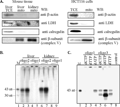
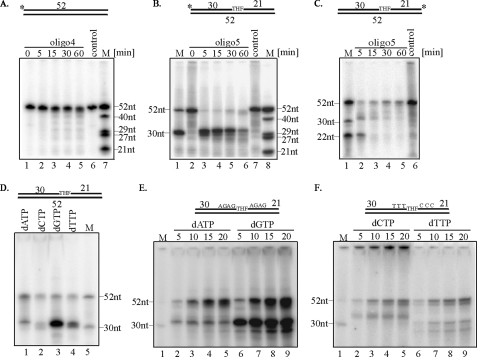
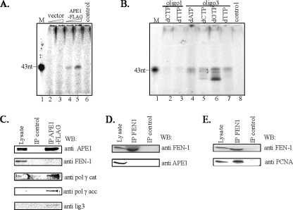
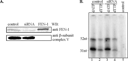
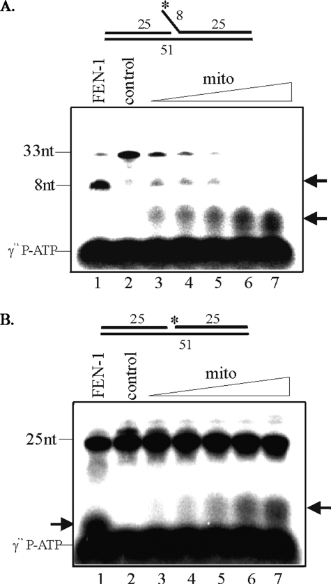
The mitochondrial genome is highly susceptible to damage by reactive oxygen species (ROS) generated endogenously as a byproduct of respiration. ROS-induced DNA lesions, including oxidized bases, abasic (AP) sites, and oxidized AP sites, cause DNA strand breaks and are repaired via the base excision repair (BER) pathway in both the nucleus and mitochondria. Repair of damaged bases and AP sites involving 1-nucleotide incorporation, named single nucleotide (SN)-BER, was observed with mitochondrial and nuclear extracts. During SN-BER, the 5'-phosphodeoxyribose (dRP) moiety, generated by AP-endonuclease (APE1), is removed by the lyase activity of DNA polymerase gamma (pol gamma) and polymerase beta in the mitochondria and nucleus, respectively. However, the repair of oxidized deoxyribose fragments at the 5' terminus after strand break would require 5'-exo/endonuclease activity that is provided by the flap endonuclease (FEN-1) in the nucleus, resulting in multinucleotide repair patch (long patch (LP)-BER). Here we show the presence of a 5'-exo/endonuclease in the mitochondrial extracts of mouse and human cells that is involved in the repair of a lyase-resistant AP site analog via multinucleotide incorporation, upstream and downstream to the lesion site. We conclude that LP-BER also occurs in the mitochondria requiring the 5'-exo/endonuclease and pol gamma with 3'-exonuclease activity. Although a FEN-1 antibody cross-reacting species was detected in the mitochondria, it was absent in the LP-BER-proficient APE1 immunocomplex isolated from the mitochondrial extract that contains APE1, pol gamma, and DNA ligase 3. The LP-BER activity was marginally affected in FEN-1-depleted mitochondrial extracts, further supporting the involvement of an unidentified 5'-exo/endonuclease in mitochondrial LP-BER.


Figures

FIGURE 1.
FIGURE 1.
Functional properties of mitochondrial extracts. A, purity of mitochondrial extracts analyzed by Western blot of total cell (TCE) or mitochondrial (mito) extracts from young mouse liver and kidney (left panel), and human HCT116 cells (right panel) probed with antibody against: lactate dehydrogenase, LDH (cytoplasmic), calregulin (ER), β-actin (cytoskeletal), β-subunit of complex V (mitochondrial). B, SN-BER of uracil-containing oligo2 duplex using two different amounts of mitochondrial extracts of liver or kidney of young mice. C, LP-BER of THF-containing oligo3 duplex using mitochondrial extracts from HCT116 cells. M, marker (43 and 30 nt). control, no extract.
FIGURE 2.
FIGURE 2.
Analysis of mitochondrial LP-BER. Time course of repair of nondamaged oligo4 duplex (A) and THF-containing oligo5 duplex (B)5′-end-labeled or (C)3′-end-labeled oligo5 duplex, by mitochondrial extract of young mouse liver. The asterisk indicates 32P-terminal label. DNA repair assay for unlabeled THF-containing oligo5 duplex (D) or oligo6 duplex (E and F) using mitochondrial extracts of young mouse liver or HCT116 cells respectively and individual [α-32P]dNTPs used to monitor repair are indicated. M, marker oligonucleotides of indicated length.
FIGURE 3.
FIGURE 3.
Characterization of HCT116 mitochondrial repair complex for LP-BER. A, repair of unlabeled THF-containing oligo3 duplex with [α-32P]dTTP and two different amounts of mitochondrial APE1-FLAG or control FLAG immunocomplex. B, monitoring of LP-BER with APE1-FLAG immunocomplex with [α-32P]dNTPs. M, marker (43 nt). control, no protein. Western analysis of mitochondrial APE1-FLAG immunocomplex (C). The absence of APE1 (D) and presence of PCNA (E) in FEN-1 immunocomplex isolated from mitochondrial and nuclear extracts, respectively. Lysate, 50 μg of mitochondrial or nuclear extracts.
FIGURE 4.
FIGURE 4.
Effect of FEN-1 down-regulation on repair by HCT116 mitochondrial extract. A, Western analysis of FEN-1 in mitochondrial extracts of HCT116 cells transfected with control or FEN-1-specific siRNA. Human recombinant FEN-1 was used as a control. The membrane was reprobed for β-subunit of ATP synthase to assure equal loading. B, repair of THF-containing oligo5 duplex with mitochondrial extracts from control and FEN-1-down-regulated cells. control, no extract.
FIGURE 5.
FIGURE 5.
Distinct mitochondrial 5′-exo/endonuclease activity in HCT116 cells. Substrates were labeled at the terminus of flap of oligo8 (A) or gap of oligo7 (B) as indicated by an asterisk, and increasing concentration of mitochondrial extracts was used in assays (lanes 3–7). Human recombinant FEN-1 was used as a positive control (lane 1). control, no extract.

Similar articles

Cited by

References

    1. Robin, E. D., and Wong, R. (1988) J. Cell. Physiol. 136507 –513 - PubMed
    1. Anderson, S., Bankier, A. T., Barrell, B. G., de Bruijn, M. H., Coulson, A. R., Drouin, J., Eperon, I. C., Nierlich, D. P., Roe, B. A., Sanger, F., Schreier, P. H., Smith, A. J., Staden, R., and Young, I. G. (1981) Nature 290457 –465 - PubMed
    1. Richter, C., Park, J. W., and Ames, B. N. (1988) Proc. Natl. Acad. Sci. U. S.A. 856465 –6467 - PMC - PubMed
    1. Mandavilli, B. S., Santos, J. H., and Van Houten, B. (2002) Mutat. Res. 509127 –151 - PubMed
    1. Sung, J. S., and Demple, B. (2006) FEBS J. 2731620 –1629 - PubMed

Publication types