Comparative Study
. 2008 Oct;29(9):1753-60.
doi: 10.3174/ajnr.A1189. Epub 2008 Jul 17.

Comparison of multidetector CT angiography and MR imaging of cervical artery dissection

Affiliations
Comparative Study

Comparison of multidetector CT angiography and MR imaging of cervical artery dissection

A T Vertinsky et al. AJNR Am J Neuroradiol. 2008 Oct.

Abstract

Background and purpose: Conventional angiography has been historically considered the gold standard for the diagnosis of cervical artery dissection, but MR imaging/MR angiography (MRA) and CT/CT angiography (CTA) are commonly used noninvasive alternatives. The goal of this study was to compare the ability of multidetector CT/CTA and MR imaging/MRA to detect common imaging findings of dissection.

Materials and methods: Patients in the data base of our Stroke Center between 2003 and 2007 with dissections who had CT/CTA and MR imaging/MRA on initial work-up were reviewed retrospectively. Two neuroradiologists evaluated the images for associated findings of dissection, including acute ischemic stroke, luminal narrowing, vessel irregularity, wall thickening/hematoma, pseudoaneurysm, and intimal flap. The readers also subjectively rated each vessel on the basis of whether the imaging findings were more clearly displayed with CT/CTA or MR imaging/MRA or were equally apparent.

Results: Eighteen patients with 25 dissected vessels (15 internal carotid arteries [ICA] and 10 vertebral arteries [VA]) met the inclusion criteria. CT/CTA identified more intimal flaps, pseudoaneurysms, and high-grade stenoses than MR imaging/MRA. CT/CTA was preferred for diagnosis in 13 vessels (5 ICA, 8 VA), whereas MR imaging/MRA was preferred in 1 vessel (ICA). The 2 techniques were deemed equal in the remaining 11 vessels (9 ICA, 2 VA). A significant preference for CT/CTA was noted for VA dissections (P < .05), but not for ICA dissections.

Conclusion: Multidetector CT/CTA visualized more features of cervical artery dissection than MR imaging/MRA. CT/CTA was subjectively favored for vertebral dissection, whereas there was no technique preference for ICA dissection. In many cases, MR imaging/MRA provided complementary or confirmatory information, particularly given its better depiction of ischemic complications.


Figures
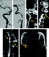
Fig 1.
Fig 1.
CT/CTA imaging of multivessel dissection (right ICA and VA) provides more detailed information than MR imaging/MRA about vessel morphology. Conventional cerebral angiographic images of the right ICA (A) and right VA (B). Curved planar reformat images from the CTA study of the right ICA (C) and right VA (D). Both demonstrate vessel wall irregularity, mild luminal narrowing, and pseudoaneurysm of the right ICA (arrows). E, Corresponding MIP image from contrast-enhanced MRA shows both dissections (arrows), but with less clear depiction than that on CTA. F, Axial T1-weighted fat-suppressed image demonstrates a crescent sign consistent with intramural hematoma around both the right ICA and VA (arrows).
Fig 2.
Fig 2.
CT/CTA and MR imaging/MRA of a left ICA dissection provide similar information about vessel morphology, but MR imaging is deemed superior for making the diagnosis, particularly because of detection of acute stroke. A, Axial T1-weighted fat-suppressed image demonstrates a crescent sign around the left ICA (arrow). B, CTA curved planar reformat shows irregularity and thickening of the left ICA wall with calcified plaque. These findings are not specific for dissection and could be due to atherosclerosis. C, Contrast-enhanced MRA MIP reformat demonstrates the similar appearance of the irregular left ICA. D and E, Noncontrast CT (D) does not show the acute infarct, which is clearly identified in the left middle cerebral artery–anterior cerebral artery watershed territory on DWI (E).
Fig 3.
Fig 3.
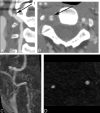
CT/CTA and MR imaging/MRA provide different advantages in this case of right ICA dissection, but diagnosis is made with equal confidence on both techniques. A and B, Noncontrast CT image (A) shows mild hypoattenuation involving the right basal ganglia (arrow), confirmed as an acute stroke with DWI (B). C, Axial source image from CTA shows expansion of the outer wall of the right ICA consistent with wall thickening as well as narrowing of the vessel lumen (arrow). D, Axial T1-weighted fat-suppressed MR image demonstrates a crescent sign (arrow) around the right ICA.
Fig 4.
Fig 4.
Example of a long-segment high-grade stenosis (string sign) identified on CTA, which appeared occluded on MR imaging/MRA. A and B, Curved planar reformatted images of the left ICA demonstrate flamelike tapering of the proximal vessel with wall thickening and long-segment high-grade stenosis (arrow). C, Axial plane from CTA shows the tiny residual lumen of the vessel (arrow). D, Contrast-enhanced MRA acquired on the same day demonstrates apparent occlusion just distal to the carotid bifurcation (arrow). E, T1-weighted fat-suppressed axial image demonstrates methemoglobin in the left ICA wall. F and G, Axial source images from 2D time-of-flight MR angiography show the T1 shinethrough of the methemoglobin, but the absence of flow-related enhancement in the left distal cervical ICA.
Fig 5.
Fig 5.
Right vertebral artery dissection with a pseudoaneurysm seen on CTA, which is not visualized on MR imaging/MRA. A, Curved planar reformatted image from CTA of the distal right vertebral artery demonstrates irregularity (arrow) as well as a small distal pseudoaneurysm. B, An axial CTA source image shows contrast within the true lumen and the pseudoaneurysm anteriorly (arrow). C, Contrast-enhanced MRA demonstrates the irregularity associated with the dissection, but not the pseudoaneurysm. D, 2D time-of-flight MRA depicts the narrowed true lumen but does not show the pseudoaneurysm, presumably due to saturation of slow-flowing blood.

References

    1. Schievink WI. Spontaneous dissection of the carotid and vertebral arteries. N Engl J Med 2001;344:898–906 - PubMed
    1. Flis CM, Jager HR, Sidhu PS. Carotid and vertebral artery dissections: clinical aspects, imaging features and endovascular treatment. Eur Radiol 2007;17:820–34 - PubMed
    1. Lee VH, Brown RD Jr, Mandrekar JN, et al. Incidence and outcome of cervical artery dissection: a population-based study. Neurology 2006;67:1809–12 - PubMed
    1. Caplan LR. Dissections of brain-supplying arteries. Nat Clin Pract Neurol 2008;4:34–42 - PubMed
    1. Pelkonen O, Tikkakoski T, Leinonen S, et al. Extracranial internal carotid and vertebral artery dissections: angiographic spectrum, course and prognosis. Neuroradiology 2003;45:71–77 - PubMed

Publication types

MeSH terms