

Molecular regulation of the hypothalamic-pituitary-adrenal axis in adult male guinea pigs after prenatal stress at different stages of gestation
- PMID: 18635650
- PMCID: PMC2652172
- DOI: 10.1113/jphysiol.2008.153684
Molecular regulation of the hypothalamic-pituitary-adrenal axis in adult male guinea pigs after prenatal stress at different stages of gestation
Abstract
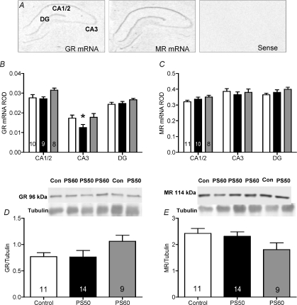
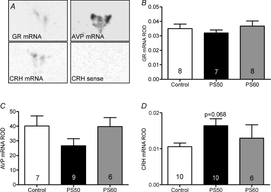
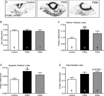
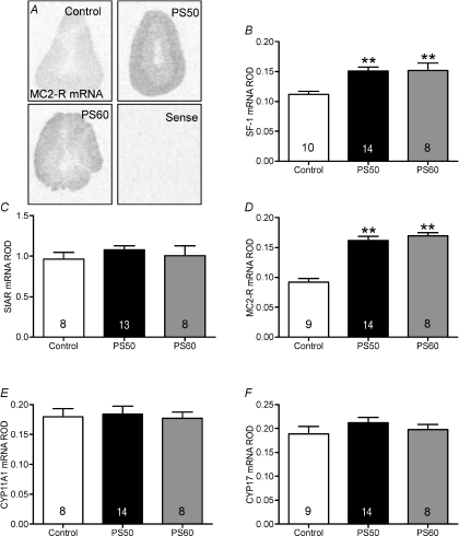
Studies in humans and animals have demonstrated that maternal stress during fetal development can lead to altered hypothalamic-pituitary-adrenal (HPA) axis function and behaviour postnatally. We have previously shown adult male guinea pigs that were born to mothers exposed to a stressor during the phase of rapid fetal brain growth (gestational days (GD) 50, 51 and 52; prenatal stress (PS)50) exhibit significantly increased basal plasma cortisol levels. In contrast, male guinea pig offspring whose mothers were exposed to stress later in gestation (GD60, 61 and 62; PS60) exhibited a significantly higher plasma cortisol response to activation of the HPA axis. In the present study, we hypothesized that the endocrine changes in HPA axis function observed in male guinea pig offspring would be reflected by altered molecular regulation of the HPA axis. Corticosteroid receptors in the hippocampus, hypothalamus and pituitary were measured, as well as corticotropin-releasing hormone (CRH), pro-opiomelanocortin (POMC) and adrenal enzymes in the paraventricular nucleus, pituitary and adrenal cortex, respectively, by in situ hybridization and Western blot. PS50 male offspring exhibited a significant reduction in glucocorticoid receptor (GR) mRNA (P <0.01) in the CA3 region of the hippocampus and significantly increased POMC mRNA (P <0.05) in the pituitary, consistent with the increase in basal HPA axis activity observed. In line with elevated activity of the HPA axis, both PS50 and PS60 male offspring exhibited significantly higher steroidogenic factor (SF)-1 (P <0.001) and melanocortin 2 receptor (MC2-R) mRNA (P <0.001) in the adrenal cortex. This study demonstrates that short periods of prenatal stress during critical windows of neuroendocrine development affect the expression of key regulators of HPA axis activity leading to the changes in endocrine function observed in prenatally stressed male offspring. Further, these changes are dependent on the timing of the maternal stressor, a pattern that is emerging in human studies.
Figures
Similar articles
-
Prenatal stress modifies behavior and hypothalamic-pituitary-adrenal function in female guinea pig offspring: effects of timing of prenatal stress and stage of reproductive cycle.Endocrinology. 2008 Dec;149(12):6406-15. doi: 10.1210/en.2008-0347. Epub 2008 Aug 28. Endocrinology. 2008. PMID: 18755800
-
Short periods of prenatal stress affect growth, behaviour and hypothalamo-pituitary-adrenal axis activity in male guinea pig offspring.J Physiol. 2005 Aug 1;566(Pt 3):967-77. doi: 10.1113/jphysiol.2005.090191. Epub 2005 Jun 2. J Physiol. 2005. PMID: 15932885 Free PMC article.
-
Prenatal glucocorticoid exposure alters hypothalamic-pituitary-adrenal function and blood pressure in mature male guinea pigs.J Physiol. 2004 Jul 1;558(Pt 1):305-18. doi: 10.1113/jphysiol.2004.063669. Epub 2004 May 14. J Physiol. 2004. PMID: 15146051 Free PMC article.
-
Maternal and fetal hypothalamic-pituitary-adrenal axes during pregnancy and postpartum.Ann N Y Acad Sci. 2003 Nov;997:136-49. doi: 10.1196/annals.1290.016. Ann N Y Acad Sci. 2003. PMID: 14644820 Review.
-
Early environmental regulation of forebrain glucocorticoid receptor gene expression: implications for adrenocortical responses to stress.Dev Neurosci. 1996;18(1-2):49-72. doi: 10.1159/000111395. Dev Neurosci. 1996. PMID: 8840086 Review.
Cited by
-
Gestational Hypoxia and Developmental Plasticity.Physiol Rev. 2018 Jul 1;98(3):1241-1334. doi: 10.1152/physrev.00043.2017. Physiol Rev. 2018. PMID: 29717932 Free PMC article. Review.
-
Epigenetic and transgenerational reprogramming of brain development.Nat Rev Neurosci. 2015 Jun;16(6):332-44. doi: 10.1038/nrn3818. Epub 2015 Apr 29. Nat Rev Neurosci. 2015. PMID: 25921815 Free PMC article. Review.
-
Cytokines and the neurodevelopmental basis of mental illness.Front Neurosci. 2013 Oct 17;7:180. doi: 10.3389/fnins.2013.00180. Front Neurosci. 2013. PMID: 24146637 Free PMC article. Review.
-
Lifetime stress experience: transgenerational epigenetics and germ cell programming.Dialogues Clin Neurosci. 2014 Sep;16(3):297-305. doi: 10.31887/DCNS.2014.16.3/tbale. Dialogues Clin Neurosci. 2014. PMID: 25364281 Free PMC article. Review.
-
Sex as a Biological Variable: Who, What, When, Why, and How.Neuropsychopharmacology. 2017 Jan;42(2):386-396. doi: 10.1038/npp.2016.215. Epub 2016 Sep 23. Neuropsychopharmacology. 2017. PMID: 27658485 Free PMC article. Review.
References
-
- Bosch OJ, Kromer SA, Neumann ID. Prenatal stress: Opposite effects on anxiety and hypothalamic expression of vasopressin and corticotropin-releasing hormone in rats selectively bred for high and low anxiety. Eur J Neurosci. 2006;23:541–551. - PubMed
-
- Bradford MM. A rapid and sensitive method for the quantitation of microgram quantities of protein utilizing the principle of protein-dye binding. Anal Biochem. 1976;72:248–254. - PubMed
-
- Clarke AS, Wittwer DJ, Abbott DH, Schneider ML. Long-term effects of prenatal stress on HPA axis activity in juvenile rhesus monkeys. Dev Psychobiol. 1994;27:257–269. - PubMed
-
- Coe CL, Kramer M, Czeh B, Gould E, Reeves AJ, Kirschbaum C, et al. Prenatal stress diminishes neurogenesis in the dentate gyrus of juvenile rhesus monkeys. Biol Psychiatry. 2003;54:1025–1034. - PubMed
Publication types
MeSH terms
LinkOut - more resources
Full Text Sources
Miscellaneous