. 2008 Nov;149(11):5706-12.
doi: 10.1210/en.2008-0717. Epub 2008 Jul 17.

Regulator of G protein signaling-4 controls fatty acid and glucose homeostasis

Affiliations

Regulator of G protein signaling-4 controls fatty acid and glucose homeostasis

Irena Iankova et al. Endocrinology. 2008 Nov.

Abstract

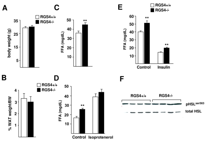
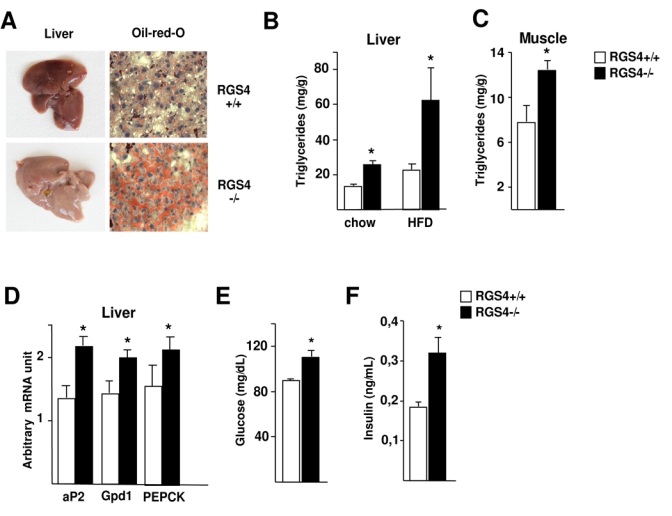
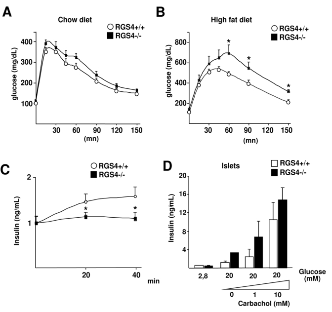
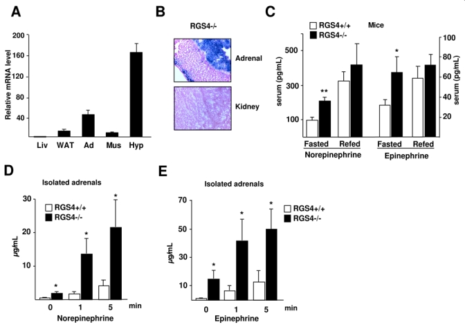
Circulating free fatty acids are a reflection of the balance between lipogenesis and lipolysis that takes place mainly in adipose tissue. We found that mice deficient for regulator of G protein signaling (RGS)-4 have increased circulating catecholamines, and increased free fatty acids. Consequently, RGS4-/- mice have increased concentration of circulating free fatty acids; abnormally accumulate fatty acids in liver, resulting in liver steatosis; and show a higher degree of glucose intolerance and decreased insulin secretion in pancreas. We show in this study that RGS4 controls adipose tissue lipolysis through regulation of the secretion of catecholamines by adrenal glands. RGS4 controls the balance between adipose tissue lipolysis and lipogenesis, secondary to its role in the regulation of catecholamine secretion by adrenal glands. RGS4 therefore could be a good target for the treatment of metabolic diseases.


Figures

FIG 1
FIG 1
Adipose tissue weight, fatty acid quantification, and in vivo lipolysis analysis in RGS4−/− mice. (A) Body weight of RGS4 +/+ and RGS4 −/− mice (RGS4+/+, n=8; RGS4−/−, n=8) was analyzed at 25 weeks. (B) White adipose tissue (WAT) weight is represented as a percentage of body weight in the same mice. (C) Quantification of serum free fatty acid (FFA) concentration in mice fasted for 16 hours. (D) Isoproterenol-induced in vivo lipolysis in 7 hour-fasted RGS4+/+ (n=6) and RGS4−/− (n=6) mice measured by FFA concentration in serum. Blood serum was collected before (control) and 15 minutes after injection of isoproterenol (10 mg/kg). (E) Analysis of in vivo lipolysis in response to insulin in overnight fasted RGS4+/+ and −/− mice. FFA serum levels were determined before (control) and 60 minutes after intraperitoneal injection of insulin (0,75 UI/kg). (F) Expression of HSL phosphorylated on Ser563 compared to the expression of total HSL was assessed by Western blot. Data are mean ± SEM. Statistically significant differences (ANOVA, p<0.05) are indicated in this, and subsequent figures by an asterisk.
FIG 2
FIG 2
Lipid content in liver and serum metabolic factors in RGS4−/− mice. (A) Representative pictures of whole livers of 25-week-old RGS4+/+ or −/− mice. Pale colour is indicative of lipid accumulation. Oil Red O-stained liver sections in the same mice demonstrate lipid content. (B) Hepatic triglyceride content in 25 week-old RGS4+/+ (n=5) and −/− (n=5) mice on a chow or high fat diet for 4 weeks. (C) Muscle triglyceride content in 25 week-old RGS4+/+ (n=5) and −/− (n=5) normal fed mice. (D) Hepatic expression levels of aP2, Gpdl, and PEPCK in normal-fed mice as determined using quantitative RT-PCR. Serum fasting glucose concentration (E) and insulin serum levels (F) were determined in 18-week-old RGS4+/ or −/− mice as indicated.
FIG 3
FIG 3
Glucose homeostasis in RGS4−/− mice. Intraperitoneal glucose tolerance test (IPGTT) in 25 week-old mice under chow diet (A) (RGS4+/+, n=8; RGS4−/−, n=8) or after 3 weeks of high fat diet (B) (RGS4 +/+, n=5; RGS4 −/−, n=5). Blood glucose concentrations were measured at the indicated time points following i.p. injection of glucose (2 g/kg). (C) Quantification of relative serum insulin levels measured by ELISA test at the indicated time points in the same mice during the IPGTT in A. (D) Insulin secretion of isolated islets from RGS4−/− or +/+ mice stimulated for 1 hour in the presence of 2.8 mM or 20 mM glucose, and increasing concentrations of carbachol. Results are relative to total insulin and protein content.
FIG 4
FIG 4
RGS4 expression during adipocyte differentiation. (A) RGS4 expression in 3T3L1 preadipocyte differentiation was determined using quantitative RT-PCR. Results were normalized by the expression level of rS9. (B) Oil Red O staining of 3T3L1 adipocytes expressing either an empty vector (control) or a vector encoding RGS4 (RGS4) 8 days after induction of differentation. (C) RGS4 mRNA expression, analysed by RT-PCR, in 3T3L1 empty vector or RGS4 vector expressing cells.
FIG 5
FIG 5
Increased catecholamine secretion in RGS4−/− mice. (A) RGS4 expression in mouse tissues (Liv=liver; WAT=white adipose tissue; Ad=adrenals; Mus=muscle; Hyp=hypothalamus). (B) Representative β-gal staining of adrenal glands and kidney sections of RGS4−/− mice as indicated. (C) Quantification of serum concentration of norepinephrine and epinephrine in overnight-fasted and 4 hour-refed 23 week-old mice (RGS4+/+, n=5, RGS4−/−, n=5). Norepinephrine (D) and epinephrine (E) concentrations were measured at the indicated time points following stimulation of acute adrenal slices (n=6) with acetylcholine (100 μM).

References

    1. Langin D. Adipose tissue lipolysis as a metabolic pathway to define pharmacological strategies against obesity and the metabolic syndrome. Pharmacol Res. 2006;53:482–91. - PubMed
    1. DeHaven J, Sherwin R, Hendler R, Felig P. Nitrogen and sodium balance and sympathetic-nervous-system activity in obese subjects treated with a low-calorie protein or mixed diet. N Engl J Med. 1980;302:477–82. - PubMed
    1. Arner P. Human fat cell lipolysis: biochemistry, regulation and clinical role. Best Pract Res Clin Endocrinol Metab. 2005;19:471–82. - PubMed
    1. Tentolouris N, Liatis S, Katsilambros N. Sympathetic system activity in obesity and metabolic syndrome. Ann N Y Acad Sci. 2006;1083:129–52. - PubMed
    1. Zimmermann R, Strauss JG, Haemmerle G, et al. Fat mobilization in adipose tissue is promoted by adipose triglyceride lipase. Science. 2004;306:1383–6. - PubMed

Publication types

MeSH terms