. 2008 Jul 18;321(5887):401-4.
doi: 10.1126/science.1158159.

Four-jointed is a Golgi kinase that phosphorylates a subset of cadherin domains

Affiliations

Four-jointed is a Golgi kinase that phosphorylates a subset of cadherin domains

Hiroyuki O Ishikawa et al. Science. .

Abstract

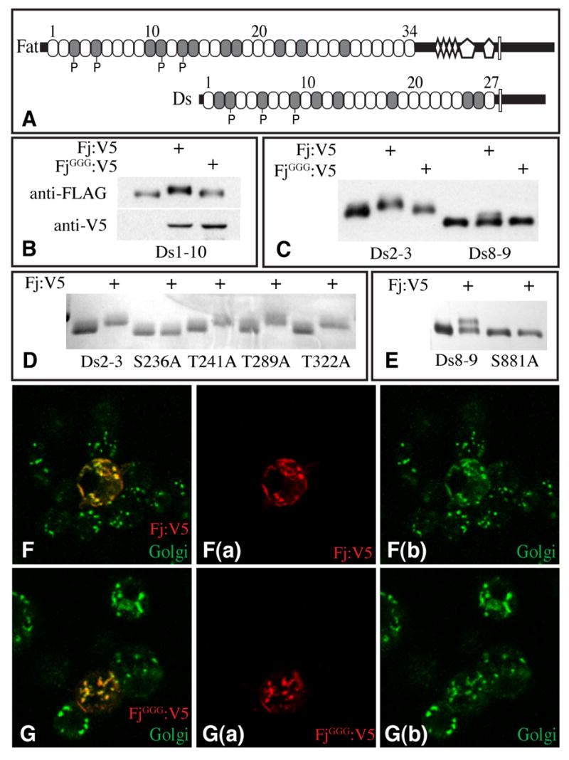
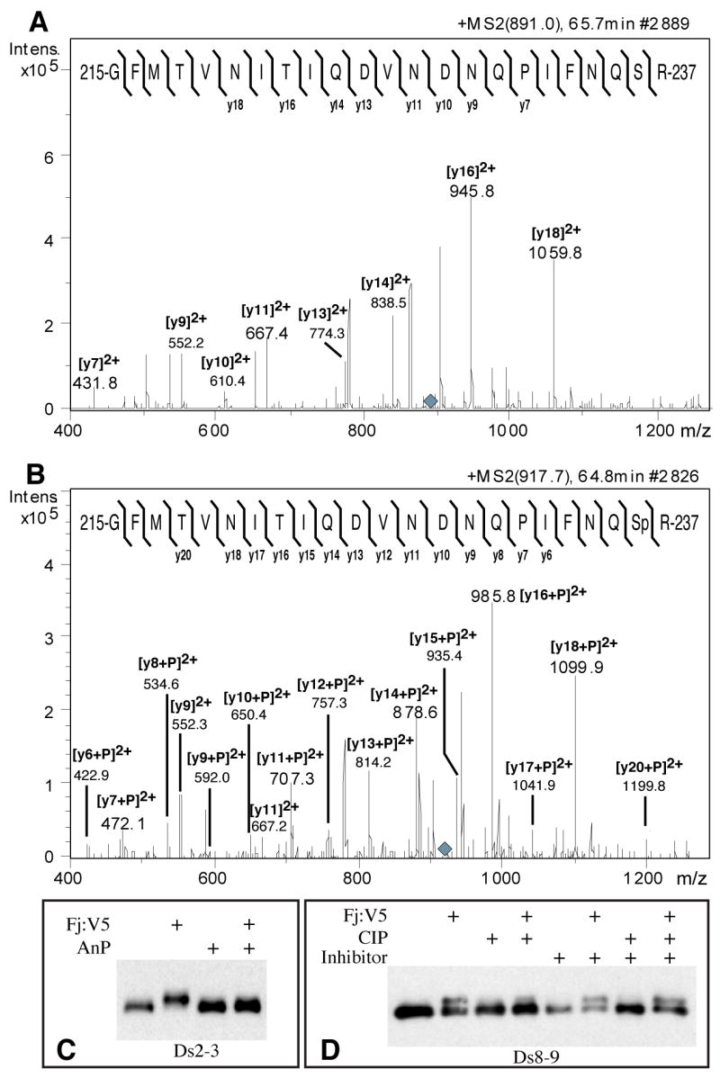
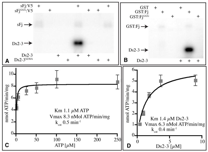
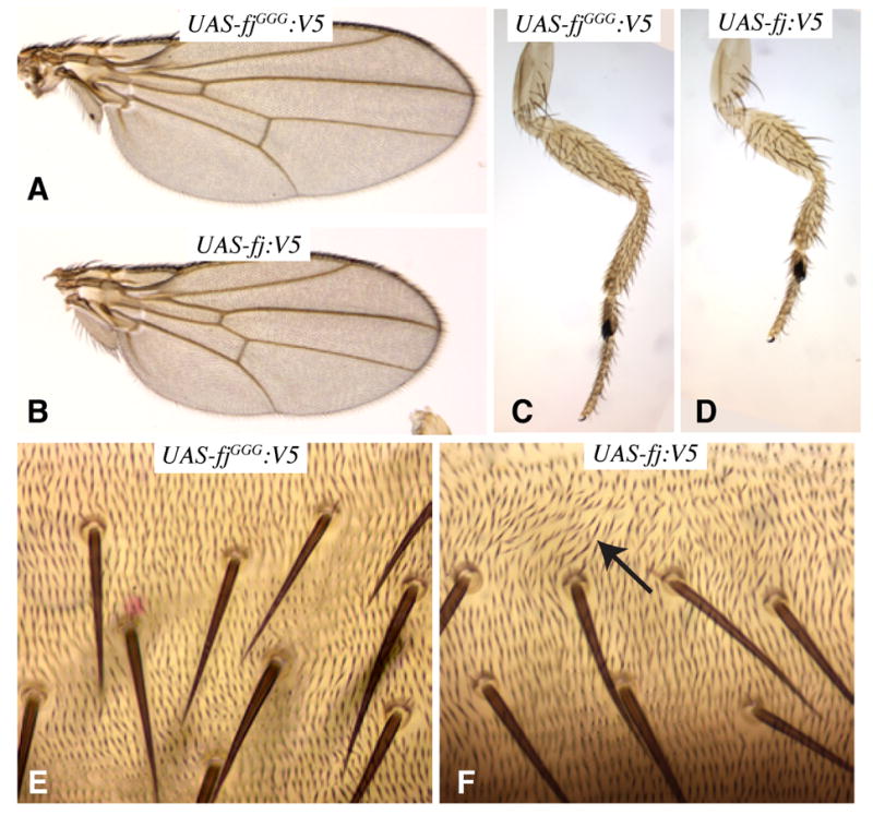
The atypical cadherin Fat acts as a receptor for a signaling pathway that regulates growth, gene expression, and planar cell polarity. Genetic studies in Drosophila identified the four-jointed gene as a regulator of Fat signaling. We show that four-jointed encodes a protein kinase that phosphorylates serine or threonine residues within extracellular cadherin domains of Fat and its transmembrane ligand, Dachsous. Four-jointed functions in the Golgi and is the first molecularly defined kinase that phosphorylates protein domains destined to be extracellular. An acidic sequence motif (Asp-Asn-Glu) within Four-jointed was essential for its kinase activity in vitro and for its biological activity in vivo. Our results indicate that Four-jointed regulates Fat signaling by phosphorylating cadherin domains of Fat and Dachsous as they transit through the Golgi.


Figures

Figure 1
Figure 1. Four-jointed-dependent mobility shift of cadherin domain polypeptides
A) Schematics of Fat and Ds. Cadherin domains (ovals), EGF domains (diamonds), LamG domains (pentagons) and transmembrane domains (rectangles) are indicated. Shading identifies cadherin domains with a Ser or Thr as the seventh amino acid, “P” identifies Cadherin domains at which Fj-mediated phosphorylation was detected. B, C, E show Western blots (anti-FLAG) of Ds Cadherin domains isolated from medium of S2 cells; cells were co-transfected with plasmids to express Fj:V5 or FjGGG:V5 as indicated. B) Ds1-10:FLAG. Fj expression is indicated by anti-V5 staining. C) Ds2-3:FLAG and Ds8-9:FLAG. D) Coomasie stain of Ds2-3:FLAG and point mutants immunoprecipitated from the medium of transfected S2 cells, where indicated cells express Fj:V5. E) Ds8-9:FLAG and Ds8-9S881A:FLAG mutant. F-G Show S2 cells transfected to express Fj:V5 (F) or FjGGG:V5 (G) and stained with anti-V5 (red) and anti-Golgi (green); panels to the right show individual stains.
Figure 2
Figure 2. Four-jointed dependent phosphorylation of Ds and Fat cadherin domains
A) MS/MS fragmentation of a tryptic peptide from Ds2-3:FLAG isolated from S2 cells, eluting on HPLC at 65.7 minutes (Supplementary Fig. S3A). The parental ion is triply charged (m/z 891.0, grey diamond). The mass and fragmentation pattern identify this as aa 215-237; the positions of identified y ions from peptide fragmentation are indicated. B) MS/MS fragmentation of a tryptic peptide from Ds2-3:FLAG isolated from Fj:V5-expressing S2 cells, eluting on HPLC at 64.8 minutes (parent ion m/z 917.7, grey diamond). The positions of identified y ions from peptide fragmentation are indicated. C) Western blot on Ds2-3:FLAG isolated from S2 cell media, indicated cells expressed Fj:V5, and/or were treated with antarctic phosphatase (AnP). D) Western blot on Ds8-9:FLAG isolated from S2 cell media, indicated cells expressed Fj:V5, and/or treated with calf intestinal alkaline phosphatase (CIP), and/or treated with phosphatase inhibitor cocktail.
Figure 3
Figure 3. Kinase activity of Four-jointed
A, B) Autoradiograms of protein gels dispalying the products of in vitro kinase reactions with the indicated enzymes (top) and substrates (bottom). The positions of Ds2-3:FLAG and sFj:V5 or GST:Fj are indicated. A) Phosphorylation of Ds2-3, but not Ds2-3S236A, by Fj in vitro. B) Kinase activity of GST:Fj isolated from E. coli. C) Reaction velocity versus ATP concentration, using 2.5 μg Ds2-3:FLAG, 10 ng sFj:V5, and 1, 5, 25, 50, 100, or 250 μM ATP for 15 min. G) Reaction velocity versus Ds2-3 concentration, using 500 μM ATP, 10 ng sFj:V5, and 0.36, 0.90, 1.8, 3.6, or 9.0 μM Ds2-3:FLAG for 15 min. Plots in C and D show average values from four experiments, error bars indicate s.e.m.
Figure 4
Figure 4. Requirement for Four-jointed kinase activity in vivo
A–F Show portions of adult flies, expressing either wild-type fj:V5 or the fjGGG:V5 mutant. A) Wing from tub-Gal4 UAS-fjGGG:V5. B) Wing from tub-Gal4 UAS-fj:V5. C) Leg from tub-Gal4 UAS-fjGGG:V5. D) Leg from tub-Gal4 UAS-fj:V5. E) Abdomen from tub-Gal4 UAS-fjGGG:V5 animal. F) Abdomen from tub-Gal4 UAS-fj:V5 animal with disturbed hair polarity (arrow).

References

    1. Strutt H, Strutt D. Bioessays. 2005;27:1218. - PubMed
    1. Pan D. Genes Dev. 2007;21:886. - PubMed
    1. Casal J, Struhl G, Lawrence P. Curr Biol. 2002;12:1189. - PubMed
    1. Cho E, Irvine KD. Development. 2004;131:4489. - PubMed
    1. Yang C, Axelrod JD, Simon MA. Cell. 2002;108:675. - PubMed

Publication types

MeSH terms