. 2008 Nov;15(11):1772-81.
doi: 10.1038/cdd.2008.109. Epub 2008 Jul 18.

The intestinal epithelium compensates for p53-mediated cell death and guarantees organismal survival

Affiliations

The intestinal epithelium compensates for p53-mediated cell death and guarantees organismal survival

Y A Valentin-Vega et al. Cell Death Differ. 2008 Nov.

Abstract

Mdm2 is the major inhibitor of the p53 tumor suppressor. Loss of Mdm2 in mice or in specific tissues of the mouse always yields p53-dependent lethal phenotypes. However, the role of Mdm2 in tissues with high turnover capacity is unknown. We have engineered mice lacking Mdm2 in the intestinal epithelium using the Cre/LoxP system. Loss of Mdm2 (Mdm2(intDelta)) results in viable animals, but neonates display multiple intestinal abnormalities such as hyperplasia, enterocyte vacuolization, and inflammation. These defects correlate with a drastic increase in p53-dependent apoptosis in highly proliferative and differentiated cells. Unexpectedly, the observed phenotypes disappear with age. The tissue selects against Mdm2-null cells and increases its proliferative capacity. Additionally, the intestinal stem and progenitor cell populations are enriched leading to an increase in crypt fission events. Enhanced proliferation is achieved by activation of the canonical Wnt and EGFR-mediated Ras/MAPK pathways. While Mdm2 is a critical inhibitor of p53 in the intestinal epithelium, the tissue employs a series of processes that compensate for cell death.


Figures
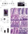
Figure 1
Figure 1
Young Mdm2intΔ mice developed multiple intestinal abnormalities. (a) PCR analysis of P3 pups show the nonrecombined floxed Mdm2 (FM) and recombined (intΔ) Mdm2 alleles. Red triangles, loxP sites; black triangles, primers; black lines indicate PCR products. E, exon; kb, kilobases, bp, base pairs. SI, small intestine; C, colon; T, tail. (b) Body size and weight of P3 littermates of different genotypes. (c and d) H&E staining of small intestines of P3 mice at original magnification × 50 (c) or × 200 (d). Black arrowhead marks an intestinal edema in an Mdm2intΔ pup. (e and f) Measurement of the intervillus width of P3 Mdm2intΔ and control mice. Yellow lines denote the width of intervillus pockets. (g) Intestinal villi of P3 mice exhibiting vacuoles in enterocytes (black arrowhead)
Figure 2
Figure 2
Mdm2 regulated p53 activity in highly proliferative and terminally differentiated intestinal epithelial cells. (a) Immunohistochemistry with p53 antibodies in small intestines of P3 mice. (b) Apoptotic cells were labeled by Caspase-3 immunofluorescence (green) and nuclei with TOPRO-3 (blue). Yellow lines outline intervillus pockets. (c) Quantification of apoptosis in intervillus pockets at P3 evaluated by scoring apoptotic bodies in H&E-stained sections (200 intervillus units scored per mouse; n≥3 per group). (d) mRNA quantification of p53-target genes by reverse transcriptase real-time PCR analysis. (n≥3 per group, asterisk means P<0.02). (e) H&E-stained small intestines of P3 Mdm2intΔ neonates without p53. V, villus region; IV, intervillus region
Figure 3
Figure 3
Intestines of Adult Mdm2intΔ mice have decreased p53 levels and recover. (a) H&E staining of intestinal jejunum of 8-week-old mice. V, villus region; C, crypt region. (b) p53 immunohistochemistry performed in intestinal jejunum of 8-week-old mice (original magnification ×600). Staining in the lamina propria (LP) is an artifact. Black arrowheads denote p53-positive cells. (c) Comparison of the apoptotic indices between P3 (black bars) and 8-week-old (gray bars) Mdm2intΔ mice. Apoptosis was measured by scoring apoptotic bodies in the jejunum of adult mice and in whole intestines of P3 mice (200 intervillus units scored per mouse; n = 3 per group). C, control group, M2intΔ, Mdm2intΔ mice. (d) Body weight gain of control (white bars), normal size Mdm2intΔ (gray bars) and small size Mdm2intΔ (black bars) mice by week
Figure 4
Figure 4
Intestines of Mdm2intΔ mice select against Mdm2-null cells. (a) β-galactosidase staining (blue) of small intestines of P3 and 8-week-old mice. (b) PCR-based recombination analysis in 3-day and 8-week-old mouse tissues (n≥3 per group) using primers A, B, and C (see Figure 1a). Asterisk marks the band amplified using primers A and B, which represents the wild-type or Mdm2- null alleles. SI, small intestine; T, tail
Figure 5
Figure 5
Mechanisms of intestinal compensation in Mdm2intΔ mice. (a) Ki-67 immunofluorescence (turquoise) in small intestines of P3 mice with cytokeratin (orange) and TOPRO-3 nuclear (dark blue; original magnification ×400). Graph represents the quantification of the Ki-67 staining (200 intervillus units scored per mouse; n =3 per group). (b) H&E staining and quantification of intervillus units of P3 mice (200 intervillus units scored per mouse; n≥4 per group; original magnification ×400). Arrowhead pinpoints a crypt fission event occurring in an Mdm2intΔ pup. (c) Immunohistochemistry using an Msi-1antibody in intestines of P3 mice. Black arrowheads mark Msi-1-positive cells in intervillus pockets. A total of 200 intervillus units were scored per mouse (original magnification ×400, n≥4 per group). Note that the pictures of both control and mutant intestines were taken at the same magnification, but the mutant intestines are larger due to the hyperplastic and hyperthrophic phenotype observed in these mutants
Figure 6
Figure 6
The canonical Wnt signaling, and EGFR-mediated Ras/MAPK pathways are active in the intestinal epithelium of Mdm2intΔ neonates. (a) Immunohistochemistry against active β-catenin. Arrowhead pinpoints intracellular accumulation of β-catenin in the intervillus region of Mdm2intΔ neonates. (b) Immunohistochemistry against CD44, a membrane protein, in the small intestine of P3 mice. (c) The EGFR-mediated Ras/MAPK signaling cascade. Amphiregulin (AR) or Epiregulin (ER) bind to EGFR at the plasma membrane (PM) and induce Ras activity. Ras binds and activates Raf, which in turn phosphorylates and activates MEK1 and MEK2. Finally, MEK1/2 phosphorylates ERK1 and ERK2, which accumulate in the nucleus. In the nucleus, ERK1/2 phosphorylates a number of transcription factors involved in cell proliferation and survival. (d) Quantification of amphiregulin and epiregulin mRNAs in 3-day-old intestines (n≥3 per group). (e) Immunohistochemistry against EGFR in P3 mouse intestines. Box shows higher magnification of the intervillus region. (f) Immunohistochemistry against p-ERK1/2 in 3-day-old small intestines. V, villus region, IV, intervillus region

References

    1. Vousden KH. p53: death star. Cell. 2000;103:691–694. - PubMed
    1. Brooks CL, Gu W. p53 ubiquitination: Mdm2 and beyond. Mol Cell. 2006;21:307–315. - PMC - PubMed
    1. Evans SC, Viswanathan M, Grier JD, Narayana M, El-Naggar AK, Lozano G. An alternatively spliced HDM2 product increases p53 activity by inhibiting HDM2. Oncogene. 2001;20:4041–4049. - PubMed
    1. Momand J, Jung D, Wilczynski S, Niland J. The MDM2 gene amplification database. Nucleic Acids Res. 1998;26:3453–3459. - PMC - PubMed
    1. Oliner JD, Kinzler KW, Meltzer PS, George DL, Vogelstein B. Amplification of a gene encoding a p53-associated protein in human sarcomas. Nature. 1992;358:80–83. - PubMed

Publication types