. 2008 Sep;9(9):930-6.
doi: 10.1038/embor.2008.136. Epub 2008 Jul 18.

The tumour suppressor CYLD is a negative regulator of RIG-I-mediated antiviral response

Affiliations

The tumour suppressor CYLD is a negative regulator of RIG-I-mediated antiviral response

Constantin S Friedman et al. EMBO Rep. 2008 Sep.

Abstract

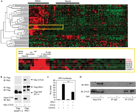
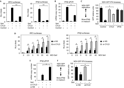
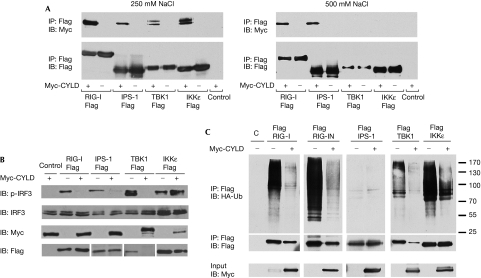
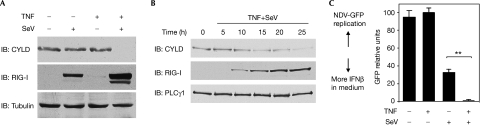
On detecting viral RNAs, the RNA helicase retinoic acid-inducible gene I (RIG-I) activates the interferon regulatory factor 3 (IRF3) signalling pathway to induce type I interferon (IFN) gene transcription. How this antiviral signalling pathway might be negatively regulated is poorly understood. Microarray and bioinformatic analysis indicated that the expression of RIG-I and that of the tumour suppressor CYLD (cylindromatosis), a deubiquitinating enzyme that removes Lys 63-linked polyubiquitin chains, are closely correlated, suggesting a functional association between the two molecules. Ectopic expression of CYLD inhibits the IRF3 signalling pathway and IFN production triggered by RIG-I; conversely, CYLD knockdown enhances the response. CYLD removes polyubiquitin chains from RIG-I as well as from TANK binding kinase 1 (TBK1), the kinase that phosphorylates IRF3, coincident with an inhibition of the IRF3 signalling pathway. Furthermore, CYLD protein level is reduced in the presence of tumour necrosis factor and viral infection, concomitant with enhanced IFN production. These findings show that CYLD is a negative regulator of RIG-I-mediated innate antiviral response.


Conflict of interest statement

The authors declare that they have no conflict of interest.

Figures

Figure 1
Figure 1
RIG-I interacts with CYLD in silico and functionally. (A) Expression profiles of 47 genes encoding DUBs (PMID: 12917690) and 31 genes encoding CARD-containing proteins, across 79 tissues and cell types performed in duplicate. Genes were clustered hierarchically and visualized with TreeView. CYLD, a member of the DUB family, and DDX58 (RIG-I), which encodes a CARD-domain-containing protein, clustered closely together on the basis of their similar expression profile overall, and are significantly enriched in immune tissues and cells as found by the Wilcoxon rank-sum test (P<0.0007 and P<0.0002, respectively). (B) A derivative of the 293 EBNA cell line was transfected with 5 μg each of Flag-RIG-I or Flag-A20ZF1-4 as a negative control, and empty vector or Myc-CYLD. Flag immunoprecipitations were performed and sequentially blotted with anti-Myc and anti-Flag. (C) IRF3 reporter activity in 293 EBNA cells co-transfected with 100 ng RIG-I and 2 μg of a plasmid encoding CYLD or GST as a negative control. The results are expressed as mean±s.d. (n=3; **P<0.01 by Student's t-test). (D) 293 EBNA cells were infected with SeV at an MOI of 10 for 24 h. Control IgG or anti-CYLD immunoprecipitations were performed and sequentially blotted with anti-RIG-I and anti-CYLD. CARD, Caspase recruitment domain; CD, cluster of differentiation; CYLD, cylindromatosis; DC, dendritic cells; DUBs, deubiquitinating enzymes; GST, glutathione S-transferase; MOI, multiplicity of infection; IB, immunoblotting; IP, immunoprecipitation; IRF3, interferon regulatory factor 3; NK, natural killer; PBMC, peripheral blood mononuclear cells; RIG-I, retinoic acid-inducible gene I; RLU, relative luciferase units; SeV, Sendai virus Cantell; siRNA, short interfering RNA; ZF, zinc fingers.
Figure 2
Figure 2
CYLD inhibits SeV-induced IFNβ production. (A) IRF3 and IFNβ promoter activity in 293 EBNA cells transfected with negative control or CYLD, and either mock-infected or infected with SeV at an MOI of 20. The results are expressed as mean±s.d. (n=3; *P<0.05, **P<0.01 by Student's t-test for panels A–F). (B) qPCR analysis of IFNβ messenger RNA in 293 EBNA cells transfected with negative control or CYLD, and either mock-infected or infected with SeV at an MOI of 20. (C) GFP expression in Vero cells infected with NDV-GFP virus after treatment with conditioned media from cells transfected with 2 μg CYLD, Ebola virus VP35 (positive control) or bacterial GST (negative control) and infected with SeV at an MOI of 10. Higher levels of GFP expression indicate lower levels of IFNβ in the media (n=3). (D) IRF3 and IFNβ promoter activity in 293 EBNA cells transfected with siRNA duplexes that are non-silencing (si-NS) or against CYLD (si-CYLD), followed by mock or SeV infection at the indicated MOI. The results are expressed as mean±s.d. (n=4). (E) qPCR analysis of IFNβ mRNA in cells transfected with si-NS or si-CYLD, followed by mock or SeV infection at an MOI of 10. (F) IFN bioassay performed with conditioned media from siRNA-transfected cells infected with SeV at an MOI of 20. CYLD, cylindromatosis; GFP, green fluorescent protein; GST, glutathione S-transferase; IFN, interferon; IRF3, interferon regulatory factor 3; qPCR, quantitative PCR; MOI, multiplicity of infection; NDV, Newcastle disease virus; SeV, Sendai virus Cantell; siRNA, short interfering RNA.
Figure 3
Figure 3
Physical and functional interactions of CYLD with IPS-1, TBK1 and IKKɛ. (A) Cells were co-transfected with 5 μg of Flag-RIG-I, Flag-IPS-1, Flag-TBK1 or Flag-IKKɛ and 5 μg of either Myc-CYLD or GST as a negative control. Flag immunoprecipitations were performed, followed by blotting with the indicated antibodies. The washes for the immunoprecipitations were performed in buffer with either 250 mM NaCl (left panel) or 500 mM NaCl (right panel). (B) 293 EBNA cells were transfected for 24 h with 1 μg CYLD or empty vector control, and 200 ng each of RIG-I, IPS-1, TBK1 or IKKɛ. Lysates were blotted for phospho-IRF3 (p-IRF3), total IRF3, Myc-CYLD and Flag-tagged RIG-I, IPS-1, TBK1 or IKKɛ. (C) 293 EBNA cells were transfected with Flag-tagged RIG-I, RIG-IN, IPS-1, TBK1 or IKKɛ, together with HA-tagged K63-Ub and either empty vector or Myc-tagged CYLD. Triton-soluble lysates were denatured in 1% SDS and immunoprecipitated with anti-Flag. The immunoprecipitations were blotted with anti-HA to detect K63-Ub and re-probed with anti-Flag. The input lysates were blotted with anti-Myc to detect CYLD. CYLD, cylindromatosis; GST, glutathione S-transferase; HA, haemagglutinin; IB, immunoblotting; IP, immunoprecipitation; K63-Ub, ubiquitin mutant with a single lysine at position 63; RIG-I, retinoic acid-inducible gene I; TBK1, TANK binding protein 1.
Figure 4
Figure 4
CYLD protein is downregulated during SeV infection in the presence of TNF. (A) 293 EBNA cells were cultured with 25 ng/ml TNF for 24 h before infection with SeV (MOI of 10) for a further 24 h. Triton-soluble lysates were sequentially blotted with antibodies specific for CYLD, RIG-I and tubulin as a loading control. (B) Cells were treated with TNF for 24 h before infection with SeV for varying periods of time. Lysates were blotted as indicated. (C) Cells were treated as in (A), and culture supernatants were collected for IFN bioassay (n=3; **P<0.01). CYLD, cylindromatosis; GFP, green fluorescent protein; IB, immunoblotting; IFN, interferon; MOI, multiplicity of infection; NDV, Newcastle disease virus; PLCγ1, phospholipase C-γ1; RIG-I, retinoic acid-inducible gene I; SeV, Sendai virus Cantell; TNF, tumour necrosis factor.

References

    1. Bignell GR et al. (2000) Identification of the familial cylindromatosis tumour-suppressor gene. Nat Genet 25: 160–165 - PubMed
    1. Brummelkamp TR, Nijman SM, Dirac AM, Bernards R (2003) Loss of the cylindromatosis tumour suppressor inhibits apoptosis by activating NF-κB. Nature 424: 797–801 - PubMed
    1. Cardenas WB, Loo YM, Gale M Jr, Hartman AL, Kimberlin CR, Martinez-Sobrido L, Saphire EO, Basler CF (2006) Ebola virus VP35 protein binds double-stranded RNA and inhibits α/β interferon production induced by RIG-I signaling. J Virol 80: 5168–5178 - PMC - PubMed
    1. Fitzgerald KA, McWhirter SM, Faia KL, Rowe DC, Latz E, Golenbock DT, Coyle AJ, Liao SM, Maniatis T (2003) IKKɛ and TBK1 are essential components of the IRF3 signaling pathway. Nat Immunol 4: 491–496 - PubMed
    1. Gack MU et al. (2007) TRIM25 RING-finger E3 ubiquitin ligase is essential for RIG-I-mediated antiviral activity. Nature 446: 916–920 - PubMed

Publication types

MeSH terms