

Diallyl sulfide inhibits PhIP-induced cell death via the inhibition of DNA strand breaks in normal human breast epithelial cells
- PMID: 18636192
- PMCID: PMC3060617
Diallyl sulfide inhibits PhIP-induced cell death via the inhibition of DNA strand breaks in normal human breast epithelial cells
Abstract
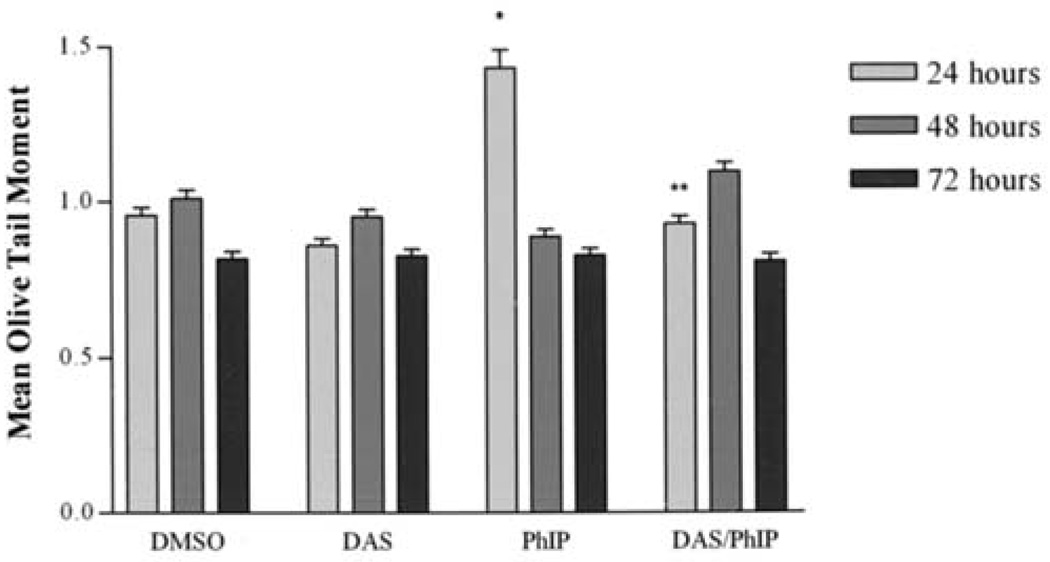
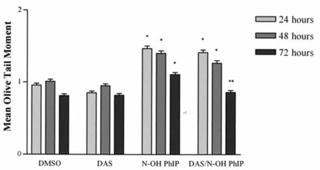
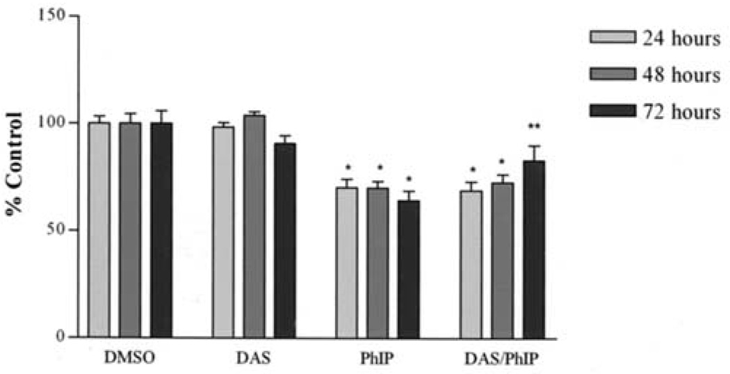
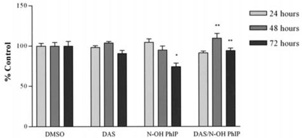
2-Amino-1-methyl-6-phenylimidazo[4,5-b]pyridine (PhIP) is associated with mammary carcinomas in animals and humans. PhIP is metabolized by CYP 1A1/1A2 and cytochrome b5 reductase, producing free radicals causing DNA strand breaks. Diallyl sulfide (DAS) prevents cancer in animals. We hypothesized that DAS will attenuate PhIP-induced DNA strand breaks and cell death. To test this hypothesis, we treated MCF-10A cells with PhIP, DAS and PhIP/DAS for 24, 48 and 72 h. DAS inhibited the PhIP-induced DNA strand breaks by 22% after 48 h and the strand breaks were completely inhibited at 72 h. PhIP reduced cell viability at each time point. However, DAS only attenuated this reduction in cell viability by 56% at 72 h. N-OH PhIP inhibited cell viability by 26% at 72 h. DAS completely attenuated this reduction in cell viability and may prevent PhIP-induced breast cancer via alterations in DNA damage and cell viability.
Figures

References
-
- World Health Organization. Cancer: diet and physical activity's impact. [Accessed May 2005]. http://www.who.int/dietphysicalactivity/publications/facts/cancer/en/
-
- Zheng W, Gustafson DR, Sinha R, Cerhan JR, Moore D, Hong CP, Anderson KE, Kushi LH, Sellers TA, Folsom AR. Well-done meat intake and the risk of breast cancer. J Natl Cancer Inst. 1998;90:4320–4324. - PubMed
-
- Nagao M, Ushijima T, Wakabayashi K, Ochiai M, Kushida H, Sugimura T, Hasegawa R, Shirai T, Ito N. Dietary carcinogens and mammary carcinogenesis. Induction of rat mammary carcinomas by administration of heterocyclic amines in cooked foods. Cancer. 1994;74:1063–1069. - PubMed
-
- Ito N, Hasegawa R, Sano M, Tamano S, Esumi H, Takayama S, Sugimura T. A new colon and mammary carcinogen in cooked food, 2-amino-1-methyl-6-phenylimidazo[4,5-b]pyridine (PhIP) Carcinogenesis. 1991;12:1503–1506. - PubMed
-
- Shirai T, Sano M, Tamano S, Takahashi M, Hirose M, Futakuchi M, Hasegawa R, Imaida K, Matsumoto K, Wakabayashi K, Sugimura T, Ito N. The prostate: a target for carcinogenicity of 2-amino-1-methyl-6-phenylimidazo[4,5-b] pyridine (PhIP) derived from cooked foods. Cancer Res. 1997;57:195–198. - PubMed
Publication types
MeSH terms
Substances
Grants and funding
LinkOut - more resources
Full Text Sources
Miscellaneous
