. 2008 Dec;217(3):643-51.
doi: 10.1002/jcp.21535.

Role of the hemopexin domain of matrix metalloproteinases in cell migration

Affiliations

Role of the hemopexin domain of matrix metalloproteinases in cell migration

Antoine Dufour et al. J Cell Physiol. 2008 Dec.

Abstract

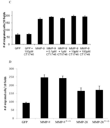
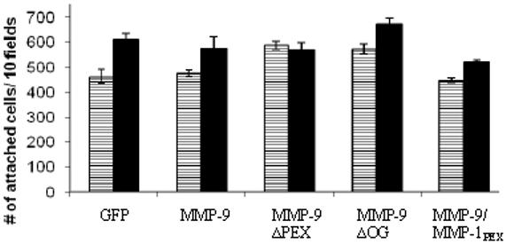
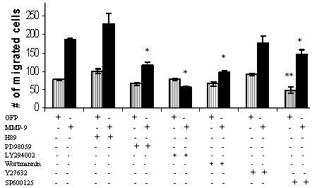
The biological functions of matrix metalloproteinases (MMPs) extend beyond extracellular matrix degradation. Non-proteolytic activities of MMPs are just beginning to be understood. Herein, we evaluated the role of proMMPs in cell migration. Employing a Transwell chamber migration assay, we demonstrated that transfection of COS-1 cells with various proMMP cDNAs resulted in enhancement of cell migration. Latent MMP-2 and MMP-9 enhanced cell migration to a greater extent than latent MMP-1, -3, -11 and -28. To examine if proteolytic activity is required for MMP-enhanced cell migration, three experimental approaches, including fluorogenic substrate degradation assay, transfection of cells with catalytically inactive mutant MMP cDNAs, and addition of hydroxamic acid-derived MMP inhibitors, were employed. We demonstrated that the proteolytic activities of MMPs are not required for MMP-induced cell migration. To explore the mechanism underlying MMP-enhanced cell migration, structure-function relationship of MMP-9 on cell migration was evaluated. By using a domain swapping approach, we demonstrated that the hemopexin domain of proMMP-9 plays an important role in cell migration when examined by a transwell chamber assay and by a phagokinetic migration assay. TIMP-1, which interacts with the hemopexin domain of proMMP-9, inhibited cell migration, whereas TIMP-2 had no effect. Employing small molecular inhibitors, MAPK and PI3K pathways were found to be involved in MMP-9-mediated cell migration. In conclusion, we demonstrated that MMPs utilize a non-proteolytic mechanism to enhance epithelial cell migration. We propose that hemopexin homodimer formation is required for the full cell migratory function of proMMP-9.


Figures

Figure 1
Figure 1
Effect of MMPs expressed in COS-1 cells on random cell migration. A, B) Expression of MMPs in COS-1 cells enhances cell migration. COS-1 cells transfected with corresponding cDNAs as indicated were examined by a Transwell chamber migration assay. Migrated cells were microscopically examined (A) and counted (B). *Significant difference; p < 0.05, as compared to COS-1 cells. C, D) Comparable expression of MMPs in transfected COS-1 cells examined by Western blotting. COS-1 cells transfected with various cDNAs were lysed and immunoblot analysis was performed using the corresponding antibodies as indicated (C). Densitometric analyses of expression levels of MMPs and tubulin, respectively, in COS-1 cells were performed. Relative expression levels of MMPs in COS-1 cells were determined based on ratios between levels of specific MMPs and the corresponding respective levels of tubulin (D).
Figure 1
Figure 1
Effect of MMPs expressed in COS-1 cells on random cell migration. A, B) Expression of MMPs in COS-1 cells enhances cell migration. COS-1 cells transfected with corresponding cDNAs as indicated were examined by a Transwell chamber migration assay. Migrated cells were microscopically examined (A) and counted (B). *Significant difference; p < 0.05, as compared to COS-1 cells. C, D) Comparable expression of MMPs in transfected COS-1 cells examined by Western blotting. COS-1 cells transfected with various cDNAs were lysed and immunoblot analysis was performed using the corresponding antibodies as indicated (C). Densitometric analyses of expression levels of MMPs and tubulin, respectively, in COS-1 cells were performed. Relative expression levels of MMPs in COS-1 cells were determined based on ratios between levels of specific MMPs and the corresponding respective levels of tubulin (D).
Figure 2
Figure 2
ProMMPs enhance cell migration of COS-1 cells. A) No enzymatic activity is detected in the conditioned medium of COS-1 cells transfected with MMP-2 and -9 cDNAs. Fluorogenic substrate peptide was incubated with APMA- or non-APMA-treated conditioned medium, as indicated, followed by measurement of the degradation products using a fluorescent plate reader. B) No effect of TIMP-1 and -2 on MMP-2, -11, and -28-enhanced cell migration. COS-1 cells transfected with a combination of MMPs and TIMP, as indicated, were examined by a Transwell chamber migration assay. Migrated cells were stained and microscopically counted. C) Addition of synthetic MMP inhibitor in MMP-transfected COS-1 cells does not interfere with MMP-9-enhanced cell migration. Synthetic MMP inhibitor CT1746 at different doses was incubated with cells transfected with MMP-9 cDNA, and Transwell migration assay was subsequently performed. Migrated cells were stained and microscopically counted. D, Constitutively inactive MMP-9 and -28 enhance cell migration as well as wild type enzymes. COS-1 cells transfected with wild-type, mutant MMP-9, or -28 were examined by the Transwell migration assay. Migrated cells were stained and microscopically counted.
Figure 2
Figure 2
ProMMPs enhance cell migration of COS-1 cells. A) No enzymatic activity is detected in the conditioned medium of COS-1 cells transfected with MMP-2 and -9 cDNAs. Fluorogenic substrate peptide was incubated with APMA- or non-APMA-treated conditioned medium, as indicated, followed by measurement of the degradation products using a fluorescent plate reader. B) No effect of TIMP-1 and -2 on MMP-2, -11, and -28-enhanced cell migration. COS-1 cells transfected with a combination of MMPs and TIMP, as indicated, were examined by a Transwell chamber migration assay. Migrated cells were stained and microscopically counted. C) Addition of synthetic MMP inhibitor in MMP-transfected COS-1 cells does not interfere with MMP-9-enhanced cell migration. Synthetic MMP inhibitor CT1746 at different doses was incubated with cells transfected with MMP-9 cDNA, and Transwell migration assay was subsequently performed. Migrated cells were stained and microscopically counted. D, Constitutively inactive MMP-9 and -28 enhance cell migration as well as wild type enzymes. COS-1 cells transfected with wild-type, mutant MMP-9, or -28 were examined by the Transwell migration assay. Migrated cells were stained and microscopically counted.
Figure 3
Figure 3
The hemopexin domain of MMP-9 is required for MMP-9-enhanced cell migration. A) Schematic diagram of wild-type and mutant MMP-9. B) Expression of wild-type and mutant MMP-9 in COS-1 cells. The conditioned medium (CM) and lysates (CL) of COS-1 cells transfected with wild-type or mutant MMP-9 cDNAs were examined by Western blotting under denaturing conditions using anti-MMP-9 monoclonal antibodies. C, D, E) Decreased cell migration in COS-1 cells transfected with MMP-9 PEX domain mutation. Migratory ability of COS-1 cells transfected with cDNAs as indicated was examined by a Transwell chamber migration assay (C, p-values reflect comparison with MMP-9 transfected cells: * p < 0.01) or phagokinetic assay (D). Cleared migratory tracks as depicted by the absence of gold particles in phagokinetic assay were measured by ImageJ NIH imaging software (E). p-values were calculated in comparison with MMP-9 transfected cells: * p < 0.01.
Figure 3
Figure 3
The hemopexin domain of MMP-9 is required for MMP-9-enhanced cell migration. A) Schematic diagram of wild-type and mutant MMP-9. B) Expression of wild-type and mutant MMP-9 in COS-1 cells. The conditioned medium (CM) and lysates (CL) of COS-1 cells transfected with wild-type or mutant MMP-9 cDNAs were examined by Western blotting under denaturing conditions using anti-MMP-9 monoclonal antibodies. C, D, E) Decreased cell migration in COS-1 cells transfected with MMP-9 PEX domain mutation. Migratory ability of COS-1 cells transfected with cDNAs as indicated was examined by a Transwell chamber migration assay (C, p-values reflect comparison with MMP-9 transfected cells: * p < 0.01) or phagokinetic assay (D). Cleared migratory tracks as depicted by the absence of gold particles in phagokinetic assay were measured by ImageJ NIH imaging software (E). p-values were calculated in comparison with MMP-9 transfected cells: * p < 0.01.
Figure 4
Figure 4
Interference of MMP-9-induced cell migration by TIMP-1, but not TIMP-2. A) Detection of MMP-9 dimer in the conditioned medium of transfected COS-1 cells. The conditioned media from COS-1 cells transfected with MMP-9 or MMP-9/MMP-1PEX cDNAs were examined by Western blotting under non-denaturing conditions using anti-MMP-9 antibodies. The homodimer was identified in COS-1 cells expressing wild-type MMP-9, but not in the MMP-9/MMP-1PEX. B) Expression of TIMP-1, but not TIMP-2 in cells interferes with MMP-9-mediated cell migration. COS-1 cells were co-transfected with MMP-9 or MMP-9/MMP-1PEX along with TIMP-1 or TIMP-2 cDNAs and cell migration was assessed by a Transwell chamber migration assay. * p < 0.01 C) Exogenous of TIMP-1, but not TIMP-2 inhibits MMP-9-induced cell migration. COS-1 cells transfected with MMP-9 cDNAs were incubated with exogenous TIMP-1 (50 nM) or TIMP-2 (50 nM) followed by the Tranwell chamber migration assay. * p < 0.01
Figure 4
Figure 4
Interference of MMP-9-induced cell migration by TIMP-1, but not TIMP-2. A) Detection of MMP-9 dimer in the conditioned medium of transfected COS-1 cells. The conditioned media from COS-1 cells transfected with MMP-9 or MMP-9/MMP-1PEX cDNAs were examined by Western blotting under non-denaturing conditions using anti-MMP-9 antibodies. The homodimer was identified in COS-1 cells expressing wild-type MMP-9, but not in the MMP-9/MMP-1PEX. B) Expression of TIMP-1, but not TIMP-2 in cells interferes with MMP-9-mediated cell migration. COS-1 cells were co-transfected with MMP-9 or MMP-9/MMP-1PEX along with TIMP-1 or TIMP-2 cDNAs and cell migration was assessed by a Transwell chamber migration assay. * p < 0.01 C) Exogenous of TIMP-1, but not TIMP-2 inhibits MMP-9-induced cell migration. COS-1 cells transfected with MMP-9 cDNAs were incubated with exogenous TIMP-1 (50 nM) or TIMP-2 (50 nM) followed by the Tranwell chamber migration assay. * p < 0.01
Figure 5
Figure 5
MMP-9 expression in transfected COS-1 cells has no effect on cell attachment. COS-1 cells transfected with GFP, MMP-9 and MMP-9 mutant cDNAs were incubated in polycarbonate wells and cell attachment was measured after 1 hour (white) and 3 hours (black). No significant differences in cell attachment were noted at either time point.
Figure 6
Figure 6
Differential effect of small molecular inhibitors on MMP-9-meidated cell migration. COS-1 cells transfected with GFP control and MMP-9 cDNAs were incubated with PD89059 (10 μM), LY294002 (10 μM), Y27632 (1 μM), H89 (10 μM), wortmannin (10 μM) or SP600125 (10 μM) for 6 hours in the Transwell chamber cell migration assay. * p < 0.05 compared to MMP-9 and ** p < 0.05 compared to GFP.

References

    1. Abecassis I, Olofsson B, Schmid M, Zalcman G, Karniguian A. RhoA induces MMP-9 expression at CD44 lamellipodial focal complexes and promotes HMEC-1 cell invasion. Exp. Cell. Res. 2003;291:363–376. - PubMed
    1. Albrect-Buehler G. The phagokinetic tracks of 3T3 cells. Cell. 1997;11:395–404. - PubMed
    1. Allan JA, Docherty AJP, Barker PJ, Huskisson NS, Reynolds JJ, Murphy G. Binding of gelatinases A and B to type-1 collagen and other matrix components. Biochem. J. 1995;309:299–306. - PMC - PubMed
    1. Bannikov GA, Karelina TV, Collier IE, Marmer BL, Goldberg GI. Substrate binding of gelatinase B induces its enzymatic activity in the presence of intact propeptide. J. Biol. Chem. 2002;277:16022–16027. - PubMed
    1. Bennett BL, Sasaki DT, Murray BW, O’Leary EC, Sakata ST, Xu W, Leisten JC, Motiwala A, Pierce S, Satoh Y, Bhagwat SS, Manning AM, Anderson DW. SP600125, an anthrapyrazolone inhibitor of Jun N-terminal kinase. Proc. Natl. Acad. Sci. U S A. 2001;98(24):13681–13686. - PMC - PubMed

Publication types

MeSH terms

Substances