

Genetic modifiers of the physical malformations in velo-cardio-facial syndrome/DiGeorge syndrome
- PMID: 18636633
- PMCID: PMC2818567
- DOI: 10.1002/ddrr.4
Genetic modifiers of the physical malformations in velo-cardio-facial syndrome/DiGeorge syndrome
Abstract
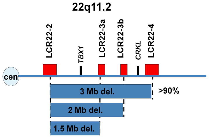
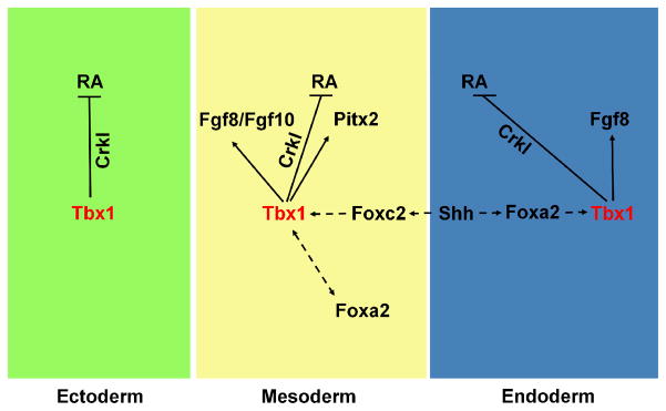
Velo-cardio-facial syndrome/DiGeorge syndrome (VCFS/DGS), the most common micro-deletion disorder in humans, is characterized by craniofacial, parathyroid, and thymic defects as well as cardiac outflow tract malformations. Most patients have a similar hemizygous 3 million base pair deletion on 22q11.2. Studies in mouse have shown that Tbx1, a T-box containing transcription factor present on the deleted region, is likely responsible for the etiology of the syndrome. Furthermore, mutations in TBX1 have been found in rare non-deleted patients. Despite having the same sized deletion, most VCFS/DGS patients exhibit significant clinical variability. Stochastic, environmental and genetic factors likely modify the phenotype of patients with the disorder. Here, we review mouse genetics studies, which may help identify possible genetic modifiers for the physical malformations in VCFS/DGS.
Figures
References
-
- Abu-Issa R, Smyth G, Smoak I, et al. Fgf8 is required for pharyngeal arch and cardiovascular development in the mouse. Development. 2002;129(19):4613–4625. - PubMed
-
- Aggarwal VS, Liao J, Bondarev A, et al. Dissection of Tbx1 and Fgf interactions in mouse models of 22q11DS suggests functional redundancy. Hum Mol Genet. 2006;15(21):3219–3228. - PubMed
-
- Bachiller D, Klingensmith J, Shneyder N, et al. The role of chordin/Bmp signals in mammalian pharyngeal development and DiGeorge syndrome. Development. 2003;130(15):3567–3578. - PubMed
-
- Bockman DE, Redmond ME, Kirby ML. Altered development of pharyngeal arch vessels after neural crest ablation. Ann N Y Acad Sci. 1990;588:296–304. - PubMed
-
- Bottcher RT, Niehrs C. Fibroblast growth factor signaling during early vertebrate development. Endocr Rev. 2005;26(1):63–77. - PubMed
Publication types
MeSH terms
Substances
Grants and funding
LinkOut - more resources
Full Text Sources
