

Synovium-derived stem cell-based chondrogenesis
- PMID: 18637024
- PMCID: PMC2772098
- DOI: 10.1111/j.1432-0436.2008.00299.x
Synovium-derived stem cell-based chondrogenesis
Abstract
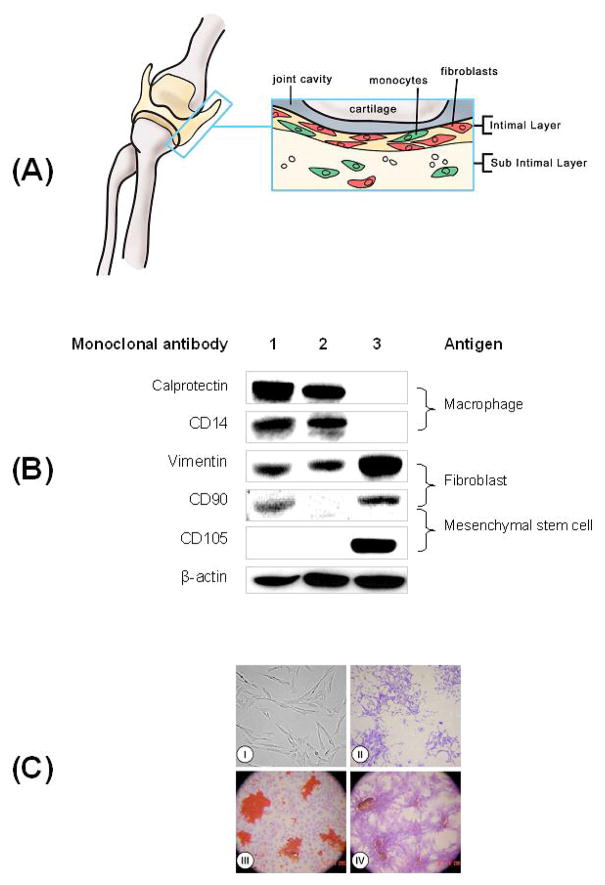
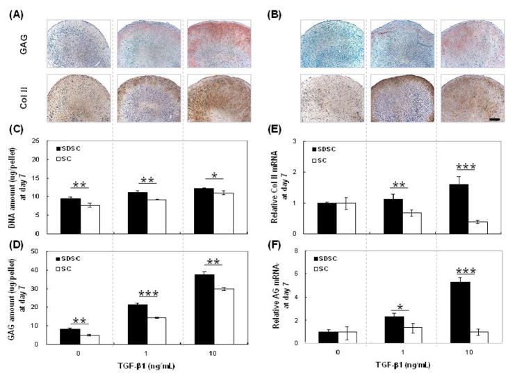
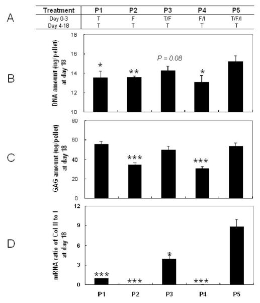
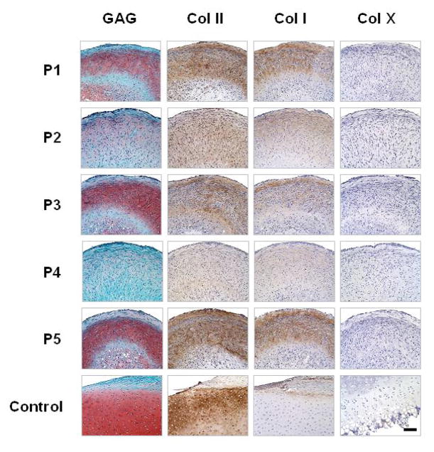
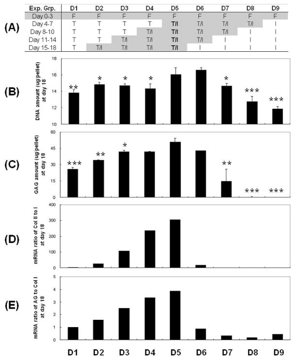
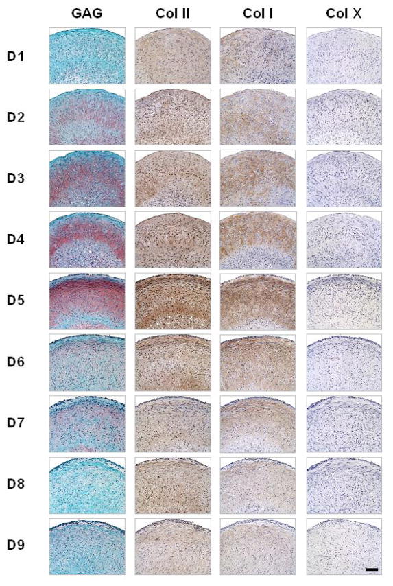
Synovium is considered a candidate source of cells for cartilage tissue engineering. Compared with mesenchymal stem cells (MSCs) from other sources, synovium-derived stem cells (SDSCs) have a higher capacity for chondrogenic differentiation. Our objective was to define cocktails of growth factors that support the growth and chondrogenic differentiation of SDSCs in chemically defined medium. We established a fast and highly selective technique of negative isolation of SDSC populations. The individual and combined effects of three growth factors-transforming growth factor-beta1 (TGF-beta1), insulin-like growth factor I (IGF-I), and basic fibroblast growth factor (FGF-2)-were evaluated in serum-free pellet cultures of SDSCs for the chondrogenesis of SDSCs using histology, biochemical analysis, and real-time RT-PCR. In vitro studies identified TGF-beta1 as the key factor for both the growth and chondrogenesis of SDSCs. The highest rates of SDSC growth were observed with the synergistic interaction of all three factors. With respect to chondrogenic differentiation of SDSCs, the interaction of TGF-beta1 and IGF-I applied simultaneously was superior to the sequential application of these two factors or any other combination of growth factors studied. Based on these findings, we propose a two-step protocol for the derivation of chondrogenic SDSCs: a cocktail of TGF-beta1, IGF-I, and FGF-2 is applied first to induce cell growth followed by a cocktail of TGF-beta1 and IGF-I applied to induce chondrogenesis.
Figures
Similar articles
-
Histone deacetylase 4 promotes TGF-beta1-induced synovium-derived stem cell chondrogenesis but inhibits chondrogenically differentiated stem cell hypertrophy.Differentiation. 2009 Dec;78(5):260-8. doi: 10.1016/j.diff.2009.08.001. Epub 2009 Aug 29. Differentiation. 2009. PMID: 19716643
-
Repair of large animal partial-thickness cartilage defects through intraarticular injection of matrix-rejuvenated synovium-derived stem cells.Tissue Eng Part A. 2013 May;19(9-10):1144-54. doi: 10.1089/ten.TEA.2012.0351. Epub 2013 Jan 15. Tissue Eng Part A. 2013. PMID: 23216161
-
Coculture of synovium-derived stem cells and nucleus pulposus cells in serum-free defined medium with supplementation of transforming growth factor-beta1: a potential application of tissue-specific stem cells in disc regeneration.Spine (Phila Pa 1976). 2009 May 20;34(12):1272-80. doi: 10.1097/BRS.0b013e3181a2b347. Spine (Phila Pa 1976). 2009. PMID: 19455002
-
Enhanced proliferation and chondrogenic differentiation of human synovium-derived stem cells expanded with basic fibroblast growth factor.Tissue Eng Part A. 2011 Apr;17(7-8):991-1002. doi: 10.1089/ten.TEA.2010.0277. Epub 2011 Feb 10. Tissue Eng Part A. 2011. PMID: 21091327
-
Synovium-derived stem cells: a tissue-specific stem cell for cartilage engineering and regeneration.Tissue Eng Part B Rev. 2012 Aug;18(4):301-11. doi: 10.1089/ten.TEB.2012.0002. Epub 2012 Apr 19. Tissue Eng Part B Rev. 2012. PMID: 22429320 Review.
Cited by
-
Human chondrocyte migration behaviour to guide the development of engineered cartilage.J Tissue Eng Regen Med. 2017 Mar;11(3):877-886. doi: 10.1002/term.1988. Epub 2015 Jan 28. J Tissue Eng Regen Med. 2017. PMID: 25627968 Free PMC article.
-
Reconstruction of an in vitro niche for the transition from intervertebral disc development to nucleus pulposus regeneration.Stem Cells Dev. 2013 Apr 15;22(8):1162-76. doi: 10.1089/scd.2012.0597. Epub 2013 Feb 15. Stem Cells Dev. 2013. PMID: 23259403 Free PMC article. Review.
-
Time-dependent processes in stem cell-based tissue engineering of articular cartilage.Stem Cell Rev Rep. 2012 Sep;8(3):863-81. doi: 10.1007/s12015-011-9328-5. Stem Cell Rev Rep. 2012. PMID: 22016073 Free PMC article.
-
Matrilin-3 chondrodysplasia mutations cause attenuated chondrogenesis, premature hypertrophy and aberrant response to TGF-β in chondroprogenitor cells.Int J Mol Sci. 2014 Aug 21;15(8):14555-73. doi: 10.3390/ijms150814555. Int J Mol Sci. 2014. PMID: 25196597 Free PMC article.
-
Tissue engineering of articular cartilage with biomimetic zones.Tissue Eng Part B Rev. 2009 Jun;15(2):143-57. doi: 10.1089/ten.TEB.2008.0563. Tissue Eng Part B Rev. 2009. PMID: 19203206 Free PMC article. Review.
References
-
- Baddoo M, Hill K, Wilkinson R, Gaupp D, Hughes C, Kopen GC, Phinney DG. Characterization of mesenchymal stem cells isolated from murine bone marrow by negative selection. J Cell Biochem. 2003;89:1235–1249. - PubMed
-
- Baker J, Liu JP, Robertson EJ, Efstratiadis A. Role of insulin-like growth factors in embryonic and postnatal growth. Cell. 1993;75:73–82. - PubMed
-
- Bruder SP, Ricalton NS, Boynton RE, Connolly TJ, Jaiswal N, Zaia J, Barry FP. Mesenchymal stem cell surface antigen SB-10 corresponds to activated leukocyte cell adhesion molecule and is involved in osteogenic differentiation. J Bone Miner Res. 1998;13:655–663. - PubMed
-
- Chamberlain G, Fox J, Ashton B, Middleton J. Mesenchymal Stem Cells: their Phenotype, Differentiation Capacity, Immunological Features and Potential for Homing. Stem Cells. 2007;25:2739–2749. - PubMed
-
- Cheifetz S, Bellon T, Cales C, Vera S, Bernabeu C, Massague J, Letarte M. Endoglin is a component of the transforming growth factor-beta receptor system in human endothelial cells. J Biol Chem. 1992;267:19027–19030. - PubMed
Publication types
MeSH terms
Substances
Grants and funding
LinkOut - more resources
Full Text Sources
Medical
