. 2009 Feb;18(2):122-9.
doi: 10.1111/j.1600-0625.2008.00771.x. Epub 2008 Jul 7.

Protein kinase C isoenzymes differentially regulate the differentiation-dependent expression of adhesion molecules in human epidermal keratinocytes

Affiliations

Protein kinase C isoenzymes differentially regulate the differentiation-dependent expression of adhesion molecules in human epidermal keratinocytes

Andrea Szegedi et al. Exp Dermatol. 2009 Feb.

Abstract

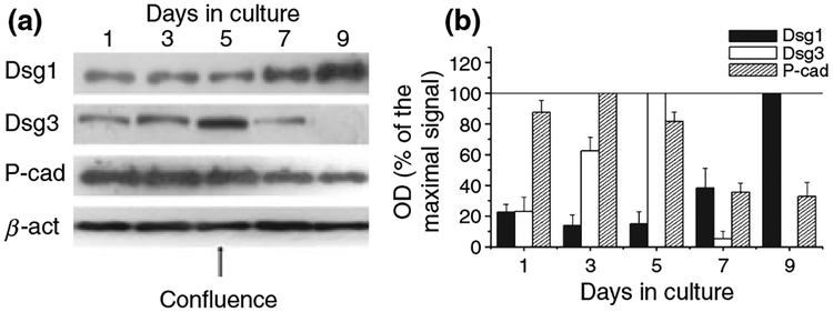
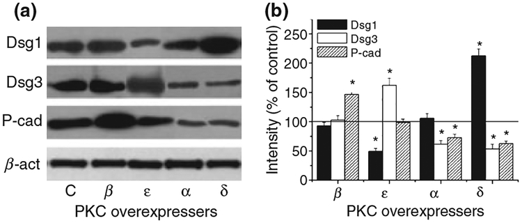
Epidermal expression of adhesion molecules such as desmogleins (Dsg) and cadherins is strongly affected by the differentiation status of keratinocytes. We have previously shown that certain protein kinase C (PKC) isoforms differentially alter the growth and differentiation of human epidermal HaCaT keratinocytes. In this paper, using recombinant overexpression and RNA interference, we define the specific roles of the different PKC isoenzymes in modulation of expression of adhesion molecules in HaCaT keratinocytes. The level of Dsg1, a marker of differentiating keratinocytes, was antagonistically regulated by two Ca-independent 'novel' nPKC isoforms; i.e. it increased by the differentiation-promoting nPKCdelta and decreased by the growth-promoting nPKCepsilon. The expression of Dsg3 (highly expressed in proliferating epidermal layers) was conversely regulated by these isoenzymes, and was also inhibited by the differentiation inducer Ca-dependent 'conventional' cPKCalpha. Finally, the expression of P-cadherin (a marker of proliferating keratinocytes) was regulated by all of the examined PKCs, also in an antagonistic manner (inhibited by cPKCalpha/nPKCdelta and stimulated by cPKCbeta/nPKCepsilon). Collectively, the presented results strongly argue for the marked, differential, and in some instances antagonistic roles of individual Ca-dependent and Ca-independent PKC isoforms in the regulation of expression of adhesion molecules of desmosomes and adherent junctions in human epidermal keratinocytes.


Figures

Figure 1.
Figure 1.
Alteration of expression of adhesion molecules during the high cell density-induced differentiation of HaCaT keratinocytes. (a) HaCaT cells were harvested at various culturing days (confluence was reached at day 5), similar amounts of proteins were subjected to SDS-PAGE, and Western immunoblotting was performed using antibodies against the adhesion molecules Dsg1, Dsg3 and P-cad. To assess equal loading, nitrocellulose membranes were stripped and re-probed with an anti-β-actin antibody (β-act). The figure is a representative of four to five experiments yielding similar results. (b) The amounts of the adhesion molecules were quantitated by densitometry (optical density; OD), the values were normalized to the OD values of β-actin, and the results were expressed as the percentage of the maximal amount of the given molecule within the experiment. Values represent the mean ± SEM of four to five independent experiments.
Figure 2.
Figure 2.
Expression pattern of adhesion molecules in HaCaT keratinocytes overexpressing individual PKC isoforms. (a) Stable transfectants of HaCaT cells overexpressing the different PKC isoforms or the control empty vector (C) were harvested at similar proliferation and differentiation stages (i.e. 80–85% confluence) and similar amounts of proteins were subjected to Western immunoblotting to detect the adhesion molecules Dsg1, Dsg3 and P-cad. To assess equal loading, nitrocellulose membranes were stripped and re-probed with an anti-β-actin antibody (β-act). The figure is a representative of four to five experiments yielding similar results. (b) The amounts of the adhesion molecules were quantitated by densitometry (optical density; OD), then normalized to those of β-actin, and finally expressed as the percentage of the control (empty vector transfected) regarded as 100%. Values represent the mean ± SEM of five to six independent experiments. Asterisks mark significant (P < 0.05) differences, defined by Student’s t-test, compared with control.
Figure 3.
Figure 3.
Effect of RNAi-driven ‘knock-down’ of individual PKC isoforms on the expression of adhesion molecules in HaCaT keratinocytes. (a) Various specific RNAi probes (Sp) against the PKC isoforms as well as the scrambled (control) RNAi probes (Sc) were introduced to the cells as described under ‘Materials and methods’ section. Forty-eight hours after transfection, cells were harvested and subjected to Western blot analysis (WB) to define the expression of the ‘targeted’ PKC isoform; the other PKC isoforms; the various adhesion molecules (Dsg1, Dsg3 and P-cad); and the endogenous control β-actin (β-act). In addition, to monitor the lack of effect of transfection, the expression of PKC isoforms was compared in non-transfected (NT) and scrambled RNAi probes (Sc) transfected cells. The figure is a representative of five to six experiments yielding similar results. (b) Time-course of efficacy of inhibition of PKC isoform expression as assessed by Western blotting at various time-points after transfection with RNAi. The amounts of the individual PKC isoforms were quantitated by densitometry (optical density; OD), then normalized to those of β-actin, and finally expressed as the percentage of the scrambled RNAi probes transfected (control) samples regarded as 100%. (c) Time-course of efficacy of inhibition of PKC isoform mRNA levels as assessed by Q-PCR at various time-points after transfection. PKC-specific transcripts were determined as described under ‘Materials and methods’ section; then, values were normalized to those of β-actin and finally expressed as the percentage of the scrambled RNAi probe transfected (control) samples regarded as 100%. (d) Western blot analysis of adhesion molecules 48 h after transfection. The amounts of the adhesion molecules were quantitated by densitometry (optical density; OD), then normalized to those of β-actin, and finally expressed as the percentage of the scrambled RNAi probes transfected (control) samples regarded as 100%. In b–d, points represent the mean ± SEM of five to six independent experiments and asterisks mark significant (P < 0.05) differences, defined by Student’s t-test, compared with the respective controls.
Figure 4.
Figure 4.
Effect of the nPKCδ inhibitor rottlerin on the expression of adhesion molecules in HaCaT keratinocytes. (a) Cells were treated with various concentrations of rottlerin for 48 h, then harvested and similar amounts of proteins were subjected to Western immunoblotting to detect the adhesion molecules Dsg1, Dsg3 and P-cad. To assess equal loading, nitrocellulose membranes were stripped and re-probed with an anti-β-actin antibody (β-act). The figure is a representative of four to five experiments yielding similar results. (b) The amounts of the adhesion molecules were quantitated by densitometry (optical density; OD), then normalized to those of β-actin, and finally expressed as the percentage of the vehicle-treated (control, C) cells regarded as 100%. Points represent the mean ± SEM of four to five independent experiments. Asterisks mark significant (P < 0.05) differences, defined by Student’s t-test, compared with control.

References

    1. Koch PJ, Franke WW. Desmosomal cadherins: another growing multigene family of adhesin molecules. Curr Opin Cell Biol 1994: 6: 682–687. - PubMed
    1. North AJ, Bardsley WG, Hyam J et al. Molecular map of the desmosomal plague. J Cell Sci 1999: 112: 4325–4336. - PubMed
    1. Amagai M, Ahmed AR, Kitajima Y et al. Are desmoglein auto-antibodies essential for the immunopathogenesis of pemphigus vulgaris, or just ‘witnesses of disease’? Exp Dermatol 2006: 15: 815–831. - PubMed
    1. Garrod DR, Merritt AJ, Nie Z. Desmosomal cadherins. Curr Opin Cell Biol 2002: 14: 537–545. - PubMed
    1. Ishikawa H, Li K, Tamai K, Sawamura D, Uitto J. Cloning of the mouse desmoglein 3 gene (Dsg3): interspecies conservation within the cadherin superfamily. Exp Dermatol 2000: 9: 229–239. - PubMed

Publication types

MeSH terms