. 2008 Jul 18:6:20.
doi: 10.1186/1477-5956-6-20.

Profiling of serum and tissue high abundance acute-phase proteins of patients with epithelial and germ line ovarian carcinoma

Affiliations

Profiling of serum and tissue high abundance acute-phase proteins of patients with epithelial and germ line ovarian carcinoma

Yeng Chen et al. Proteome Sci. .

Abstract

Background: Acute-phase response involves the simultaneous altered expression of serum proteins in association to inflammation, infection, injury or malignancy. Studies of the acute-phase response usually involve determination of the levels of individual acute-phase serum proteins. In the present study, the acute-phase response of patients with epithelial (EOCa) and germ-line (GOCa) ovarian carcinoma was investigated using the gel-based proteomic approach, a technique which allowed the simultaneous assessment of the levels of the acute-phase serum high abundance proteins. Data obtained were validated using ELISA and immunostaining of biopsy samples.

Results: Enhanced expression of clusterin (CLU), alpha1-antitrypsin, haptoglobin and leucine rich glycoprotein was detected in all patients. However, the levels of alpha1-antichymotrypsin (ACT) was only enhanced in EOCa patients, while patients with GOCa were typically characterized by elevated levels of ceruloplasmin but lower levels of alpha2-HS glycoprotein. The enhanced expression of CLU in EOCa and GOCa patients and up-regulated expression of ACT specifically in EOCa patients were confirmed by ELISA. Immunohistochemical staining of biopsy samples of EOCa and GOCa patients demonstrated correlation of the acute-phase protein expression.

Conclusion: Patients with EOCa and GOCa demonstrated distinctive aberrant expression of serum and tissue high abundance acute-phase proteins compared to negative control women.


Figures

Figure 1
Figure 1
Typical 2-DE serum protein profiles of the negative control women and patients with GOCa and EOCa. Unfractionated serum samples of patients and negative controls were subjected to 2-DE and silver staining. Panel A demonstrates a typical representative 2-DE serum protein profile of negative control women. Panels B and C demonstrate typical representative unfractionated serum protein profiles of the patients with GOCa and EOCa, respectively. The aberrantly expressed proteins are marked in brackets for panels B and C. For all panels, the acidic sides of the 2-DE gels are to the left and relative molecular mass declines from the top.
Figure 2
Figure 2
Percentage of volume contribution of 2-DE detectable serum proteins of control women (Con) and patients with EOCa and GOCa. Percentage of volume contribution (%vol) of protein spots was analyzed by ImageMaster™ 2D Platinum Software Version 5. Analysis was restricted to twelve clusters of protein spots.
Figure 3
Figure 3
Analysis of serum protein expression by competitive ELISA. Competitive ELISA was preformed using sheep/goat anti-human ACT, AHS, CLU, CPL, AAT and HAP as primary antibodies. Analyses were performed in triplicate. Relative expression of serum proteins in GOCa and EOCa patients is defined as percentage (%) of up- or down regulation from that of negative controls.
Figure 4
Figure 4
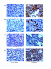
Immunohistochemical analysis of protein expression in ovarian tissues of normal donors and ovarian carcinoma patients. Immunohistochemical staining was performed using antisera to (A) CPL, (B) CLU, (C) ACT, and (D) AAT. Panels A and B demonstrate immunohistochemical stains showing strong positive CPL and CLU expression in ovarian cancer (OCa) tissues but negative expression in normal ovarian tissues (internal positive control). Panels C and D demonstrate immunohistochemical stains showing moderate positive ACT and strong positive AAT expression in OCa tissues, respectively, but weak positive expression in normal ovarian tissues.

References

    1. Gabay J, Kushner I. Acute-phase proteins and other systemic responses to inflammation. N Engl J Med. 1999;340:448–454. doi: 10.1056/NEJM199902113400607. - DOI - PubMed
    1. Adkins JN, Varnum SM, Auberry KJ, Moore RJ, Angell NH, Smith RD, Springer DL, Pounds JG. Toward a human blood serum proteome – Analysis by multidimensional separation coupled with mass spectrometry. Mol Cell Proteomics. 2002;1:947–955. doi: 10.1074/mcp.M200066-MCP200. - DOI - PubMed
    1. Wang YY, Cheng P, Chan DW. A simple affinity spin tube filter method for removing high-abundant common proteins or enriching low-abundant biomarkers for serum proteomic analysis. Proteomics. 2003;3:243–248. doi: 10.1002/pmic.200390036. - DOI - PubMed
    1. Pieper R, Gatlin CL, Makusky AJ, Russo PS, Schatz CR, Miller SS, Su Q, McGrath AM, Estock MA, Parmar PP, Zhao M, Huang ST, Zhou J, Wang F, Esquer-Blasco R, Anderson NL, Taylor J, Steiner S. The human serum proteome: Display of nearly 3700 chromatographically separated protein spots on two-dimensional electrophoresis gels and identification of 325 distinct proteins. Proteomics. 2003;3:1345–1364. doi: 10.1002/pmic.200300449. - DOI - PubMed
    1. Veenstra TD, Conrads TP, Hood BL, Avellino AM, Ellenbogen RG, Morrison RS. Biomarkers: Mining the biofluid proteome. Mol Cell Proteomics. 2005;4:409–418. doi: 10.1074/mcp.M500006-MCP200. - DOI - PubMed