Review
. 2008 Nov 30;163(1-3):44-53.
doi: 10.1016/j.resp.2008.06.019. Epub 2008 Jun 28.

Cell wounding and repair in ventilator injured lungs

Affiliations
Review

Cell wounding and repair in ventilator injured lungs

Richard A Oeckler et al. Respir Physiol Neurobiol. .

Abstract

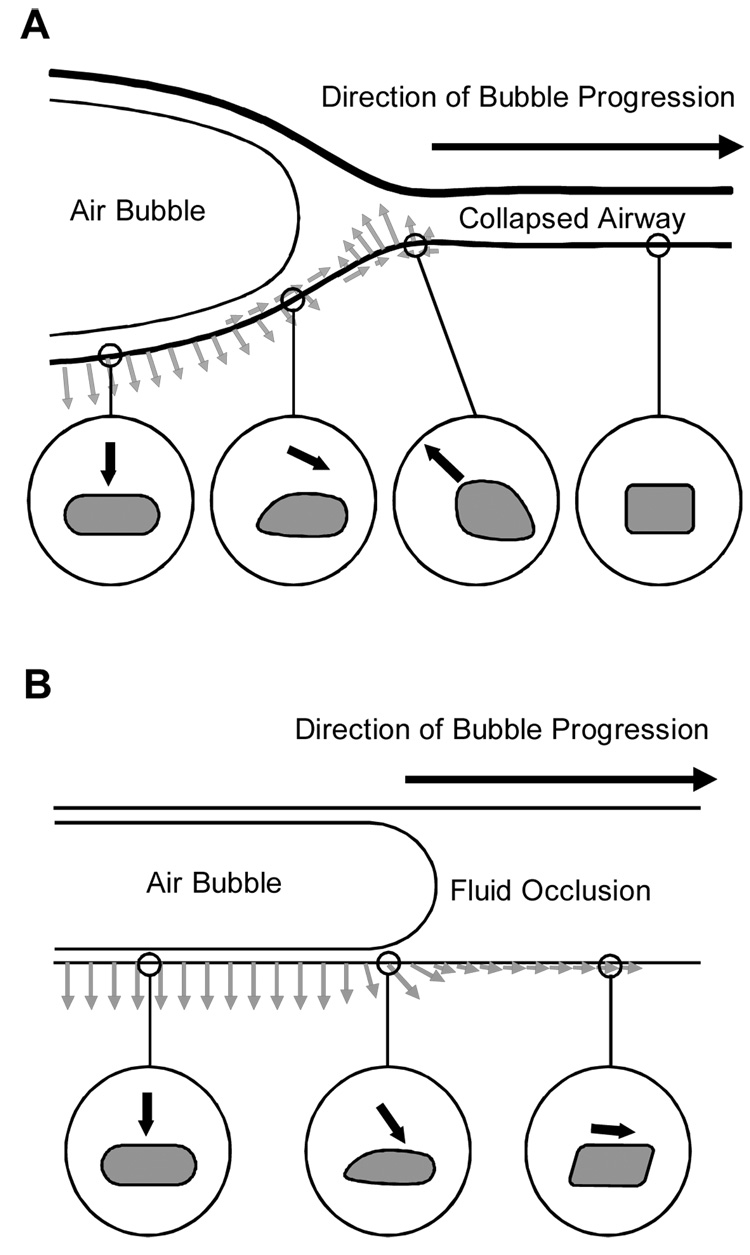
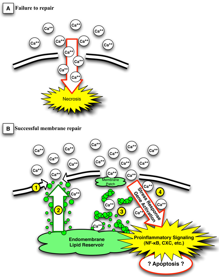
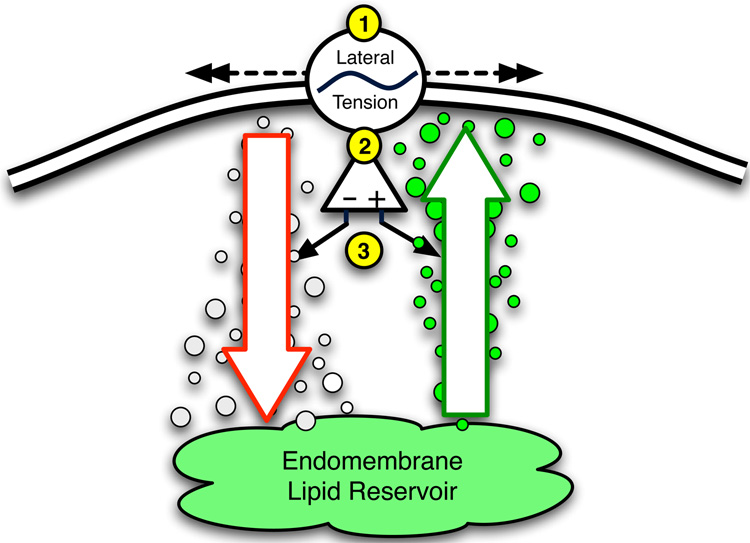
Acute lung injury (ALI) is a common, frequently hospital-acquired condition with a high morbidity and mortality. The stress associated with invasive mechanical ventilation represents a potentially harmful exposure, and attempts to minimize deforming stress through low tidal ventilation have proven efficacious. Lung cells are both sensors and transducers of deforming stress, and are frequently wounded in the setting of mechanical ventilation. Cell wounding may be one of the drivers of the innate immunologic and systemic inflammatory response associated with mechanical ventilation. These downstream effects of mechanotransduction have been referred to collectively as "Biotrauma". Our review will focus on cellular stress failure, that is cell wounding, and the mechanisms mediating subsequent plasma membrane repair, we hold that a better mechanistic understanding of cell plasticity, deformation associated remodeling and repair will reveal candidate approaches for lung protective interventions in mechanically ventilated patients. We will detail one such intervention, lung conditioning with hypertonic solutions as an example of ongoing research in this arena.


Figures

Figure 1
Figure 1
Schematic of alveolar septae contributing to acinar surface area compensation in response to volume-induced deformations. Ventilation at normal tidal volumes result in little change in acinar volume, whereas once volume increases such that septal unfolding is complete, wall stress will increase and acinar volume will change, potentially resulting in overdistension injury.
Figure 2
Figure 2
The baby lung concept and regional overexpansion. White circles represent functional aerated or recruitable units. Black circles represent non-recruitable units. In the healthy lung (top), distribution of tidal volume across lung units results in normal, non-injurious ventilation whereas in the heterogeneously injured lung (bottom), nonuniform distribution of tidal volume occurs to preferentially recruitable units, causing overexpansion and potential injury to basement and plasma membranes.
Figure 3
Figure 3
Hypothetical stresses imparted on the epithelial cells of an airway during reopening (Reprinted with permission from Bilek et al(Bilek et al. 2003)). (A) A collapsed compliant airway shown to the right is forced open by a finger of air moving from left to right. A dynamic wave of stresses is imparted on the airway tissues as the bubble progresses. The circles show the cycle of stresses an airway epithelial cell might experience during reopening. The cell far downstream is nominally stressed. As the bubble approaches, the cell is a pulled up and toward the bubble. As the bubble passes, the cell is pushed away from the bubble. After the bubble has passed, the cell is pushed outward. (B) A fluid occlusion in a rigid narrow channel is cleared by the progression of a finger of air moving from left to right. A dynamic wave of stresses is imparted on the pulmonary epithelial cells lining the channel wall. The circles show the cycle of stresses that the cells might experience during reopening. Far downstream the cell is pushed forward and slightly out. As the bubble approaches, a sudden rise in pressure and a peak in shear stress occurs, pushing the cell forward and outward with much greater force. After the bubble has passed, the cell is pushed outward.
Figure 4
Figure 4
Propidium iodide imaging of a lung section. Propidium iodide (red color) is impermeable to the plasma membrane (PM). It is able to enter the cell after a PM disruption such as seen under conditions of cellular injury. Once in the cell, it intercalates with nucleic acid, fluorescing red after excitiation at the proper wavelength. The detection of PI necessarily means a PM break has occurred. See text for details.
Figure 5
Figure 5
The two possible fates of a damaged cell. In panel A, the plasma membrane is not resealed, calcium freely enters the cell, and cell death ensues by cellular necrosis. In panel B, the 3 mechanisms of plasma membrane repair are illustrated. Lateral lipid flow (#1) can fill the smallest defects, and is the primary mechanism in the human red blood cell. Lipid trafficking to the membrane (#2) accounts for the resealing and repair in most smaller membrane disruptions, while the formation of membrane plugs by coalescence of lipid vesicles in the presence of calcium influx fills larger membrane gaps (#3). Membrane disruption and ion flux (#4) can activate important stress response genes and proinflammatory signaling cascades such as NF-kappaB and CXC chemokines. This in turn may lead to prolonged response to the membrane defect or even apoptotic changes, which may result in damage in excess of that caused by necrotic cell death alone (see text for discussion).
Figure 6
Figure 6
Schematic representation of deformation-induced lipid trafficking (DILT). Deformations of the plasma membrane (PM) result in changes in lateral PM tension. Increases in lateral tension lead to an increase in total PM content through a net increase in exocytic lipid trafficking from the endomembrane to PM, while endocytosis and a decrease in total PM predominates under conditions of decreased lateral tension.
Figure 7
Figure 7
Measuring plasma membrane tension via optically trapped lipid tethers. The top panel represents a schematic diagram of a plasma membrane (PM) lipid tether being pulled from a cell, while the bottom panel is a representative tracing of the force vs. displacement curve measured in realtime. A 0.5–1.0 micron, coated bead is optically trapped and inserted into the PM (#1). The bead acts as a handle, and as it is moved away from the cell, a lipid “tether.” Note the initial force increase to pull this tether from the PM (#1, lower panel). As the bead is pulled further toward #2, force plateaus over a finite displacement confirming the presence of a “lipid reservoir.” As the reservoir is used up, no further lipid can be trafficked to the membrane to buffer the tether and force begins to increase (#3) until the bead (and tether) are no longer able to be held by the optical trap. The recoil of the tether pulls the bead back to the PM surface (#1). See text for full discussion.

References

    1. The Acute Respiratory Distress Syndrome Network. Ventilation with lower tidal volumes as compared with traditional tidal volumes for acute lung injury and the acute respiratory distress syndrome. N Engl J Med. 2000;342:1301–1308. - PubMed
    1. Abraham E. Neutrophils and acute lung injury. Crit Care Med. 2003;31:S195–S199. - PubMed
    1. Bachofen H, Schurch S. Alveolar surface forces and lung architecture. Comp Biochem Physiol A Mol Integr Physiol. 2001;129:183–193. - PubMed
    1. Bilek AM, Dee KC, Gaver DP., 3rd Mechanisms of surface-tension-induced epithelial cell damage in a model of pulmonary airway reopening. J Appl Physiol. 2003;94:770–783. - PubMed
    1. Caruso P, Meireles SI, Reis LF, Mauad T, Martins MA, Deheinzelin D. Low tidal volume ventilation induces proinflammatory and profibrogenic response in lungs of rats. Intensive Care Med. 2003;29:1808–1811. - PubMed