. 2008 Sep;180(3):1154-60.
doi: 10.1016/j.juro.2008.04.140. Epub 2008 Jul 18.

Histopathological assessment of prostate cancer bone osteoblastic metastases

Affiliations

Histopathological assessment of prostate cancer bone osteoblastic metastases

Martine P Roudier et al. J Urol. 2008 Sep.

Abstract

Purpose: Many patients with prostate cancer have bone metastases that appear osteoblastic on radiography, and yet these patients are at increased risk for fracture. The discrepancy between the radiological and clinical aspects of those events is not well understood. We better characterized the histopathology of bone processes in prostate cancer bone metastases.

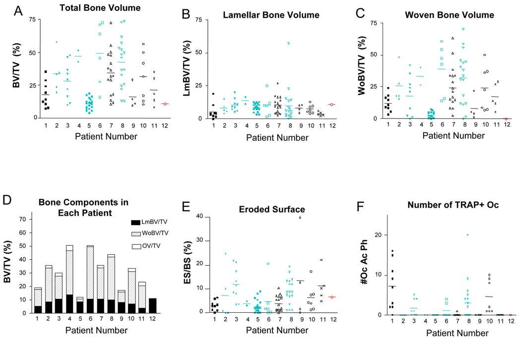
Materials and methods: Histomorphometry was used to evaluate multisite bone biopsies in 12 patients who died with multiple bone metastases, of whom 7 had received bisphosphonate therapy.

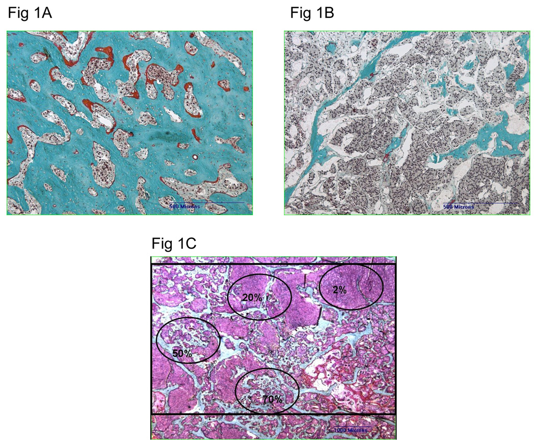
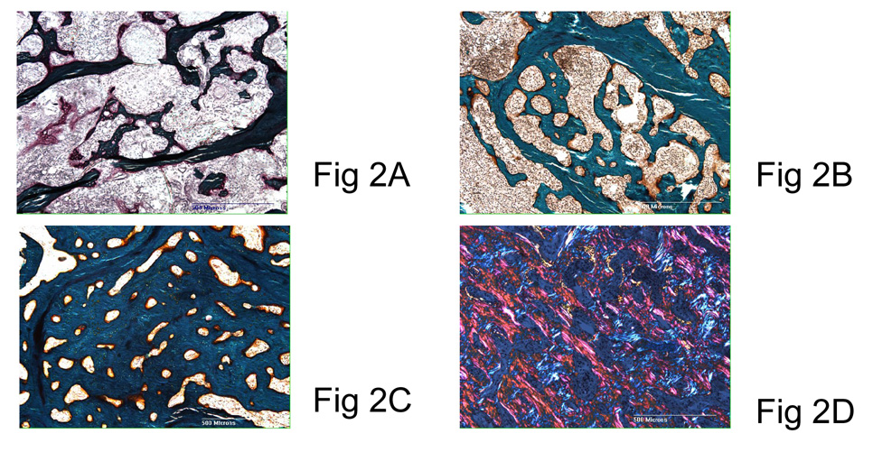
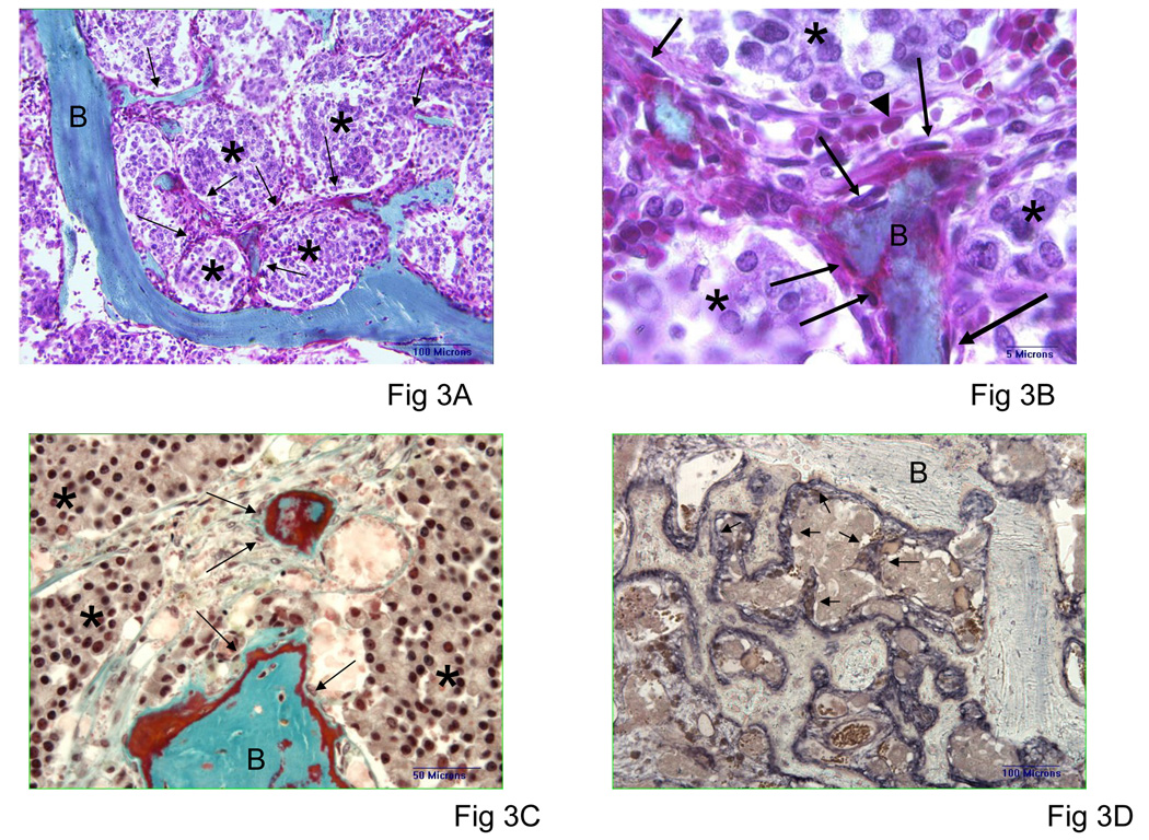
Results: Bone histomorphometry revealed a wide spectrum of cancer induced bone changes in different metastatic sites in individual patients, ranging from a pronounced osteodense to a pronounced osteopenic type. Each metastatic lesion was associated with various amounts of resorption. Decreased bone volume was seen in half of all biopsies. Osteodense lesions were largely composed of under mineralized woven bone, which increases the frailty of new bone. Interestingly woven bone was produced by alkaline phosphatase spindle-shaped cells arising from the connective stroma surrounding tumor cells. The bone response generally was similar in bisphosphonate treated patients and those who did not receive bisphosphonate.

Conclusions: Despite the osteoblastic nature of bone metastases in prostate cancer, the osteolytic-osteopenic bone lesions found in each clinically osteoblastic case may explain the frequent fractures observed in these cases. In addition, the finding that woven bone formed directly from the tumor stroma and not from the adjacent bone surface supports further research into the mechanisms of abnormal bone formation in prostate cancer bone metastases.


Figures

Fig. 1
Fig. 1
Spectrum of histological patterns observed in prostate cancer bone metastases from one patient: (A) Osteodense biopsy and (B) osteopenic biopsy taken from two different anatomical sites in the same patient. In all biopsies, bone marrow is entirely filled with prostate cancer cells. Magnification 5x. (C) Low magnification (1.25x) of a metastatic biopsy showing heterogeneity of bone volumes and remodeling types within a single biopsy; bone is green, prostate cancer is pink, and osteoid is red. The black rectangle on the photograph represents the area measured in each biopsy and corresponds to 15 consecutive fields in the image analysis system. Note the difference between approximative bone volumes indicated in the 5 black ovals drawn over the photograph. Goldner’s-Masson-Trichrome stain.
Fig. 2
Fig. 2
Photomicrographs of tumor-bearing bone biopsies showing progression of the osteodense/osteoblastic pattern of prostate cancer bone metastases. (A) Tumor bearing lesion with early irregular tumor-induced bone trabeculae arising from the tumor-infiltrated bone marrow space and bridging normal-looking native bone trabeculae. Note the osteolysis of native trabeculae. (B) Thickening of tumor-induced irregular bone trabeculae. Network of large tumor-induced trabeculae with small remains of barely visible native bone trabeculae. Note association of a large osteolytic pattern remodeling native and new bone trabeculae. (C) Osteodense osteoblastic pattern consisting of thick new bone trabeculae filling most of the bone marrow space, note the persistence of one native bone trabeculae and the absence of lytic component. Goldner’s-Masson-Trichrome stain. Magnification 5x. (D) Aspect of a large osteodense tumor-bearing biopsy (same as shown in Fig1A) observed with polarization. The dense bone formed between the native trabeculae network was totally woven bone without lamellar bone. Goldner’s-Masson-Trichome stain. Magnification 5x.
Fig. 3
Fig. 3
Formation of tumor-induced bone in prostate cancer bone metastases (undecalcified sections). (A) Early new bone formation arising from tumor stroma (arrows). Lamellar bone and new bone are blue/green (letter B). The bone formation arises from stroma walls of the tumor (tumor is marked by an asterix). Goldner’s-Masson-Trichrome stain. Magnification 20x. (B) Higher magnification of Fig 3A showing detail of spindle-shaped cells (arrows) close to osteoid and capillary (short arrow). Goldner’s-Masson-Trichrome stain. Magnification 40x. (C) Spindle cells (arrows) forming woven bone are alkaline phosphatase positive (black); NBT/BCIP. Magnification 20x.
Fig. 4
Fig. 4
Histomorphometric measurements of biopsies from the 12 patients: (A) bone volume/tissue volume; (B) lamellar bone volume/tissue volume (LmBV); (C) woven bone volume/tissue bone volume (WoBV/TV); (D) percentage of bone components in each patient: lamellar bone in dark green (LmBV/TV), woven bone in light green (WoBV/TV), and osteoid bone volume in orange (OV/TV); (E) eroded surface/bone surface; and (F) osteoclast number. Patients indicated in blue were treated with bisphosphonates. Patient 12 had only non tumor-bearing biopsies; data from one non-tumor bearing biopsy are displayed. Bars represent median values.
Fig. 5
Fig. 5
TRAP-positive osteoclasts in bone metastases. TRAP-toluidine blue staining. Magnification 10x. (A) Bisphosphonate-treated patients; TRAP-positive osteoclasts are large and pale. (B) Non–bisphosphonate-treated patients; TRAP-positive osteoclasts are small and bright red.
Fig. 6
Fig. 6
Comparison of (A) bone volume/tissue volume, (B) woven bone volume/tissue volume, (C) osteoid volume/tissue volume, (D) eroded surface/bone surface, (E) osteoclast surface/bone surface, and (F) osteoclast number in bisphosphonate-treated and non–bisphosphonate-treated patients. There was no significant difference between any of the bone criteria.

References

    1. Roudier MP, Vesselle H, True LD, Higano CS, Ott SM, King SH, et al. Bone histology at autopsy and matched bone scintigraphy findings in patients with hormone refractory prostate cancer: the effect of bisphosphonate therapy on bone scintigraphy results. Clinical and Experimental Metastasis. 2003;20:171. - PubMed
    1. Wymenga LF, Groenier K, Schuurman J, Boomsma JH, Elferink RO, Mensink HJ. Pretreatment levels of urinary deoxypyridinoline as a potential marker in patients with prostate cancer with or without bone metastasis. BJU International. 2001;88:231. - PubMed
    1. Plebani M, Bernardi D, Zaninotto M, De Paoli M, Secchiero S, Sciacovelli L. New and traditional serum markers of bone metabolism in the detection of skeletal metastases. Clinical Biochemistry. 1996;29:67. - PubMed
    1. Berruti A, Dogliotti L, Gorzegno G, Torta M, Tampellini M, Tucci M, et al. Differential patterns of bone turnover in relation to bone pain and disease extent in bone in cancer patients with skeletal metastases. Clinical Chemistry. 1999;45:1240. see comment. - PubMed
    1. Clarke NW, McClure J, George NJ. Morphometric evidence for bone resorption and replacement in prostate cancer. British Journal of Urology. 1991;68:74. - PubMed

Publication types

Substances