. 2008 Oct;62(2):167-76.
doi: 10.1016/j.diagmicrobio.2008.06.005. Epub 2008 Jul 17.

Reconsideration of viral protein immunoblotting for differentiation of human herpes simplex viruses

Affiliations

Reconsideration of viral protein immunoblotting for differentiation of human herpes simplex viruses

Robert N Bowles et al. Diagn Microbiol Infect Dis. 2008 Oct.

Abstract

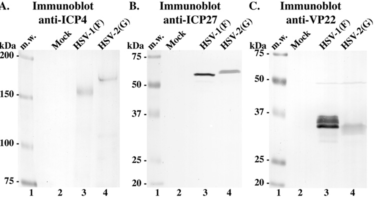
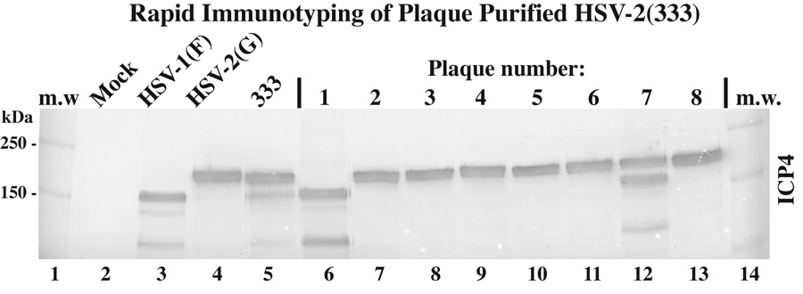
Herpes simplex virus type 1 (HSV-1) and type 2 (HSV-2) are ubiquitous human pathogens that infect their hosts for life and reactivate to cause disease at or near the initial site of infection. As the incidence of genital HSV-1 infections increase, there is an increased demand for valid viral typing diagnostics. In this report, we reconsidered and developed a triple-phase immune-typing procedure that compares differences in electrophoretic mobilities of viral ICP4, ICP27, and VP22 proteins between HSV-1 and HSV-2 strains. We isolated and immunotyped 5 primary HSV-1 strains derived from orofacial, ocular, and genital areas along with 2 primary HSV-2 strains from the genital area. Advantages of this methodology include its general technical simplicity, sensitivity, and ability to definitively type HSV. It is anticipated that this methodology will be useful in distinguishing viruses obtained in clinical cultures.


Figures

Fig. 1
Fig. 1
Immunoblots of HSV-1 and HSV-2 ICP4, ICP27, and VP22 proteins. Lysates of mock-, HSV-1(F)-, and HSV-2(G)-infected cells were separated in three independent denaturing gels (9.3, 15, and 17.5%, respectively), transferred onto nitrocellulose, and probed for (Panel A) ICP4, (Panel B) ICP27, and (Panel C) VP22. Locations (kDa) of molecular mass markers (m.w.) are indicated in the left margins.
Fig. 2
Fig. 2
Schematic representation of the protocol for obtaining clinical isolates (Top Panel). Specific details of the procedure used for obtaining the HSV clinical isolates used in this study are outlined in the text. The complete pathways (filled arrowheads) yields a plaque-purified isolate, while the detoured open arrowhead route bypasses this step. Photographic images of plaques produced by HSV-1, HSV-2 and HSV(TC) viruses (A–C). Confluent Vero cells were infected with (Panel A) HSV-1(F), (Panel B) HSV-2(G), or (Panel C) HSV(TC) at 37°C for approximately 2 days before staining with Geisma dye. Comparison of the three photographs demonstrates similar plaque morphologies.
Fig. 3
Fig. 3
ICP4, ICP27, and VP22 immunoblot typing of HSV(TC). Lysates of mock-, HSV-1(F)-, HSV-2(G)-, and HSV(TC)-infected cells were separated in three independent denaturing gels (9.3, 15, and 17.5%, respectively), transferred onto nitrocellulose, and probed for (Panel A) ICP4, (Panel B) ICP27, and (Panel C) VP22. Locations (kDa) of molecular mass markers (m.w.) are indicated in the left margins.
Fig. 4
Fig. 4
Immunoblot differentiation of HSV(TC) (D). In this case, mock-, HSV-1(F)-, HSV-2(G)-, and HSV(TC)-infected cell lysates were run on a single 15% denaturing gel, transferred onto nitrocellulose which was cut into three pieces, and immunoblotted for ICP4, ICP27, and VP22. Locations (kDa) of molecular mass markers (m.w.) are indicated in the left margin.
Fig. 5
Fig. 5
Immunoblot typing of ocular samples (A). Lysates of mock-, HSV-1(F)-, HSV-2(G)-, HSV(MS)-, and HSV(MB)-infected cells were run on a single 15% denaturing gel, transferred onto nitrocellulose, cut into two pieces, and probed for ICP4 and ICP27. Locations (kDa) of molecular mass markers (m.w.) are indicated in the left margin.
Fig. 6
Fig. 6
Immunoblot typing of genital samples (B). Lysates of mock-, HSV-1(F)-, HSV-1(TC)-, HSV-2(G)-, HSV-2(333)-, HSV(C2)-, HSV(C3)-, HSV(C4)-, and HSV(C9)-infected cells were run on a single 15% denaturing gel and immunoblotted for ICP4, ICP27, and VP22. Locations (kDa) of molecular mass markers (m.w.) are indicated in the right margin.
Fig. 7
Fig. 7
Immunoblot typing of plaque purified HSV-2(333) (C). Lysates of mock-, HSV-1(F)-, HSV-2(G)-, HSV(333)-, and eight isolated plaque-infected cells were immunoblotted for ICP4. Locations (kDa) of molecular mass markers (m.w.) are indicated in the left margin.

References

    1. Adelson ME, Feola M, Trama J, Tilton RC, Mordechai E. Simultaneous detection of herpes simplex virus types 1 and 2 by real-time PCR and Pyrosequencing. J Clin Virol. 2005;33(1):25–34. - PubMed
    1. Baker DA. Herpes simplex virus infections. Curr Opin Obstet Gynecol. 1992;4(5):676–681. - PubMed
    1. Batterson W, Roizman B. Characterization of the herpes simplex virion-associated factor responsible for the induction of alpha genes. J Virol. 1983;46(2):371–377. - PMC - PubMed
    1. Blaho JA, Mitchell C, Roizman B. Guanylylation and adenylylation of the alpha regulatory proteins of herpes simplex virus require a viral beta or gamma function. J Virol. 1993;67(7):3891–3900. - PMC - PubMed
    1. Blaho JA, Mitchell C, Roizman B. An amino acid sequence shared by the herpes simplex virus 1 alpha regulatory proteins 0, 4, 22, and 27 predicts the nucleotidylylation of the UL21, UL31, UL47, and UL49 gene products. J Biol Chem. 1994;269(26):17401–17410. - PubMed

Publication types

MeSH terms

Substances