Review
. 2009 Jan-Feb;130(1-2):10-7.
doi: 10.1016/j.mad.2008.06.002. Epub 2008 Jul 1.

Antagonistic pleiotropy and p53

Affiliations
Review

Antagonistic pleiotropy and p53

Erica Ungewitter et al. Mech Ageing Dev. 2009 Jan-Feb.

Abstract

George Williams' antagonistic pleiotropy theory of aging proposes that cellular damage and organismal aging are caused by pleiotrophic genes, or genes with multiple phenotypic effects [Williams, G.C., 1957. Pleiotropy, natural selection, and the evolution of senescence. Evolution 11, 398-411]. According to this theory, genes that exhibit antagonistic pleiotropy increase the odds of successful reproduction early in life, but have deleterious effects later in life. The tumor suppressor p53 confers protection against cancer (and death) by interrupting the abnormal proliferation of cells. When control of proliferation is applied to normal stem cells, however, it can impair tissue homeostasis and accelerate aging. We use data from recently developed models of accelerated aging in mice to determine if the deleterious effects of p53 on aging reflect antagonistic pleiotropy of the p53 gene or are attributable to genes that can modify p53 activity but are evolving independently.


Figures

Figure 1
Figure 1. p53 protein isoform structure
The amino acid positions denoted above each isoform correspond to the murine protein sequence. Arrows mark the regions containing important sites for phosphorylation in the N-terminus or acetylation in the C-terminus for each isoform. Functional domains are coded by the following colors: activator domain 1 (AD1), yellow; activator domain 2 (AD2) and the proline-rich domain (PRD), red; DNA-binding domain (DBD) and nuclear localization signal (NLS), blue; tetramerization domain (TD), grey; and the C-terminal basic domain (BD), green. (A) Full-length p53 initiates at codon 1. (B) Δ40p53 initiates at codon 41 and is lacking most of AD1. (C) Δ133p53 initiates at codon 133 and lacks both AD1 and AD1, as well as the PRD and part of the DBD. (D) The mutant protein p53m initiates at codon 246. It is similar in structure to the naturally-occurring Δ133p53 isoform, but lacks an additional 113 amino acids from the DBD.
Figure 2
Figure 2. Target gene activation and suppression by p53 (see text for full description)
The p53 protein is represented in red and post-translational modifications to the protein are shown with yellow (phosphorylated serine) or green (acetylated lysine) symbols. A) p53 binds directly to the p21 promoter (grey) as a tetramer to activate p21 expression. Phosphorylation of the N-terminus and acetylation of K2 in the C-terminus of p53, as well as direct binding with the HAT CBP (orange) is required for this interaction. B) p53 binds to TBP (the TATA binding subunit of TFIID; purple) and prevents transcription complex assembly on the Igf-1R promoter. This suppresses IGF-1R expression (OFF). C) Acetylation of p53 on a different lysine residue (K1) prevents p53 from interacting with TBP and allows transcription of the IGF-1R (ON).
Figure 3
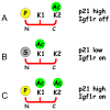
Figure 3. Acetylation has differential effects on p53 trans-activation and transsuppression (see text for full description)
The p53 protein is represented in red and post-translational modifications to the protein are shown with yellow (phosphorylated serine) or green (acetylated lysine) symbols. A) Following N-terminal phosphorylation of p53, K2 is acetylated. This activates p21 and suppresses Igf-1R expression. B) In the absence of N-terminal p53 phosphorylation, K1 is acetylated. This results in normal (basal) p21 expression, but loss of trans-suppression (increased expression) of the Igf-1R. C) When both K1 and K2 of p53 are acetylated, there is expression of both p21 and the Igf-1R.
Figure 4
Figure 4. Coordinate regulation of cell proliferation by p53 and SIRT1
Model illustrating the relationship between p53 protein level, acetylation status, aging and tumor suppression. The activity of p53 (x-axis) is affected by both the level of the protein (blue) and its acetylation status (red). Cell proliferation (y-axis) is a function of p53 activity. Cell proliferation is high when p53 activity is low, either because the protein is scarce or because it is inactivated by SIRT1. In this situation, the risk of cancer is high. Conversely, when p53 activity is elevated due to high levels of acetylated protein, proliferation is too low to maintain tissue homeostasis and longevity is compromised. When SIRT1 activity is diminished, for example during aging, p53 will remain in its acetylated and activated state, so that even low levels of protein can lead to deleterious effects on gene expression.

References

    1. Abramovitch S, Glaser T, Ouchi T, Werner H. BRCA1-Sp1 interactions in transcriptional regulation of the IGF-IR gene. FEBS Lett. 2003;541:149–54. - PubMed
    1. Antebi A. Genetics of aging in Caenorhabditis elegans. PLoS Genet. 2007;3:1565–71. - PMC - PubMed
    1. Avantaggiati ML, Ogryzko V, Gardner K, Giordano A, Levine AS, Kelly K. Recruitment of p300/CBP in p53-dependent signal pathways. Cell. 1997;89:1175–84. - PubMed
    1. Baserga RL. Haystacks and needles. Hum Pathol. 2000;31:275–6. - PubMed
    1. Baur JA, Pearson KJ, Price NL, Jamieson HA, Lerin C, Kalra A, Prabhu VV, Allard JS, Lopez-Lluch G, Lewis K, Pistell PJ, Poosala S, Becker KG, Boss O, Gwinn D, Wang M, Ramaswamy S, Fishbein KW, Spencer RG, Lakatta EG, Le Couteur D, Shaw RJ, Navas P, Puigserver P, Ingram DK, de Cabo R, Sinclair DA. Resveratrol improves health and survival of mice on a high-calorie diet. Nature. 2006;444:337–42. - PMC - PubMed

Publication types

Substances