. 2008 Sep 19;283(38):26071-80.
doi: 10.1074/jbc.M803618200. Epub 2008 Jul 18.

Caenorhabditis elegans gelsolin-like protein 1 is a novel actin filament-severing protein with four gelsolin-like repeats

Affiliations

Caenorhabditis elegans gelsolin-like protein 1 is a novel actin filament-severing protein with four gelsolin-like repeats

Tuula Klaavuniemi et al. J Biol Chem. .

Abstract

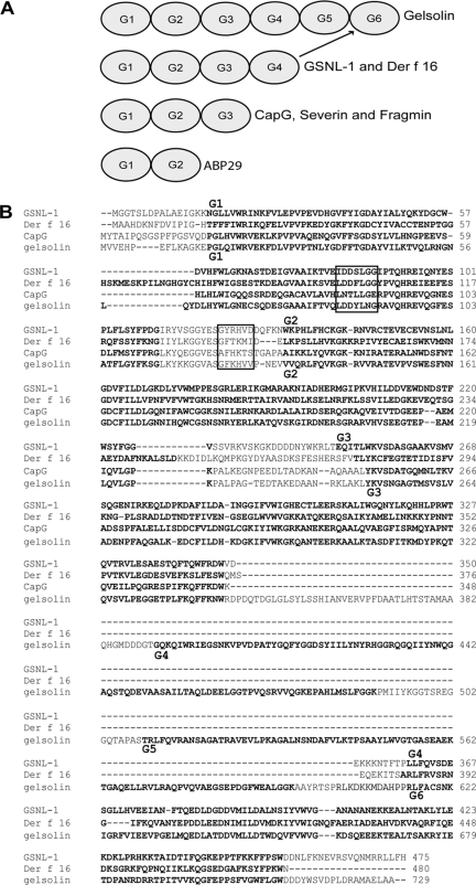
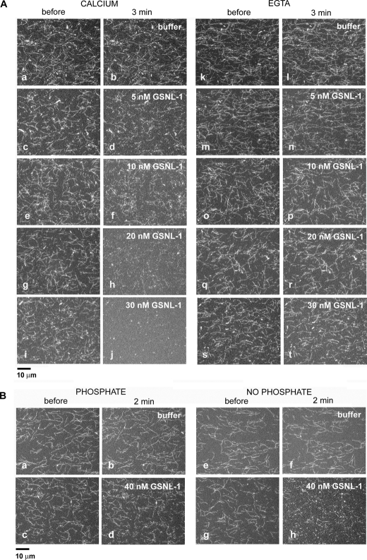
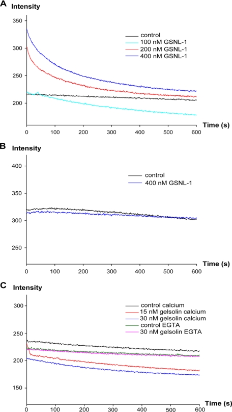
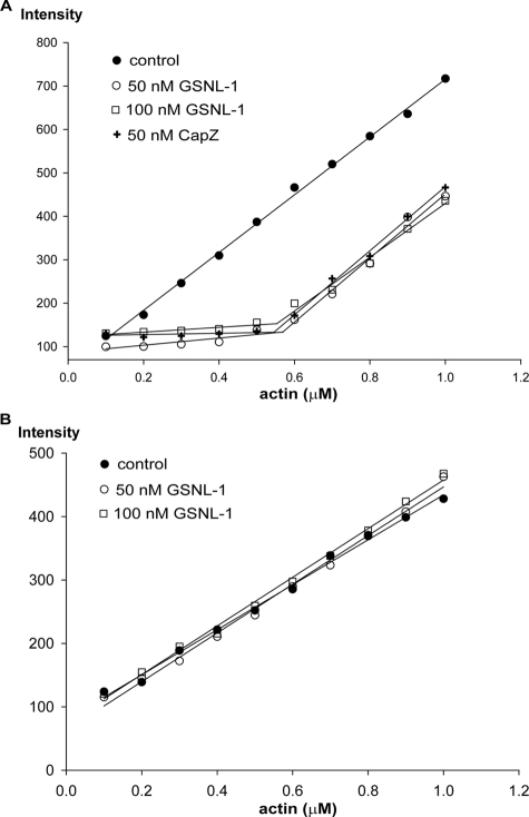
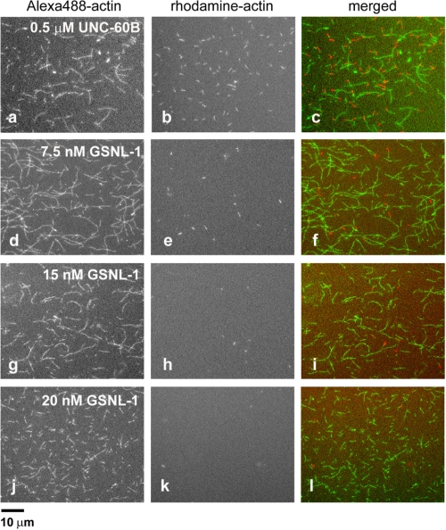
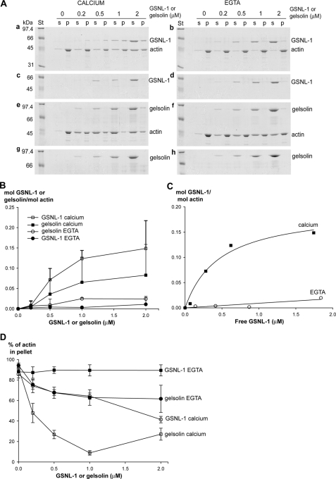
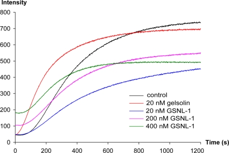
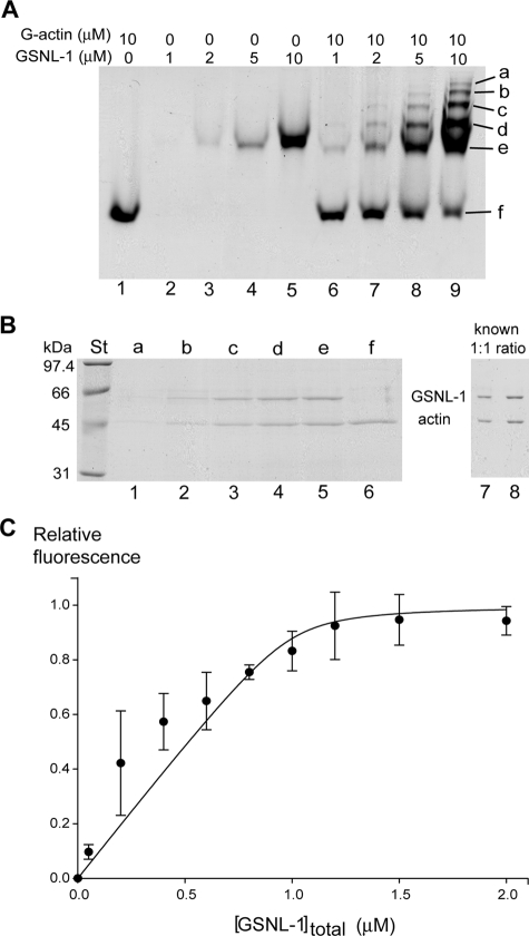
The gelsolin family of proteins is a major class of actin regulatory proteins that sever, cap, and nucleate actin filaments in a calcium-dependent manner and are involved in various cellular processes. Typically, gelsolin-related proteins have three or six repeats of gelsolin-like (G) domain, and each domain plays a distinct role in severing, capping, and nucleation. The Caenorhabditis elegans gelsolin-like protein-1 (gsnl-1) gene encodes an unconventional gelsolin-related protein with four G domains. Sequence alignment suggests that GSNL-1 lacks two G domains that are equivalent to fourth and fifth G domains of gelsolin. In vitro, GSNL-1 severed actin filaments and capped the barbed end in a calcium-dependent manner. However, unlike gelsolin, GSNL-1 remained bound to the side of F-actin with a submicromolar affinity and did not nucleate actin polymerization, although it bound to G-actin with high affinity. These results indicate that GSNL-1 is a novel member of the gelsolin family of actin regulatory proteins and provide new insight into functional diversity and evolution of gelsolin-related proteins.


Figures

FIGURE 1.
FIGURE 1.
GSNL-1 is an unconventional member of the gelsolin family. A, schematic representation of domain structures of gelsolin-related proteins including gelsolin, C. elegans GSNL-1, mite Der f 16, CapG, Dictyostelium severin, Physarum fragmin, and Lilium ABP29. Gelsolin-like (G) domains are numbered from G1 to G6 in the order of appearance from the N termini but not necessarily representing sequence homology. Indeed, G4 of GSNL-1 is most closely related to G6 of gelsolin. B, sequence alignment of GSNL-1 (GenBank™ accession number NM_073047) mite Der f 16 (GenBank™ accession number AF465625), human CapG (GenBank™ accession number M94345), and human gelsolin (GenBank™ accession number NM_198252). G domains as predicted by SMART (60) are highlighted in bold. Sequences that are important for severing activity of gelsolin (37) are indicated by boxes. G domains of GSNL-1 and gelsolin are numbered on the top and the bottom of the alignment, respectively.
FIGURE 2.
FIGURE 2.
GSNL-1 severs actin filaments. A, Alexa488-labeled actin filaments were tethered to a glass coverslip and treated with buffer alone (a, b, k, and l) or varying concentrations of GSNL-1 in the same buffer (cj and m–t) for 3 min. The buffer in a–j contained 0.1 mm CaCl2, and the buffer in k–t contained 5 mm EGTA. The filaments were observed before (a, c, e, g, i, k, m, o, q, and s) and 3 min after the incubation (b, d, f, h, j, l, n, p, r, and t), and micrographs of the same fields were taken. Bar, 10 μm. B, Alexa488-labeled actin filaments were preincubated with buffer containing 10 mm potassium phosphate (a and c) or no phosphate (e and g). Then buffer alone containing 10 μm CaCl2 and no phosphate (b and f) or the same buffer containing 40 nm GSNL-1(d and h) was infused and incubated for 2 min. The filaments were observed before (a, c, e, and g) and after the incubation (b, d, f, and h), and micrographs of the same fields were taken. Bar, 10 μm.
FIGURE 3.
FIGURE 3.
Effect of GSNL-1 on actin depolymerization. CapZ-capped actin filaments (CapZ: actin = 1:100) were diluted to 0.5 μm actin in the presence of 1 μm latrunculin A, and varying concentrations of GSNL-1 or gelsolin and changes in the intensity of light scattering were monitored for 10 min. GSNL-1 in the presence of 0.1 mm CaCl2 (A), GSNL-1 in the absence of calcium (5 mm EGTA) (B), and gelsolin in the presence and absence of calcium (C) were examined.
FIGURE 4.
FIGURE 4.
Effect of GSNL-1 on the critical concentration of actin. Varying concentrations (0.1–1 μm) of G-actin (20% pyrene-labeled) were polymerized alone or in the presence of 50 or 100 nm GSNL-1 or 50 nm CapZ. After 18 h at room temperature, the final pyrene fluorescence was measured. A, actin alone or actin with GSNL-1 or CapZ in the presence of 0.1 mm CaCl2. B, actin alone or actin with GSNL-1 in the absence of calcium (5 mm EGTA).
FIGURE 5.
FIGURE 5.
Direct observation of barbed-end capping by GSNL-1. Alexa488-labeled actin filaments were incubated with 0.5 μm UNC-60B or 7.5, 15, or 20 nm GSNL-1 for 3 min and then incubated with 0.4 μm rhodamine-labeled G-actin for 5 min. Alexa488-actin (a, d, g, and j), rhodamine-actin (b, e, h, and k), and merged images (Alexa488 in green and rhodamine in red) (c, f, i, and l) are shown. Bar, 10 μm.
FIGURE 6.
FIGURE 6.
GSNL-1 remains bound to F-actin. 5 μm F-actin was incubated with GSNL-1 or gelsolin (0–2 μm) for 1 h at room temperature and ultracentrifuged (436,000 × g, 15 min), and the supernatants (s) and the pellets (p) were analyzed by SDS-PAGE (A). The same experiments were performed for GSNL-1 and gelsolin in the absence of F-actin to estimate actin-independent precipitation. A, experiments were performed in the presence of 0.1 mm CaCl2 (a, c, e, and g) or EGTA (0. 1 mm for GSNL-1 and 1 mm for gelsolin) (b, d, f, and h). Molecular weight standards (St) are shown on the left of each gel. B–D, the band intensities were quantified, and the results are shown. B, molar ratios of GSNL-1 or gelsolin to actin in the pellets are plotted as a function of total concentrations of GSNL-1 or gelsolin in the presence and absence of calcium. Actin-independent sedimentation of GSNL-1 or gelsolin were quantified and subtracted from the experimental data in the presence of actin. Data are the average ± S.D. (for clarity, negative error bars are not shown) of three experiments. C, the results of GSNL-1 are re-plotted as a function of free GSNL-1 assuming that GSNL-1 in the supernatants was free. D, percentages of actin in the pellet fractions are plotted as a function of total concentration of GSNL-1 or gelsolin in the presence or absence of calcium. Data are average ± S.D. of three experiments.
FIGURE 7.
FIGURE 7.
GSNL-1 does not nucleate actin polymerization. 2.5 μm G-actin (20% pyrene-labeled) was incubated with GSNL-1 or gelsolin at room temperature for 5 min, and then salt was added to initiate polymerization at time 0 (final actin concentration was 2 μm).
FIGURE 8.
FIGURE 8.
GSNL-1 binds to G-actin. A, interaction between GSNL-1 and G-actin was examined by nondenaturing gel electrophoresis. 10 μm G-actin alone (lane 1), GSNL-1 alone (1, 2, 5, and 10 μm) (lanes 2–5), and 10 μm G-actin with GSNL-1 (1, 2, 5, and 10 μm) (lanes 6–9) were incubated for 30 min at room temperature, and the samples were analyzed by nondenaturing gel electrophoresis. Six major bands (a–f) were further analyzed by SDS-PAGE in B. B, the proteins from the bands a–f detected in the mixture of 10 μm GSNL-1 and 10 μm G-actin in nondenaturing electrophoresis (A, lane 9) were extracted and separated on SDS-PAGE (lanes 1–6). The band intensities were compared with a known 1:1 mixture (1 and 2 μl) of GSNL-1 (1 μm) and G-actin (1 μm) (lanes 7 and 8). Molecular weight standards (lane St) are shown on the left. C, binding of GSNL-1 to G-actin was monitored by changes in the fluorescence of pyrene. 1 μm G-actin (20% pyrene-labeled) was incubated with varying concentrations of GSNL-1 at room temperature for 30 min. Relative fluorescence values were plotted as a function of total GSNL-1 concentrations and fitted to an equation as described under “Experimental Procedures” to calculate a Kd value for GSNL-1 interaction with monomeric actin. Data are the average ± S.D. of three experiments.

Similar articles

Cited by

References

    1. Ono, S. (2007) Int. Rev. Cytol. 258 1-82 - PubMed
    1. Southwick, F. S. (2000) Proc. Natl. Acad. Sci. U. S. A. 97 6936-6938 - PMC - PubMed
    1. Ono, S., Minami, N., Abe, H., and Obinata, T. (1994) J. Biol. Chem. 269 15280-15286 - PubMed
    1. Vartiainen, M. K., Mustonen, T., Mattila, P. K., Ojala, P. J., Thesleff, I., Partanen, J., and Lappalainen, P. (2002) Mol. Biol. Cell 13 183-194 - PMC - PubMed
    1. Agrawal, P. B., Greenleaf, R. S., Tomczak, K. K., Lehtokari, V. L., Wallgren-Pettersson, C., Wallefeld, W., Laing, N. G., Darras, B. T., Maciver, S. K., Dormitzer, P. R., and Beggs, A. H. (2007) Am. J. Hum. Genet. 80 162-167 - PMC - PubMed

Publication types

LinkOut - more resources