. 2008 Jul 29;105(30):10402-7.
doi: 10.1073/pnas.0804102105. Epub 2008 Jul 18.

Protein phosphatase 1 regulates the phosphorylation state of the polarity scaffold Par-3

Affiliations

Protein phosphatase 1 regulates the phosphorylation state of the polarity scaffold Par-3

Andreas Traweger et al. Proc Natl Acad Sci U S A. .

Abstract

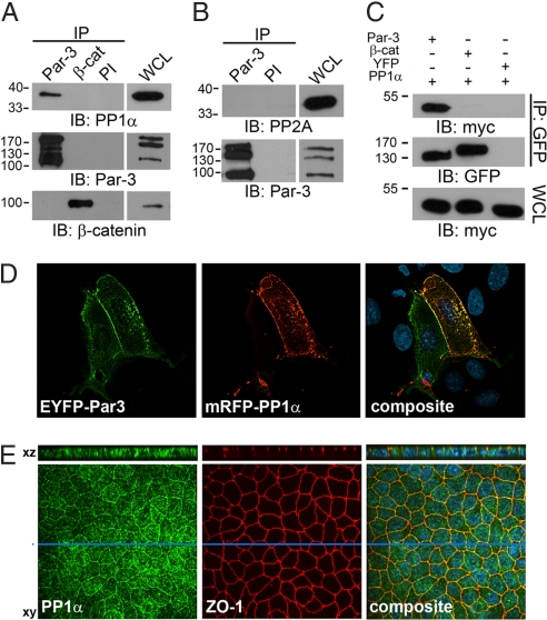
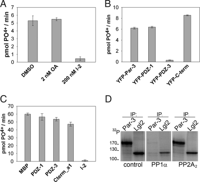
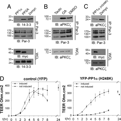
Phosphorylation of the polarity protein Par-3 by the serine/threonine kinases aPKCzeta/iota and Par-1 (EMK1/MARK2) regulates various aspects of epithelial cell polarity, but little is known about the mechanisms by which these posttranslational modifications are reversed. We find that the serine/threonine protein phosphatase PP1 (predominantly the alpha isoform) binds Par-3, which localizes to tight junctions in MDCKII cells. PP1alpha can associate with multiple sites on Par-3 while retaining its phosphatase activity. By using a quantitative mass spectrometry-based technique, multiple reaction monitoring, we show that PP1alpha specifically dephosphorylates Ser-144 and Ser-824 of mouse Par-3, as well as a peptide encompassing Ser-885. Consistent with these observations, PP1alpha regulates the binding of 14-3-3 proteins and the atypical protein kinase C (aPKC) zeta to Par-3. Furthermore, the induced expression of a catalytically inactive mutant of PP1alpha severely delays the formation of functional tight junctions in MDCKII cells. Collectively, these results show that Par-3 functions as a scaffold, coordinating both serine/threonine kinases and the PP1alpha phosphatase, thereby providing dynamic control of the phosphorylation events that regulate the Par-3/aPKC complex.


Conflict of interest statement

The authors declare no conflict of interest.

Figures

Fig. 1.
Fig. 1.
Par-3 interacts with protein phosphatase 1. (A and B) Endogenous Par-3 and β-catenin immunoprecipitated from mouse E13 lysates were probed with anti PP1α or anti-PP2A antibodies. Preimmune (PI) serum was used as a control. (C) YFP immunoprecipitates (IPs) from MDCKII cells expressing 2xmyc-PP1α and YFP-Par-3, YFP-β-catenin, or YFP were immunoblotted (IB) with anti-myc (Top) or anti-GFP (Middle) antibodies. Whole-cell lysates (WCLs) were probed for equal expression of 2xmyc-PP1α by using an anti-myc antibody (Bottom). (D) EYFP-Par-3 and mRFP-PP1α codistribute in MDCKII cells at the plasma membrane. (E) Endogenous PP1α codistributes with the tight junction marker protein ZO-1 at sites of cell–cell contact in MDCKII cells. Enface images and confocal XZ-sections (section indicated by blue bar) are shown.
Fig. 2.
Fig. 2.
PP1 binds directly to multiple regions of Par-3. (A) Purified PP1α was incubated with amylose resin-immobilized MBP-fusion proteins as indicated. MBP alone was used as a control. Bound PP1α was detected by immunoblotting with anti-PP1α antibody. (B) 293T cells coexpressing wild-type Par-3-PDZ1 or RVXF-mutated YFP-Par-3-PDZ1 (Top) and 2xmyc-PP1α were lysed and YFP-fusion proteins were immunoprecipitated with an anti-GFP antibody. Immunoprecipitates (IP) were probed with anti-myc or anti-GFP antibodies and whole-cell lysates (WCLs) with anti-myc antibody.
Fig. 3.
Fig. 3.
Par-3-bound PP1α is active and dephosphorylates Par-3 in vitro. (A) Endogenous Par-3 immunocomplexes collected from MDCKII cells were tested for the release of free phosphate from a substrate in the presence of okadaic acid (2 nM), the PP1-specific inhibitor Inhibitor-2 (200 nM), or DMSO. (B) Lysates prepared from 293T cells coexpressing 2xmyc-PP1α and various regions of Par-3 fused to YFP were incubated with anti-GFP antibody and resulting complexes were monitored for phosphatase activity. (C) Purified PP1α (100 ng) was incubated with 1 μg of various regions of Par-3 purified as MBP-fusion proteins and phosphatase activity was monitored. Each value is the mean of triplicate measurements (n = 3). Error bars represent 1 SD. (D) Purified PP1α, but not PP2A2, dephosphorylates aPKC-phosphorylated Par-3 in vitro (see Methods for details). Additionally, PP1α is less reactive toward the polarity protein Lgl2.
Fig. 4.
Fig. 4.
PP1α dephosphorylates S144 and S824, but not S25 of mouse Par-3. (A) Shown are MRM elution profiles of the peptides containing phosphorylated S144 with (Left) or without (Right) PP1α treatment. (B) The phosphorylated peptide containing S824 is not detectable after phosphatase-treatment (Left) as evidenced by multiple reaction monitoring. (C) Shown are the extracted ion current profiles (XIC) of the nonphosphorylated S25-peptides (Left) and the phosphorylated S25-peptides (Right) before and after treatment with PP1α. (D) The change in phosphorylation of S25 is calculated as the ratio of the integrated ion current profiles of nonphosphorylated/phosphorylated peptides. Shown are the ratios of phosphatase-treated (right-hand bar) and untreated samples (left-hand bar). Error bars represent 1 SD (n = 2).
Fig. 5.
Fig. 5.
PP1α influences the binding of aPKCζ and 14-3-3 to Par-3 and impacts TJ formation. (A) Endogenous Par-3 was immunoprecipitated (IP) in the presence of Calyculin A from 293T cells transiently expressing 2xmyc-PP1α, 2xmyc-PP2A, or 2xmyc and the binding of endogenous 14-3-3 was monitored by immunoblotting (IB). Whole-cell lysates (WCLs) were analyzed for the expression of 14-3-3 and the exogenously expressed proteins. (B) Endogenous Par-3 was immunoprecipitated from 293T cells treated for 30 min with 5 μM Tautomycetin or 100 nM okadaic acid and bound aPKCζ was detected by immunoblotting (IB). Whole-cell lysates were monitored for endogenous aPKCζ expression levels. (C) 293T cells were transiently transfected with 2xmyc-PP1α (H248K) or 2xmyc expression constructs and endogenous Par-3-immunocomplexes were probed with an anti-aPKCζ antibody. The expression of myc-tagged proteins and endogenous aPKCζ was monitored on whole-cell lysates. (D) YFP-PP1α (H248K) expression (Right), or YFP-expression as a control (Left), was induced in MDCK-T23 cells by the removal of doxycycline and the kinetics of TEER establishment was monitored for 24 h after a Ca2+ switch. Three independent experiments using two different cell lines were performed. The means of one representative experiment are shown (n = 3). Error bars represent 1 SD.
Fig. 6.
Fig. 6.
Model for PP1 as a functional regulator of the Par-3 scaffold. Par-3 diffusing to the lateral cortex is phosphorylated by Par-1 (EMK1/MARK2), resulting in the recruitment of 14-3-3. aPKC subsequently dissociates from Par-3 and the complex is released from the cortex. As soon as Par-3 escapes the active range of Par-1 (indicated by gray area), PP1 dephosphorylates Par-3, allowing the re-formation of a functional Par/aPKC complex at the apical domain of epithelial cells. In addition, PP1 stabilizes the interaction between aPKC and Par-3 by dephosphorylating Par-3 at S824.

References

    1. Goldstein B, Macara IG. The par proteins: Fundamental players in animal cell polarization. Dev Cell. 2007;13:609–622. - PMC - PubMed
    1. Kemphues K. PARsing embryonic polarity. Cell. 2000;101:345–348. - PubMed
    1. Ohno S. Intercellular junctions and cellular polarity: The PAR-aPKC complex, a conserved core cassette playing fundamental roles in cell polarity. Curr Opin Cell Biol. 2001;13:641–648. - PubMed
    1. Joberty G, Petersen C, Gao L, Macara IG. The cell-polarity protein Par6 links Par3 and atypical protein kinase C to Cdc42. Nat Cell Biol. 2000;2:531–539. - PubMed
    1. Lin D, et al. A mammalian PAR-3-PAR-6 complex implicated in Cdc42/Rac1 and aPKC signalling and cell polarity. Nat Cell Biol. 2000;2:540–547. - PubMed

Publication types