. 2008 Jul 29;105(30):10541-6.
doi: 10.1073/pnas.0802008105. Epub 2008 Jul 18.

Differential requirements for Alix and ESCRT-III in cytokinesis and HIV-1 release

Affiliations

Differential requirements for Alix and ESCRT-III in cytokinesis and HIV-1 release

Jez G Carlton et al. Proc Natl Acad Sci U S A. .

Abstract

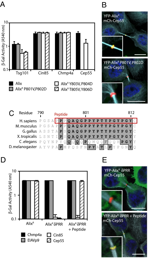
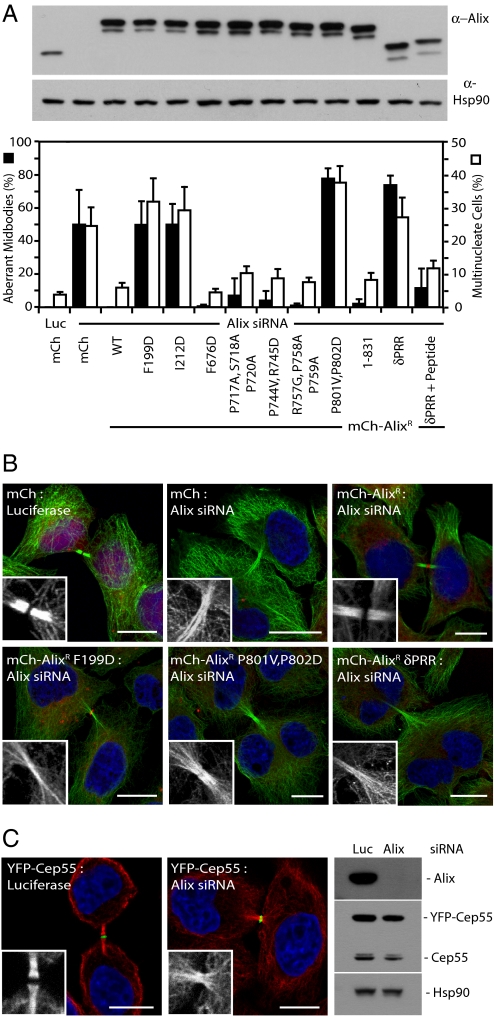
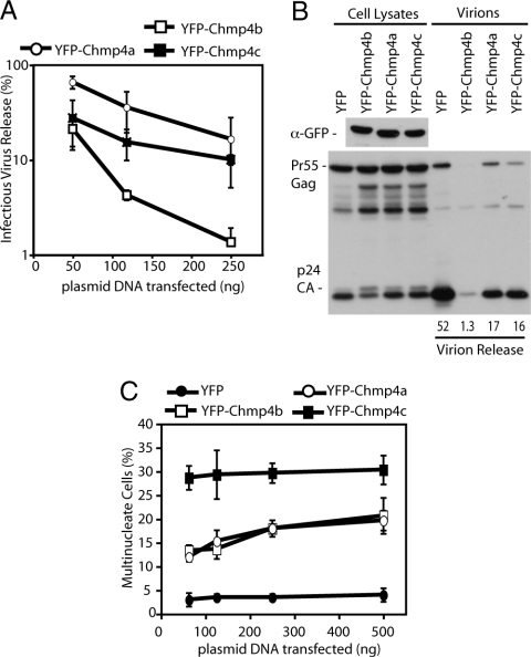
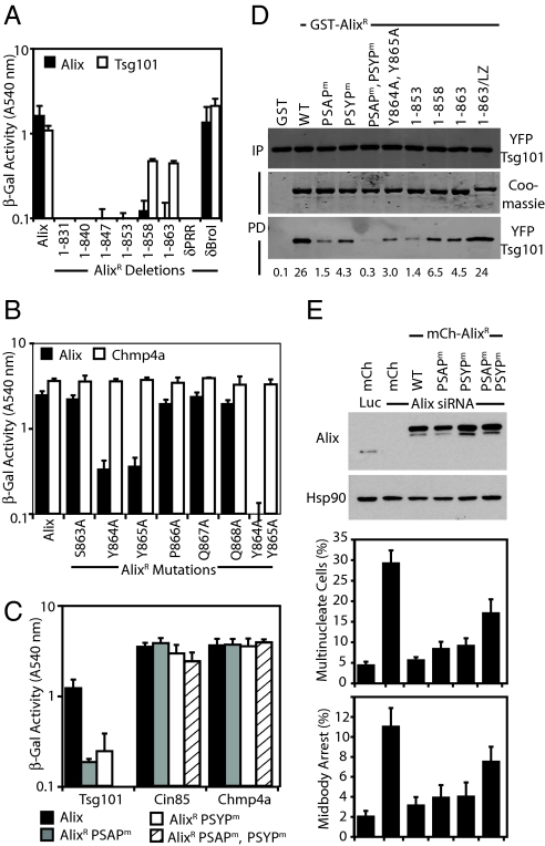
The ESCRT machinery functions in topologically equivalent membrane fission events, namely multivesicular body formation, the terminal stages of cytokinesis and HIV-1 release. Here, we show that the ESCRT-III-binding protein Alix is recruited to the midbody of dividing cells through binding Cep55 via an evolutionarily conserved peptide. Disruption of Cep55/Alix/ESCRT-III interactions causes formation of aberrant midbodies and cytokinetic failure, demonstrating an essential role for these proteins in midbody morphology and cell division. We also show that the C terminus of Alix encodes a multimerization activity that is essential for its function in Alix-dependent HIV-1 release and for interaction with Tsg101. Last, we demonstrate that overexpression of Chmp4b and Chmp4c differentially inhibits HIV-1 release and cytokinesis, suggesting possible reasons for gene expansion within the mammalian Class E VPS pathway.


Conflict of interest statement

The authors declare no conflict of interest.

Figures

Fig. 1.
Fig. 1.
Cep55 recruits Alix to the midbody through binding a conserved peptide within Alix's PRR. (A) Alix mutants were fused to the VP16-activation domain and tested for interaction with the indicated proteins fused to Gal4 DNA-binding domain by yeast two-hybrid assay (n = 3 ± SD). (B) HeLa cells were transfected with plasmids encoding mCh-Cep55 and either YFP-AlixR or YFP-AlixR P801V, P802D, fixed and stained with α-tubulin. (Scale bars: 10 μm.) (C) Sequence alignment of the Cep55-binding region of the Alix-PRR with the Cep55-binding peptide highlighted in red. (D) Alix, AlixR δPRR, and AlixR δPRR plus peptide were fused to the VP16 DNA-activation domain and tested for interactions with the indicated Gal4 DNA-binding domain fusions by yeast two-hybrid assay (n = 3 ± SD). (E) HeLa cells were transfected with plasmids encoding mCh-Cep55 and either YFP-AlixR or YFP-AlixR δPRR and AlixR δPRR plus peptide, fixed, and stained with α-tubulin. (Scale bars: 10 μm.)
Fig. 2.
Fig. 2.
Recruitment of Alix and ESCRT-III to the midbody is essential for normal midbody morphology. (A) HeLa cells stably transduced with retroviral vectors encoding mCh or mCh-AlixR fusions were treated with siRNA targeting Luciferase or Alix, and resolved cell lysates were analyzed with α-Alix or α-Hsp90 antibodies. Alternatively, cells were fixed, stained with α-tubulin, and scored for aberrant midbodies (n = 3 ±SD) and multinucleation (n > 4 ±SD). (B) Representative micrographs of aberrant midbodies from A, enlargement depicting α-tubulin channel. (Scale bars: 10 μm.) (C) Clonal HeLa cells stably expressing YFP-Cep55 were treated with siRNA targeting Luciferase or Alix. Cells were fixed and stained with α-tubulin. Enlargements depict the α-tubulin channel. (Scale bars: 10 μm.) Alternatively, cells were lysed and analyzed with α-Alix, α-Cep55, and α-Hsp90 antisera.
Fig. 3.
Fig. 3.
Differential requirements for Chmp4 isoforms in retroviral budding and cytokinesis. (A and B) Cells (293T) were cotransfected with plasmids encoding HIV-1 pNL/HXB and the indicated amount of YFP-Chmp4 constructs. Resolved cell lysates and released virions were examined by Western blot analysis with α-Gag antisera; lysates were also examined by Western blot analysis with α-GFP antisera. Quantification of virion release via infrared imaging is given. β-Galactosidase assay was performed on HeLa-TZM-bl cells infected with 293T supernatant (n = 3 ±SD). (C) HeLa cells were transfected with the indicated amounts of YFP-Chmp4 constructs. Cells were fixed, stained with α-tubulin, and scored for multinucleation (n = 3 ±SD).
Fig. 4.
Fig. 4.
Activities in the C terminus of Alix. (A and B) The indicated Alix constructs fused to the VP16-activation domain were tested for interactions with Alix or Tsg101 fused to the Gal4 DNA-binding domain by yeast two-hybrid assay (n = 3 ±SD). (C) The indicated Alix mutants were fused to the VP16-activation domain and tested for interactions with Tsg101, Cin85, and Chmp4a fused to the Gal4 DNA-binding domain by yeast two-hybrid assay (n = 3 ±SD). (D) Coprecipitation assays were performed on lysates of 293T cells cotransfected with plasmids encoding YFP-Tsg101, Myc-Vps28, and the indicated GST-AlixR fusion proteins. Resolved cells lysates (input, IP) and normalized bead-bound fractions (pull down, PD) were analyzed with α-GFP antisera. PD fractions were analyzed by Coomassie staining to visualize GST-AlixR expression, and relative binding was quantified by infrared imaging. (E) HeLa cells stably transduced with retroviral vectors encoding mCh or the mCh-AlixR fusions were treated with siRNA targeting Luciferase or Alix. Cells were fixed, stained with α-tubulin, and scored for midbody arrest and multinucleation (n = 3 ±SD). Resolved cell lysates were analyzed with α-Alix and α-Hsp90 antibodies.
Fig. 5.
Fig. 5.
Alix multimerization is required for Alix-dependent HIV-1 release. Cells (293T) were cotransfected with plasmids encoding the indicated HA-tagged AlixR constructs and an HIV-1 pNL/HXB p6(P7L, P10L) plasmid. Resolved cell lysates and released virions were examined by Western blot analysis with α-Gag antisera. Quantification of virion release (VR) via infrared imaging is given. (n = 3). Lysates were also examined by Western blot analysis with α-HA antisera. β-Galactosidase assay was performed on HeLa-TZM-bl cells infected with 293T supernatants (n = 3 ±SD).

References

    1. Glotzer M. Animal cell cytokinesis. Annu Rev Cell Dev Biol. 2001;17:351–386. - PubMed
    1. Eggert US, Mitchison TJ, Field CM. Animal cytokinesis: From parts list to mechanisms. Annu Rev Biochem. 2006;75:543–566. - PubMed
    1. Barr FA, Gruneberg U. Cytokinesis: Placing and making the final cut. Cell. 2007;131:847–860. - PubMed
    1. Fabbro M, et al. Cdk1/Erk2- and Plk1-dependent phosphorylation of a centrosome protein, Cep55, is required for its recruitment to midbody and cytokinesis. Dev Cell. 2005;9:477–488. - PubMed
    1. Martinez-Garay I, Rustom A, Gerdes HH, Kutsche K. The novel centrosomal associated protein CEP55 is present in the spindle midzone and the midbody. Genomics. 2006;87:243–253. - PubMed

Publication types

MeSH terms