. 2008 Dec;49(12):5353-65.
doi: 10.1167/iovs.07-1666. Epub 2008 Jul 18.

Specialized podosome- or invadopodia-like structures (PILS) for focal trabecular meshwork extracellular matrix turnover

Affiliations

Specialized podosome- or invadopodia-like structures (PILS) for focal trabecular meshwork extracellular matrix turnover

Mini Aga et al. Invest Ophthalmol Vis Sci. 2008 Dec.

Abstract

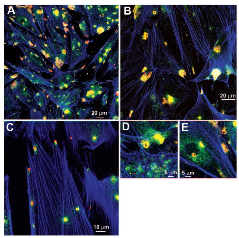
Purpose: There are distinctive areas of colocalization of matrix metalloproteinase (MMP)-2 and -14 on trabecular meshwork (TM) cells that resemble podosomes or invadopodia. Studies were conducted to determine whether TM cells exhibit podosome- or invadopodia-like structures (PILS) and whether they produce focal extracellular matrix (ECM) turnover.

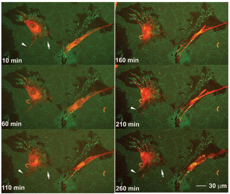
Methods: Porcine and human TM cells and perfused anterior segment organ cultures were studied. Localization of PILS components on TM cells and in sections from anterior segments was determined by immunohistochemistry and confocal microscopy. Cells were grown on type I collagen labeled with fluorescein isothiocyanate (FITC) for degradation analysis. Confocal time lapse images were taken of labeled TM cells on FITC-collagen.

Results: Immunostaining for MMP-2, MMP-14, and the typical PILS components cortactin, caldesmon, alpha-actinin, N-WASP, Arp-3, and cdc42 colocalized on these distinctive structures. Integrin-alphaV and -beta1, fibronectin, and versican colocalized with PILS components. TM cells on FITC-conjugated collagen developed focal regions of degradation. Time-lapse imaging showed dramatic and controlled movement of TM cell processes during this ECM degradation and fragment internalization. MMP-2, MMP-14, and cortactin colocalized at regions that appear to be PILS on cells within the outflow pathway in sections of human anterior segments.

Conclusions: TM cells exhibit areas where PILS components colocalize with MMP-2 and -14. Similar structures are found in sections, suggesting that PILS occur in situ in the outflow pathway. The collagen degradation suggests that PILS may serve as focal sites for targeted ECM turnover, an event linked to modifications of aqueous outflow resistance and intraocular pressure homeostasis.


Conflict of interest statement

Disclosure: M. Aga, None; J.M. Bradley, None; K.E. Keller, None; M.J. Kelley, None; T.S. Acott, None

Figures

Figure 1
Figure 1
Immunohistochemical localization of MMP-14 and -2. Porcine TM cells, cultured on collagen type I–coated membranes, were made serum free 48 hours before mechanical stretching for 24 hours. Images show immunostaining for MMP-14 (red), MMP-2 (green), and F-actin (phalloidin, blue) for several experiments at different magnification levels (AE).
Figure 2
Figure 2
Immunohistochemical localization of cortactin and MMP-2. Porcine TM cells, cultured on collagen type I–coated membranes were made serum free 48 hours before mechanical stretching for 24 hours. (AI) Immunostaining for cortactin (green) and MMP-2 (red). Scale bar, 10 μm.
Figure 3
Figure 3
Immunohistochemical localization of α-actinin, caldesmon, N-WASP, and cdc42 with cortactin and F-actin in cultured TM cells. Porcine TM cells, cultured on collagen type I–coated membranes, were made serum free 48 hours before mechanical stretching for 24 hours. Images show immunostaining for (A) α-actinin (green), cortactin (red), and F-actin (phalloidin, blue); (B) caldesmon (red), cortactin (green), and F-actin (phalloidin, blue); (C) N-WASP (red), cortactin (green), and F-actin (phalloidin, blue); and (D) cdc42 (green), cortactin (red), and F-actin (phalloidin, blue). Insets: various higher resolution images of structures.
Figure 4
Figure 4
Immunohistochemical localization of Arp, cortactin, N-WASP, and cdc42 in cultured TM cells. Porcine TM cells, cultured on collagen type I–coated membranes, were made serum free 48 hours before mechanical stretching for 24 hours. Images show immunostaining for (A) and (B) Arp-3 (green), cortactin (red), and F-actin (phalloidin, blue); (C) and (D) Arp-3 (green), N-WASP (red), and F-actin (phalloidin, blue); and (E) cdc42 (green), N-WASP (red), and F-actin (phalloidin, blue).
Figure 5
Figure 5
Immunochemical localization of integrin-β1 and -αV with cortactin and F-actin in cultured TM cells. Porcine TM cells, cultured on collagen type I–coated membranes, were made serum free 48 hours before mechanical stretching for 24 hours. Images show immunostaining for (A) integrin-β1 (red), cortactin (green), and F-actin (phalloidin, blue); (B) integrin-β1 (red) and cortactin (green); (C) integrin-αV (red), cortactin (green), and F-actin (phalloidin, blue); and (D) integrin-αV (red) and cortactin (green).
Figure 6
Figure 6
Immunohistochemical localization of fibronectin, MMP-2, versican and cortactin on cultured TM cells. Porcine TM cells, cultured on collagen type I–coated membranes, were made serum free 48 hours before mechanical stretching for 24 hours. Images show immunostaining for (A) MMP-2 (red) and fibronectin (green); (B) MMP-2 (red), fibronectin (green), and F-actin (phalloidin, blue); (C) versican (red), cortactin (green) and F-actin (phalloidin, blue); (D) versican (red) and F-actin (phalloidin, blue); (E) versican (red), cortactin (green), and F-actin (phalloidin, blue); (F) versican neoepitope (red), cortactin (green), and F-actin (phalloidin, blue); and (G) versican neoepitope (red) and cortactin (green).
Figure 7
Figure 7
Immunohistochemical localization of MMP-2, MMP-14, and cortactin in the TM of human anterior segment organ cultures. Human anterior segments were perfused at 8.9 mm Hg for 48 hours before processing for immunohistochemistry. Confocal images show immunostaining for (A) MMP-14 (red) and cortactin (green); (B) control with no primary but both secondary antibodies: (C) MMP-2 (red) and cortactin (green); and (D) MMP-14 (red) and cortactin (green). Schlemm's canal (SC) is as labeled with the TM to the right in each case.
Figure 8
Figure 8
Time-lapse confocal images of TM cells degrading FITC-labeled collagen type I. Porcine TM cells were plated on FITC-labeled collagen type I–coated chamber slides and allowed to adhere overnight. The dye (red) was then incubated with cells for 1 hour, and time lapse confocal images were collected at six focal levels every 10 minutes for 4 hours. The cells were maintained at 37°C in 5% CO2 and 100% humidity throughout. Images were selected showing two cells at 50-minute intervals from one focal plane (Z-stack). Arrowheads and arrows: regions where cellular projections extended, widened, and then withdrew, leaving regions of diminished FITC-labeling in the last frames. Full QuickTime movies of all time points and focal planes for this and several other experiments are included online as Movies S1–S4, http://www.iovs.org/cgi/content/full/49/12/5353/DC1.

References

    1. Quigley HA. Open-angle glaucoma. N Engl J Med. 1993;328:1097–1106. - PubMed
    1. Quigley HA. Number of people with glaucoma worldwide. Br J Ophthalmol. 1996;80:389–393. - PMC - PubMed
    1. Shields MB. Textbook of Glaucoma. 4th. Baltimore: Williams & Wilkins; 1998.
    1. Boland MV, Quigley HA. Risk factors and open-angle glaucoma: classification and application. J Glaucoma. 2007;16:406–418. - PubMed
    1. Bill A, Maepea O. Mechanisms and routes of aqueous humor drainage. In: Albert DM, Jakotiec FM, editors. Principles and Practices of Ophthalmology. Philadelphia: WB Saunders; 1994. pp. 206–226.

Publication types