. 2008 Aug 1;181(3):1728-36.
doi: 10.4049/jimmunol.181.3.1728.

An endogenous prostaglandin enhances environmental phthalate-induced apoptosis in bone marrow B cells: activation of distinct but overlapping pathways

Affiliations

An endogenous prostaglandin enhances environmental phthalate-induced apoptosis in bone marrow B cells: activation of distinct but overlapping pathways

Stephanie L Bissonnette et al. J Immunol. .

Abstract

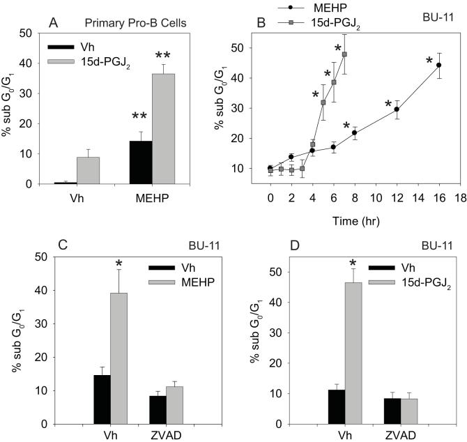
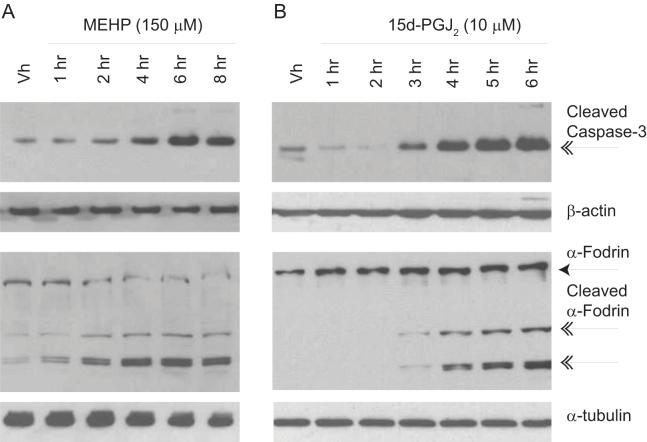
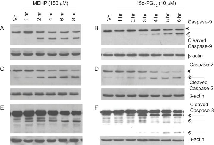
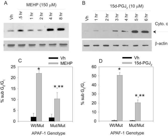
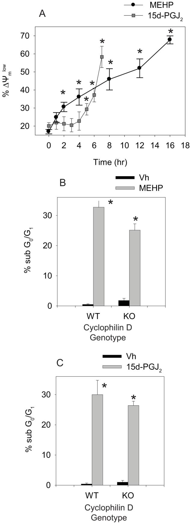
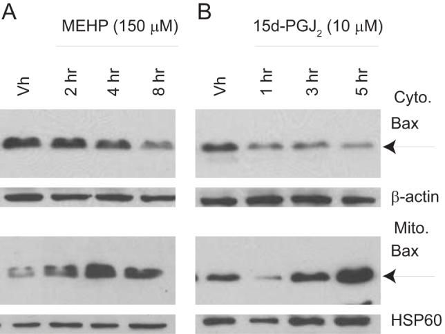
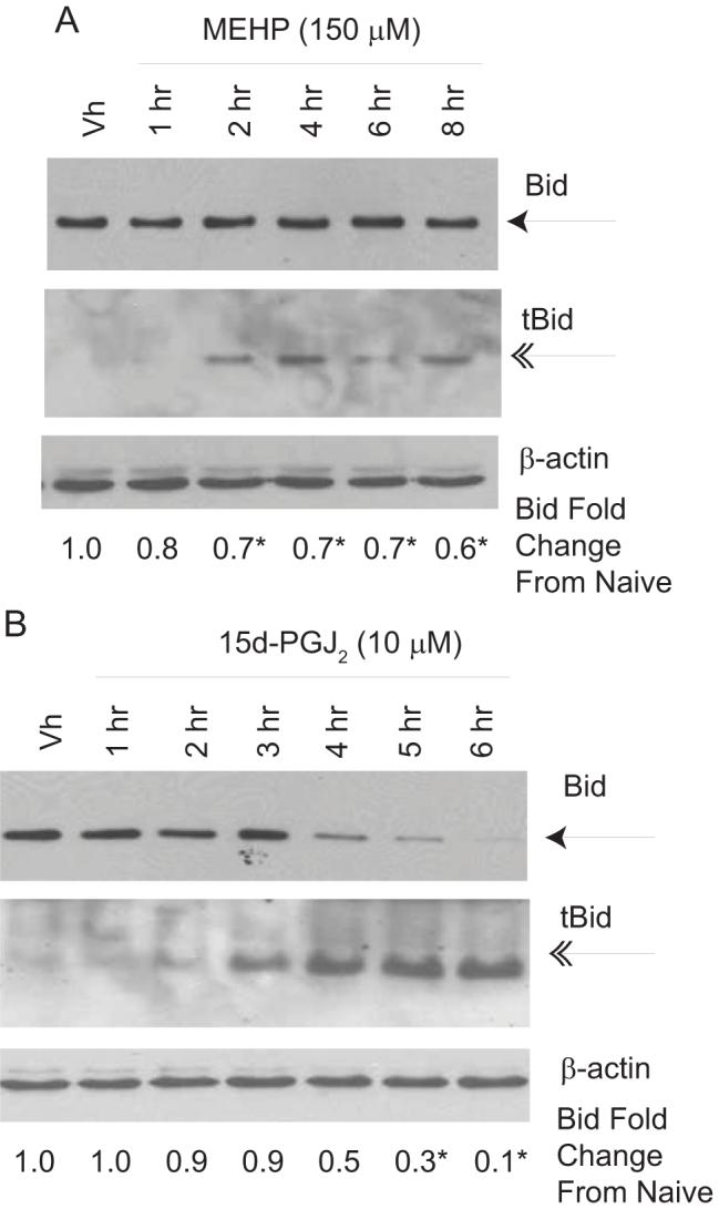
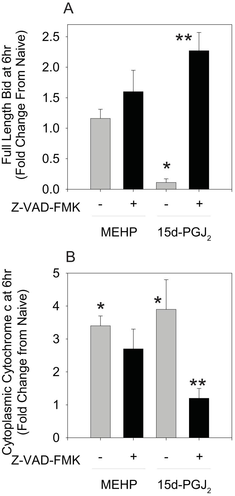
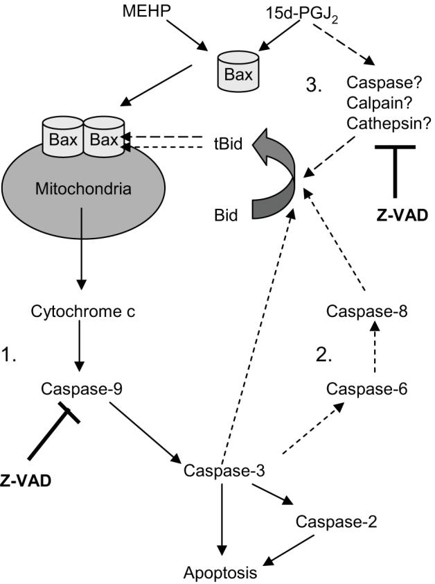
Phthalate esters are ubiquitous environmental contaminants that are produced for a variety of common industrial and commercial purposes. We have shown that mono-(2-ethylhexyl) phthalate (MEHP), the toxic metabolite of di-(2-ethylhexyl) phthalate, induces bone marrow B cell apoptosis that is enhanced in the presence of the endogenous prostaglandin 15-deoxy-Delta((12, 14))-PGJ(2) (15d-PGJ(2)). Here, studies were performed to determine whether 15d-PGJ(2)-mediated enhancement of MEHP-induced apoptosis represents activation of an overlapping or complementary apoptosis pathway. MEHP and 15d-PGJ(2) induced significant apoptosis within 8 and 5 h, respectively, in a pro/pre-B cell line and acted cooperatively to induce apoptosis in primary pro-B cells. Apoptosis induced with each chemical was accompanied by activation of a combination of initiator caspases (caspases-2, -8, and -9) and executed by caspase-3. Apoptosis induced with MEHP and 15d-PGJ(2) was reduced in APAF1 null primary pro-B cells and accompanied by alteration of mitochondrial membranes, albeit with different kinetics, indicating an intrinsically activated apoptosis pathway. Significant Bax translocation to the mitochondria supports its role in initiating release of cytochrome c. Both chemicals induced Bid cleavage, a result consistent with a truncated Bid-mediated release of cytochrome c in an apoptosis amplification feedback loop; however, significantly more Bid was cleaved following 15d-PGJ(2) treatment, potentially differentiating the two pathways. Indeed, Bid cleavage and cytochrome c release following 15d-PGJ(2) but not MEHP treatment was profoundly inhibited by Z-VAD-FMK, suggesting that 15d-PGJ(2) activates apoptosis via two pathways, Bax mobilization and protease-dependent Bid cleavage. Thus, endogenous 15d-PGJ(2)-mediated enhancement of environmental chemical-induced apoptosis represents activation of an overlapping but distinct signaling pathway.


Figures

Figure 1
Figure 1
MEHP and 15d-PGJ2 cooperatively induce apoptosis in cultured pro/pre-B cells and primary pro-B cells. (A) Suspension cultures of primary pro-B cells prepared from wildtype C57BL/6 mice were treated with Vh, MEHP (150 μM) and/or 15d-PGJ2 (2 μM) for 32 hr. (B) Suspension cultures of BU-11 cells were treated with ethanol:DMSO (50:50, Vh, 0.1%), MEHP (150 μM) or 15d-PGJ2 (10 μM) for the times indicated. (C-D) Suspension cultures of BU-11 cells pre-treated for 30 min with DMSO (Vh, 0.1%) or Z-VAD-FMK (30 μM) and then treated with ethanol:DMSO (50:50, Vh, 0.1%), MEHP (150 μM) or 15d-PGJ2 (10 μM) for 16 or 6 hr, respectively. Cell death was analyzed by hypotonic PI staining followed by flow cytometry. Data are presented as means ± SE from at least 3 independent experiments or 4 independently prepared and maintained pools of primary pro-B cells. *Statistically greater than Vh-treated (p<0.05, ANOVA, Dunnett’s). **Statistically different from all other treatment groups (p<0.05, ANOVA, Tukey-Kramer).
Figure 2
Figure 2
MEHP (A) and 15d-PGJ2 (B) treatment activate caspase-3 in cultured pro/pre-B cells. Suspension cultures of BU-11 cells were treated with ethanol:DMSO (50:50, Vh, 0.1%), MEHP (150 μM) or 15d-PGJ2 (10 μM) for the times indicated. Cytoplasmic extracts were prepared and analyzed for formation of caspase-3 fragments (17 kDa) and cleaved α-fodrin (120, 150 kDa) by immunoblotting. Representative data from at least 3 independent experiments are presented.
Figure 3
Figure 3
MEHP and 15d-PGJ2 treatment activates caspases in the intrinsic and extrinsic pathways. Suspension cultures of BU-11 cells were treated with ethanol:DMSO (50:50, Vh, 0.1%), MEHP (150 μM) or 15d-PGJ2 (10 μM) for the times indicated. Cytoplasmic extracts were prepared and analyzed for caspase-9 (A-B; 37, 39 kDa), caspase-2 (C-D; 32, 33 kDa), and caspase-8 (E-F; 43/41, 18 kDa) fragments by immunoblotting. Representative data from at least 3 independent experiments are presented.
Figure 4
Figure 4
MEHP and 15d-PGJ2 induce cytochrome c release in cultured pro/pre-B cells, and death is attenuated in APAF1fog mutant primary pro-B cells. (A-B) Suspension cultures of BU-11 cells were treated with ethanol:DMSO (50:50, Vh, 0.1%), MEHP (A; 150 μM) or 15d-PGJ2 (B; 10 μM) for the times indicated. Cytochrome c release was analyzed by immunoblotting of cytoplasmic extracts from digitonin-permeablized cells. (C-D) Suspension cultures of primary pro-B cells isolated from mice hetero- or homozygous for the APAF1fog mutation were treated with ethanol:DMSO (50:50, Vh, 0.1%), MEHP (C; 200 μM, 16hr) or 15d-PGJ2 (D; 10 μM, 8hr). Cell death was analyzed by hypotonic PI staining followed by flow cytometry. The percentage of apoptotic cells measured in a naïve population for each pool was subtracted prior to the data analysis. Data are presented as means ± SE from at least 3 independent experiments or 4 independently prepared and maintained pools of primary pro-B cells. *Statistically greater than Vh-treated (p<0.05, ANOVA, Tukey-Kramer). **Statistically less than wildtype, MEHP- or 15d-PGJ2-treated (p<0.05, ANOVA, Tukey-Kramer).
Figure 5
Figure 5
MEHP and 15d-PGJ2 induce loss of mitochondrial membrane potential, but lack of cyclophilin D does not protect against cell death. (A) Suspension cultures of BU-11 cells were treated with ethanol:DMSO (50:50, Vh, 0.1%), MEHP (150 μM) or 15d-PGJ2 (10 μM) for the times indicated. Mitochondrial membrane potential was analyzed by JC-1 staining followed by flow cytometry. (B-C) Suspension cultures of primary pro-B cells isolated from wildtype B6129SF2/J and Ppidtm1.1Mmos mice were treated with Vh, MEHP (200 μM, 16hr) or 15d-PGJ2 (10 μM, 8hr). Cell death was analyzed by hypotonic PI staining followed by flow cytometry. The percentage of apoptotic cells measured in a naïve population for each pool was subtracted prior to the data analysis. Data are presented as means ± SE from at least 3 independent experiments or 4 independently prepared and maintained pools of primary pro-B cells. *Statisically greater than Vh-treated (p<0.05, ANOVA, Dunnett).
Figure 6
Figure 6
MEHP (A) and 15d-PGJ2 (B) induce translocation of Bax to the mitochondria. Suspension cultures of BU-11 cells were treated with ethanol:DMSO (50:50, Vh, 0.1%), MEHP (150 μM) or 15d-PGJ2 (10 μM) for the times indicated. Cytoplasmic and mitochondrial fractions were prepared from digitonin-permeabilized cells and analyzed for Bax expression by immunoblotting. Representative data from at least 3 independent experiments are presented.
Figure 7
Figure 7
MEHP (A) and 15d-PGJ2 (B) induce Bid cleavage. Suspension cultures of BU-11 cells were treated with ethanol:DMSO (50:50, Vh, 0.1%), MEHP (150 μM) or 15d-PGJ2 (10 μM) for the times indicated. Cytoplasmic extracts were prepared and analyzed for full length (22 kDa) and cleaved Bid (15 kDa) by immunoblotting. Expression levels of full-length Bid were quantified as described in the Methods. Data are presented as means ± SE from at least 3 independent experiments. *Statistically less than Vh-treated (p<0.05, ANOVA, Dunnett’s).
Figure 8
Figure 8
Z-VAD-FMK suppresses 15d-PGJ2-, but not MEHP-, induced Bid cleavage (A) and cytochrome c release (B). Following a 30 min pretreatment with Vh or Z-VAD-FMK (30 μM) and treatment with Vh, 15d-PGJ2 (10 μM), or MEHP for 6 hr, cytoplasmic extracts from digitonin-permeabilized cells were prepared and analyzed by immunoblotting for full-length Bid and cytochrome c. Protein expression levels were quantified as described in the Methods. Data are presented as means ± SE from at least 3 independent experiments. *Statistically different than Vh-treated (p<0.05, ANOVA, Tukey-Kramer). **Statistically different than 15d-PGJ2 alone (p<0.05, ANOVA, Tukey-Kramer).
Figure 9
Figure 9
Hypothesized pathways of MEHP- and 15d-PGJ2-induced apoptosis.

References

    1. Blount BC, Silva MJ, Caudill SP, Needham LL, Pirkle JL, Sampson EJ, Lucier GW, Jackson RJ, Brock JW. Levels of seven urinary phthalate metabolites in a human reference population. Env. Health Persp. 2000;108:979. - PMC - PubMed
    1. Koch HM, Drexler H, Angerer J. An estimation of the daily intake of di(2-ethylhexyl)phthalate (DEHP) and other phthalates in the general population. Int. J. Hyg. Environ. Health. 2003;206:77. - PubMed
    1. Wittassek M, Heger W, Koch HM, Becker K, Angerer J, Kolossa-Gehring M. Daily intake of di(2-ethylhexyl)phthalate (DEHP) by German children -- A comparison of two estimation models based on urinary DEHP metabolite levels. Int J Hyg Environ Health. 2007;210:35. - PubMed
    1. Plonait SL, Nau H, Maier RF, Wittfoht W, Obladen M. Exposure of newborn infants to di-(2-ethylhexyl)-phthalate and 2-ethylhexanoic acid following exchange transfusion with polyvinylchloride catheters. Transfusion. 1993;33:598. - PubMed
    1. Tickner JA, Schettler T, Guidotti T, McCally M, Rossi M. Health risks posed by use of di-2-ethylhexyl phthalate (DEHP) in PVC medical devices: a critical review. Am. J. Ind. Med. 2001;39:100. - PubMed

Publication types

MeSH terms