. 2008 Aug 1;181(3):1746-52.
doi: 10.4049/jimmunol.181.3.1746.

The agonists of TLR4 and 9 are sufficient to activate memory B cells to differentiate into plasma cells in vitro but not in vivo

Affiliations

The agonists of TLR4 and 9 are sufficient to activate memory B cells to differentiate into plasma cells in vitro but not in vivo

Katharina Richard et al. J Immunol. .

Abstract

Memory B cells can persist for a lifetime and be reactivated to yield high affinity, isotype switched plasma cells. The generation of memory B cells by Ag immunization requires adjuvants that generally contain TLR agonists. However, requirements for memory B cell activation and the role of TLRs in this activation are not well understood. In this study, we analyzed the response of memory B cells from immunized mice to TLR9 and 4 agonists CpG oligodeoxynucleotides (ODN) and LPS. Mouse memory B cells express both TLR9 and 4, and respond to both CpG ODN and LPS in vitro by differentiating into high affinity IgG secreting plasma cells. In contrast, neither CpG ODN nor LPS alone is sufficient to activate memory B cells in vivo. Ag is required for the clonal expansion of Ag-specific memory B cells, the differentiation of memory B cells to high affinity IgG secreting plasma cells, and the recall of high affinity Ab responses. The Ag-specific B cells that have not yet undergone isotype switching showed a relatively higher expression of TLR4 than memory B cells, which was reflected in a heightened response to LPS, but in both cases yielded mostly low affinity IgM secreting plasma cells. Thus, although memory B cells are sensitive to TLR agonists in vitro, TLR agonists alone appear to have little affect on B cell memory in vivo.


Figures

Figure 1
Figure 1. Differentiation of B cells to antigen-specific ASCs in response to TLR4, 7, and 9 agonists in vitro
Splenic B cells were isolated from mice 5 days (A–D) or 6 weeks (E–J) after the second immunization and cultured with different concentrations of CpG ODN (A, C, E, and H), LPS (B, D, F, and I), or Gardiquimod (G and J) for 5 days. The numbers of NP-specific IgG (A–B and E–G) and IgM (C–D and H–J) ASCs were determined by ELISPOT using plates coated either with NP30-BSA for all NP-specific ASCs (Total) or NP5-BSA for high affinity NP-specific ASCs (High affinity). Shown are the representative results of three independent experiments.
Figure 2
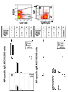
Figure 2. The frequencies of NP-specific memory B cells in the spleen of immunized mice
Splenic B cells were isolated from unimmunized mice (nonimmune) and mice 5 days and 6 weeks post the second immunization with NP-KLH and Ribi adjuvant. Cells were stained with FITC-anti-IgD, FITC-anti-IgM, PE-anti-CD138, PerCP-Cy5.5-anti-B220 antibodies, and APC-NP19 and analyzed using a flow cytometer. Shown are representative data from 8 independent experiments.
Figure 3
Figure 3. B cells with the memory phenotype differentiated into antigen-specific, high affinity IgG ASCs in culture with CpG ODN and LPS
(A) Splenic B cells from NP-KLH immunized mice were sorted into four populations: B220 IgM IgD NP CD138+ (I, plasma), B220+ IgM IgD NP+ CD138 (II, memory), B220+IgM+IgD+ NP+ CD138 (III, NP-binding, unswitched), and B220+ IgM+ IgD+ NP CD138 (IV, unspecific, unswitched). Naïve B cells (V) were isolated from the spleen of unimmunized mice. The frequencies of high affinity and total NP-specific IgG (B–D) and IgM (E–F) ASCs were determined by ELISPOT before (B and E) and after culture in CpG ODN (2 µg/ml) (C and F) or LPS (5 µg/ml) (D and G) for 5 days. Shown are representative results from three independent experiments.
Figure 4
Figure 4. Expression levels of TLR9 and TLR4 in different subpopulations of splenic B cells
Splenic B cells were isolated from unimmunized mice (nonimmune) and mice that were immunized with NP-KLH plus Ribi adjuvant. The splenic B cells were preincubated with the FcγR blocking antibody, followed by PerCP-Cy5-anti-B220, FITC-anti-IgD, FITC-anti-IgM antibodies, APC-NP19, and PE-anti-TLR4-MD2 antibodies for staining TLR4. For staining TLR9, the cells were washed, fixed, permeabilized, and stained with anti-TLR9 antibody and a pacific blue-conjugated secondary antibody. The cells were analyzed by flow cytometry. B220high B cell populations of nonimmune mice (V), NP-specific memory B cells (II), NP-binding, unswitched B cells (III), and non-specific, unswitched B cells (IV) of immunized mice were gated, and their TLR4 (right panels)- and TLR9 (left panels)-staining levels are shown. Shown are representative results from 5 independent experiments.
Figure 5
Figure 5. Clonal expansion and differentiation of memory B cells in response to immunization of antigen or TLR agonists alone or in combination
C57BL/6 mice were twice immunized with NP-KLH plus Ribi adjuvant 4 weeks apart and rested for 6 weeks. The mice were immunized for the third time with NP-KLH (400 µg/ml), Ribi adjuvant, CpG ODN (50 µg/mouse) or LPS (50 µg/mouse) alone or in combination. Controls included nonimmune mice and mice that did not receive the third immunization (None). (A) Serum was collected from one day before and five days after the challenge and analyzed for NP-specific high affinity and total IgG titers. Shown are average titers (±SE, n = 3~25). The serum titers of mice challenged with NP-KLH plus the adjuvant were significantly higher than those of mice that did not receive the third immunization or received the adjuvant alone for the third immunization. ***, p ≤ 0.001. *, p ≤ 0.05. (B–C) B cells and plasma cells were enriched and frequencies of NP-specific IgG ASCs were determined by ELISPOT before (B) and after (C) cultured with CpG ODN (2 µg/ml) in vitro for 5 days. (D) The percentage of NP-specific memory B cells among the splenic B cells was determined by flow cytometry, as described in Figure 2. Shown are the representative results of four independent experiments.

References

    1. McHeyzer-Williams LJ, McHeyzer-Williams MG. Antigen-specific memory B cell development. Annu Rev Immunol. 2005;23:487. - PubMed
    1. Lanzavecchia A, Bernasconi N, Traggiai E, Ruprecht CR, Corti D, Sallusto F. Understanding and making use of human memory B cells. Immunol Rev. 2006;211:303. - PMC - PubMed
    1. Anderson SM, Tomayko MM, Shlomchik MJ. Intrinsic properties of human and murine memory B cells. Immunol Rev. 2006;211:280. - PubMed
    1. Hofer T, Muehlinghaus G, Moser K, Yoshida T, H EM, Hebel K, Hauser A, Hoyer B, E OL, Dorner T, Manz RA, Hiepe F, Radbruch A. Adaptation of humoral memory. Immunol Rev. 2006;211:295. - PubMed
    1. Klein U, Rajewsky K, Kuppers R. Human immunoglobulin (Ig)M+IgD+ peripheral blood B cells expressing the CD27 cell surface antigen carry somatically mutated variable region genes: CD27 as a general marker for somatically mutated (memory) B cells. J Exp Med. 1998;188:1679. - PMC - PubMed

Publication types

MeSH terms