

Genetic analysis of SH2D4A, a novel adapter protein related to T cell-specific adapter and adapter protein in lymphocytes of unknown function, reveals a redundant function in T cells
- PMID: 18641339
- PMCID: PMC2613811
- DOI: 10.4049/jimmunol.181.3.2019
Genetic analysis of SH2D4A, a novel adapter protein related to T cell-specific adapter and adapter protein in lymphocytes of unknown function, reveals a redundant function in T cells
Abstract
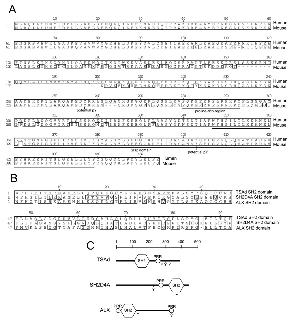
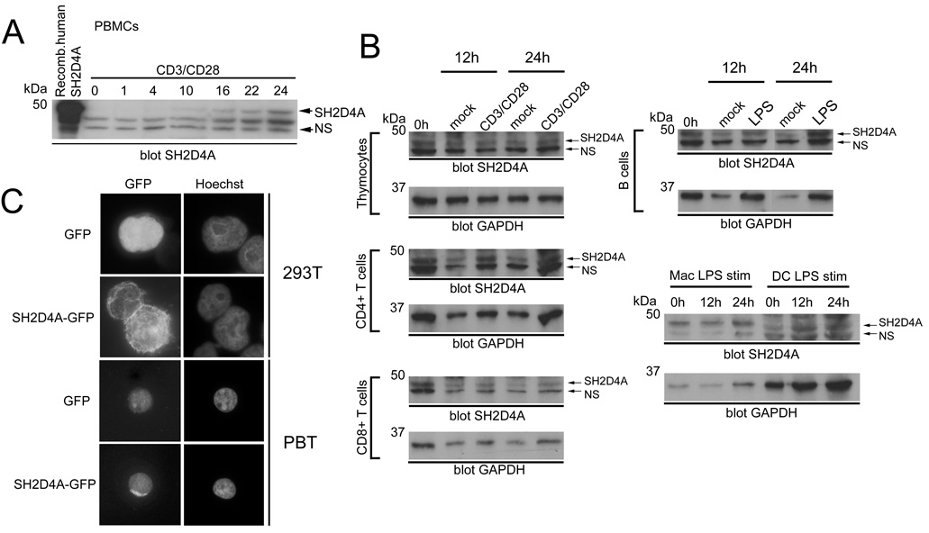
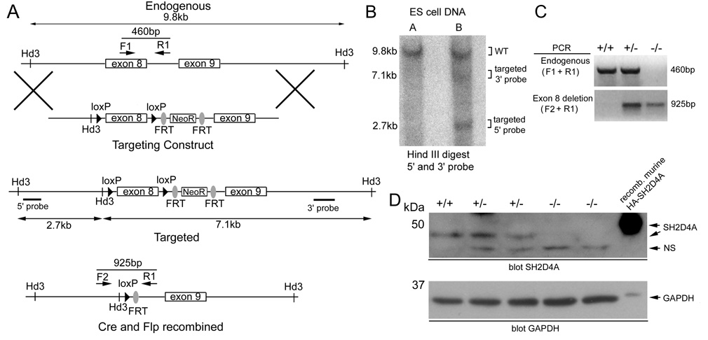
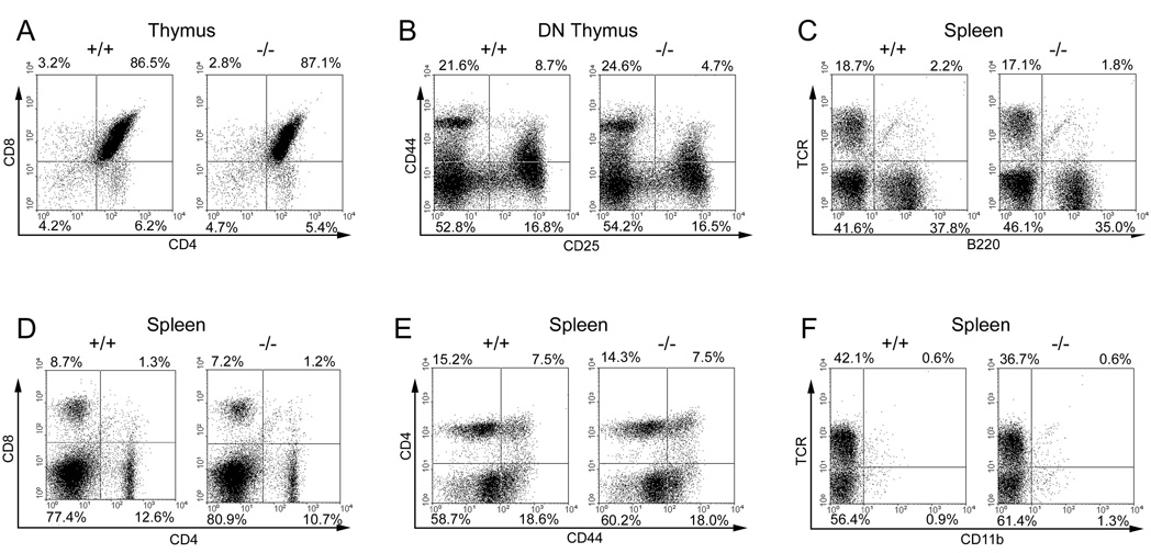
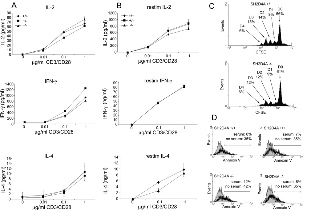
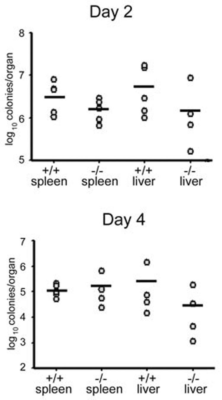
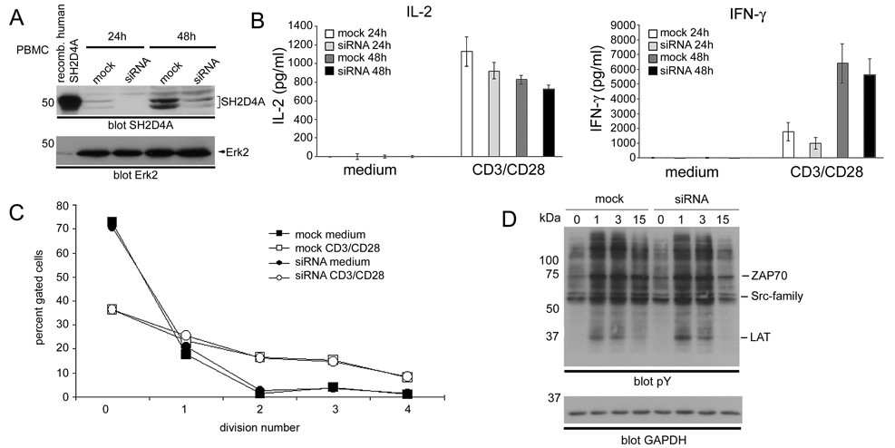
T cell-specific adapter (TSAd) protein and adapter protein in lymphocytes of unknown function (ALX) are two related Src homology 2 (SH2) domain-containing signaling adapter molecules that have both been shown to regulate TCR signal transduction in T cells. TSAd is required for normal TCR-induced synthesis of IL-2 and other cytokines in T cells and acts at least in part by promoting activation of the LCK protein tyrosine kinase at the outset of the TCR signaling cascade. By contrast, ALX functions as a negative-regulator of TCR-induced IL-2 synthesis through as yet undetermined mechanisms. In this study, we report a novel T cell-expressed adapter protein named SH2D4A that contains an SH2 domain that is highly homologous to the TSAd protein and ALX SH2 domains and that shares other structural features with these adapters. To examine the function of SH2D4A in T cells we produced SH2D4A-deficient mice by homologous recombination in embryonic stem cells. T cell development, homeostasis, proliferation, and function were all found to be normal in these mice. Furthermore, knockdown of SH2D4A expression in human T cells did not impact upon their function. We conclude that in contrast to TSAd and ALX proteins, SH2D4A is dispensable for TCR signal transduction in T cells.
Conflict of interest statement
Figures
Similar articles
-
The T-cell-specific adapter protein family: TSAd, ALX, and SH2D4A/SH2D4B.Immunol Rev. 2009 Nov;232(1):240-54. doi: 10.1111/j.1600-065X.2009.00829.x. Immunol Rev. 2009. PMID: 19909368 Review.
-
Essential role of the T cell-specific adapter protein in the activation of LCK in peripheral T cells.J Exp Med. 2006 Feb 20;203(2):281-7. doi: 10.1084/jem.20051637. Epub 2006 Jan 30. J Exp Med. 2006. PMID: 16446380 Free PMC article.
-
T cell-specific adapter protein inhibits T cell activation by modulating Lck activity.J Immunol. 2000 Sep 15;165(6):2927-31. doi: 10.4049/jimmunol.165.6.2927. J Immunol. 2000. PMID: 10975797
-
The p95-100 kDa ligand of the T cell-specific adaptor (TSAd) protein Src-homology-2 (SH2) domain implicated in TSAd nuclear import is p97 Valosin-containing protein (VCP).Immunol Lett. 2005 Mar 15;97(2):235-43. doi: 10.1016/j.imlet.2004.10.021. Epub 2004 Nov 24. Immunol Lett. 2005. PMID: 15752563
-
The emerging role of the T cell-specific adaptor (TSAd) protein as an autoimmune disease-regulator in mouse and man.Immunol Lett. 2005 Mar 15;97(2):165-70. doi: 10.1016/j.imlet.2004.10.019. Epub 2004 Nov 18. Immunol Lett. 2005. PMID: 15752554 Review.
Cited by
-
Integrative genomic identification of genes on 8p associated with hepatocellular carcinoma progression and patient survival.Gastroenterology. 2012 Apr;142(4):957-966.e12. doi: 10.1053/j.gastro.2011.12.039. Epub 2011 Dec 24. Gastroenterology. 2012. PMID: 22202459 Free PMC article.
-
A Novel 1.0 Mb Duplication of Chromosome 8p22-21.3 in a Patient With Autism Spectrum Disorder.Child Neurol Open. 2015 Jul 3;2(2):1-6. doi: 10.1177/2329048X15580673. eCollection 2015 Apr-Jun. Child Neurol Open. 2015. PMID: 35187197 Free PMC article.
-
The Ras GTPase-activating protein neurofibromin 1 promotes the positive selection of thymocytes.Mol Immunol. 2013 Oct;55(3-4):292-302. doi: 10.1016/j.molimm.2013.03.005. Epub 2013 Mar 20. Mol Immunol. 2013. PMID: 23522726 Free PMC article.
-
BEX4 inhibits the progression of clear cell renal cell carcinoma by stabilizing SH2D4A, which is achieved by blocking SIRT2 activity.Oncogene. 2025 Mar;44(10):665-678. doi: 10.1038/s41388-024-03235-6. Epub 2024 Dec 5. Oncogene. 2025. PMID: 39639172
References
-
- Simeoni L, Kliche S, Lindquist J, Schraven B. Adaptors and linkers in T and B cells. Curr Opin Immunol. 2004;16:304–313. - PubMed
-
- Samelson LE. Signal transduction mediated by the T cell antigen receptor: the role of adapter proteins. Annu Rev Immunol. 2002;20:371–394. - PubMed
-
- Spurkland A, Brinchmann JE, Markussen G, Pedeutour F, Munthe E, Lea T, Vartdal F, Aasheim HC. Molecular cloning of a T cell-specific adapter protein (TSAd) containing an Src homology (SH) 2 domain and putative SH3 and phosphotyrosine binding sites. J Biol Chem. 1998;273:4539–4546. - PubMed
-
- Choi YB, Kim CK, Yun Y. Lad, an adapter protein interacting with the SH2 domain of p56lck, is required for T cell activation. J Immunol. 1999;163:5242–5249. - PubMed
Publication types
MeSH terms
Substances
Grants and funding
LinkOut - more resources
Full Text Sources
Other Literature Sources
Molecular Biology Databases
Miscellaneous
