. 2008 Jul 1;7(13):2056-70.
doi: 10.4161/cc.7.13.6240.

Regulating the angiogenic balance in tissues

Affiliations

Regulating the angiogenic balance in tissues

Elizabeth A Pollina et al. Cell Cycle. .

Abstract

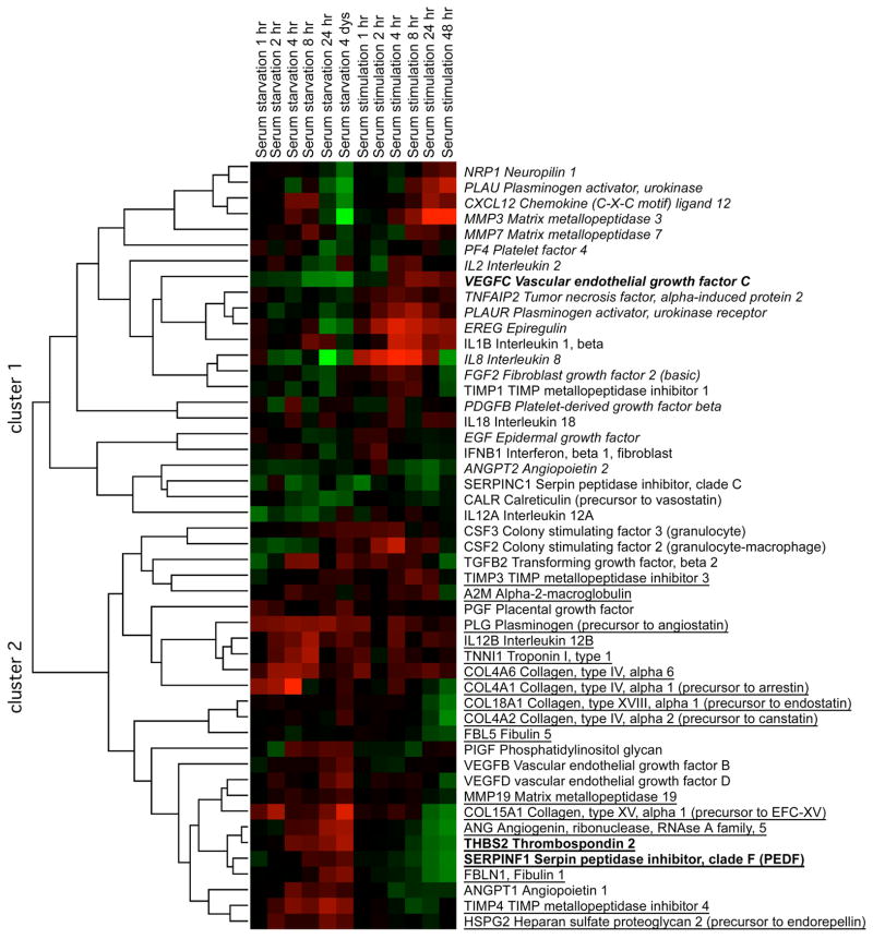
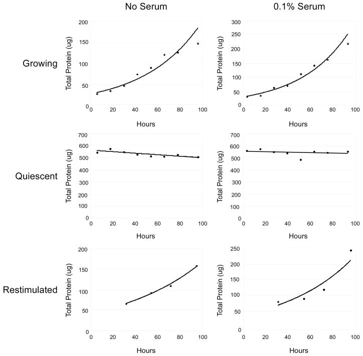
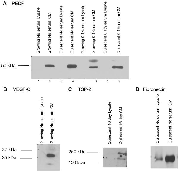
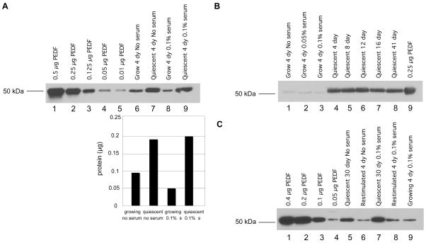
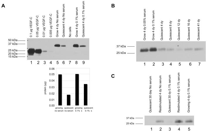
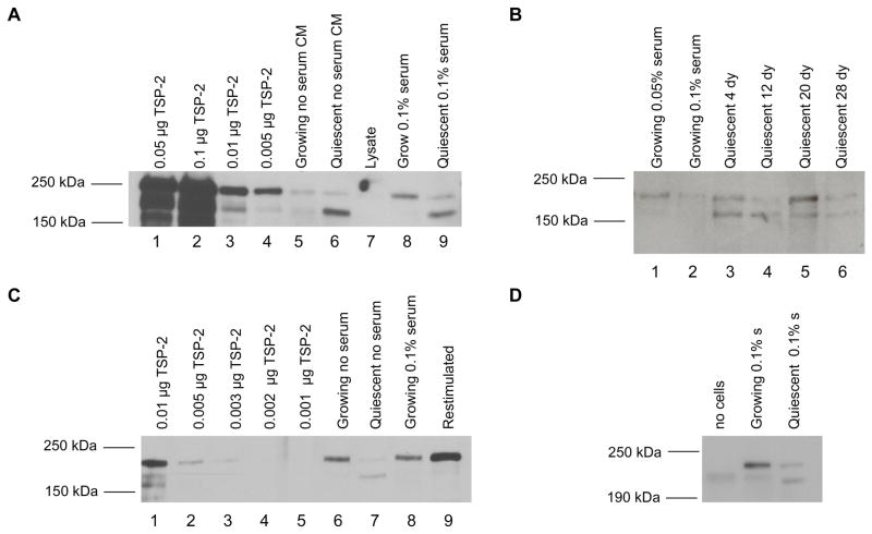
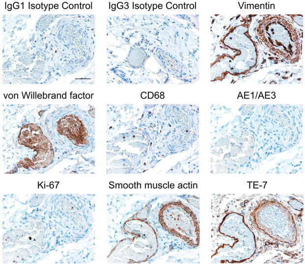
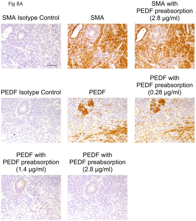
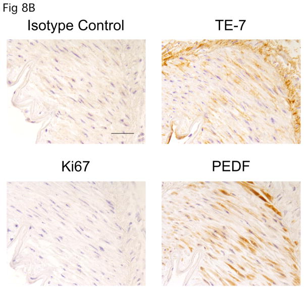
A balance between angiogenesis inducers and inhibitors in the microenvironment controls the rate of new blood vessel formation. We hypothesized that fibroblasts, an important cellular constituent of the tissue stroma, secrete molecules that contribute to this balance. We further hypothesized that fibroblasts secrete molecules that promote angiogenesis when they are in a proliferative state and molecules that inhibit angiogenesis when they are not actively cycling (quiescent). Microarray analysis revealed that angiogenesis inducers and inhibitors are regulated as fibroblasts transition into a quiescent state and reenter the cell cycle in response to changes in serum. To assess whether changes in transcript levels result in changes in the levels of secreted proteins, we collected conditioned medium from proliferating and quiescent fibroblasts and performed immunoblotting for selected proteins. Secreted protein levels of the angiogenesis inhibitor pigment epithelium derived factor (PEDF) were higher in quiescent than proliferating fibroblasts. Conversely, proliferating fibroblasts secreted increased levels of the angiogenesis inducer vascular endothelial growth factor-C (VEGF-C). For the angiogenesis inhibitor thrombospondin-2, quiescent cells secreted a prominent 160 kDa form in addition to the 200 kDa form secreted by proliferating and restimulated fibroblasts. Using immunohistochemistry we discovered that fibroblasts surround blood vessels and that the angiogenesis inhibitor PEDF is expressed by quiescent fibroblasts in uterine tissue, supporting a role for PEDF in maintaining quiescence of the vasculature. This work takes a new approach to the study of angiogenesis by examining the expression of multiple angiogenesis regulators secreted from a key stromal cell, the fibroblast.


Figures

FIGURE 1
FIGURE 1
Transcript levels of angiogenesis factors correlate with the proliferative state of fibroblasts Fibroblasts growing in 5% serum were switched to 0.1% serum, sampled for microarray analysis over a time course for four days, then restimulated with 5% serum and sampled for microarrays. During the serum withdrawal time course (columns 1–6), RNA from the proliferating cells cultured in 5% serum was used as a comparison for measuring transcript levels in serum-starved cells sampled at different timepoints. The ratio of transcript levels in serum-starved to proliferating samples for each gene in each sample is indicated in color. Red indicates upregulation in serum-starved compared with proliferating cells; green indicates downregulation. For the serum restimulation time course (column 7–12), four-day serum-starved quiescent cells served as controls for comparison with stimulated cells. Red indicates elevated expression in serum-stimulated cells compared with quiescent cells; green indicates downregulation. Angiogenesis factors were identified based on published lists, and were filtered to include proteins for which expression changed more than 1.5-fold at a time point. The filtered list was hierarchically clustered by average linkage using Cluster 3.0 and visualized using Java Treeview. Cluster 1 contains genes downregulated with quiescence and upregulated with serum stimulation. Genes in cluster 1 are enriched for angiogenesis inducers, which are indicated in italics. Genes in cluster 2 are upregulated with quiescence and downregulated with serum stimulation. Genes in cluster 2 are enriched in angiogenesis inhibitors, which are indicated by underlining. The three genes chosen for further analysis of protein level changes are in indicated in bold.
FIGURE 2
FIGURE 2
Protein concentration as a function of time in proliferating, quiescent and restimulated cells Proliferating, quiescent, and restimulated cells were plated as described in Materials and Methods either in the absence of serum or in the presence of 0.1% serum. Protein content was determined for one plate at each of the indicated timepoints after plating. Regression curves that best fit the data are shown, and the equations are given in Supplementary Table 1. Proliferating and restimulated cells were modeled on the assumption of exponential growth while quiescent cultures were constant (0.1% serum) or decreased in protein content slightly over time (no serum). The integrated areas under these curves were used for normalization of the volume of conditioned medium for analysis and are given in Supplementary Table 1.
FIGURE 3
FIGURE 3
Angiogenesis regulators are secreted into the conditioned medium A. Conditioned medium and cellular lysates were collected from proliferating and quiescent fibroblasts maintained in no serum or 0.1% serum for four days. Conditioned medium samples were normalized based on the integral method. Precipitated conditioned medium or 22 μg of protein lysates collected from the same cultures were separated on SDS-PAGE gels and immunoblotted for PEDF. PEDF was detected in the conditioned medium but not the lysate under all tested conditions. A background band at about 75 kDa was consistently observed in samples with serum. B. Conditioned medium and cellular lysates were collected and separated as in A. Membranes were immunoblotted with an anti-VEGF-C antibody. Processed VEGF-C was observed in conditioned medium but not cellular lysates. In some samples, preprocessed VEGF-C of approximately 58 kDa was detected in the cellular lysate (not shown). C. Medium conditioned for four days and cellular lysates were collected from cells maintained in quiescence in the absence of serum for 16 days. Precipitated conditioned medium and lysates were separated and immunoblotted with an anti-thrombospondin-2 antibody raised in goats. TSP-2 was detected in conditioned medium but not cellular lysates. D. Conditioned medium and lysates were collected from quiescent cells grown in the absence of serum. Samples were separated and immunoblotted with an anti-fibronectin antibody. Fibronectin was detected in both the conditioned medium and cellular lysates as expected for an extracellular matrix protein.
FIGURE 4
FIGURE 4
Quiescent fibroblasts secrete increased levels of the angiogenesis inhibitor PEDF A. Medium was conditioned for 4 days with proliferating or quiescent fibroblasts with no serum or 0.1% serum. Conditioned medium was normalized based on the integral method. Samples and recombinant protein were separated on SDS-PAGE gels and analyzed by immunoblotting for PEDF levels. The graph below indicates band intensity in the proliferating and quiescent lanes based on analysis with the ImageJ software. In no serum or 0.1% serum, quiescent fibroblasts secreted higher levels of PEDF than proliferating fibroblasts. B. PEDF is elevated in fibroblasts maintained in quiescence up to 41 days. Conditioned medium was collected from cells growing in no serum, 0.05% serum, or 0.1% serum. Conditioned medium was also collected from cells that had been quiescent with no serum for 4, 8, 12, 16 or 41 days. Medium was conditioned for four days for each sample. In this experiment, volumes of precipitated media were normalized by the protein content ratio of 16-day quiescent cultures versus 4-day growing cultures. Medium was precipitated, separated and immunoblotted for PEDF as in A. C. PEDF levels decrease when quiescent cells are stimulated to reenter the cell cycle. Cells maintained in a quiescent state by contact inhibition in the absence of serum for 30 days were replated sparsely and stimulated to reenter the cell cycle by addition of PDGF. Medium conditioned in quiescent cells or restimulated cells for four days was collected and normalized by the integral method. Conditioned medium from quiescent and restimulated cells was precipitated, separated by SDS-PAGE, and analyzed by immunoblotting as in Fig. 4A.
FIGURE 5
FIGURE 5
Proliferating fibroblasts secrete increased levels of the angiogenesis inducer VEGF-C A. Samples of proliferating and quiescent conditioned medium, and medium that was not incubated with cells, were prepared as in Fig. 4A. Volumes were normalized according to the integral method. Medium and recombinant proteins were separated on SDS-PAGE gels and analyzed by immunoblotting for VEGF-C levels. Recombinant protein migrated as the fully processed 21 kDa form while the predominant species in the conditioned medium was an approximately 31 kDa partially processed form. For conditioned medium from cells growing in 0.1% serum, both preprocessed and fully processed forms of VEGF-C were detected. The amount of VEGF-C in each sample was determined by comparison with the standard curve and is indicated in the graph below. B. VEGF-C levels are reduced in cells maintained in quiescence for a long period of time. Conditioned medium was collected and precipitated from samples as in Fig. 4B. Medium was precipitated, separated and immunoblotted for VEGF-C as in Fig. 5A. C. VEGF-C levels increased in conditioned medium from quiescent cells induced to reenter the cell cycle. Conditioned medium was collected from proliferating cells, quiescent cells and cells maintained in a quiescent state for 30 days and then stimulated to reenter the cell cycle. Samples were normalized according to the integral method. Precipitated conditioned medium and recombinant protein were separated and immunoblotted with an anti-VEGF-C antibody.
FIGURE 6
FIGURE 6
Quiescent and proliferating fibroblasts secrete different forms of thrombospondin-2 A. TSP-2 migrates differently in conditioned medium from proliferating and quiescent fibroblasts. Conditioned medium was collected from proliferating and quiescent cells and normalized according to the integral method. Conditioned media samples were precipitated, separated on SDS-PAGE gels and immunoblotted with an anti-thrombospondin-2 antibody raised in goats. The 160 kDa band was prominent in conditioned medium from quiescent cells in no serum or 0.1% serum, but not in conditioned medium from proliferating cells. B. Long-term quiescent cells continue to express the 160 kDa TSP-2 band. Conditioned medium was collected from fibroblasts growing in 0.05% serum or 0.1% serum, or from fibroblasts maintained in a quiescent state with no serum for 4, 12, 20 or 28 days. A mouse monoclonal anti-TSP2 antibody was used for immunoblotting. The quiescence-specific 160 kDa band was present in samples maintained in quiescence for up to 28 days. Samples in this blot were normalized based on the amount of protein present at the time of collection. C. Expression of the 160 kDa band is reduced when quiescent fibroblasts are restimulated to enter the cell cycle. Conditioned medium was precipitated from fibroblasts in proliferating, quiescent and restimulated conditions according to the integral method. Samples and indicated amounts of recombinant protein were separated on SDS-PAGE gels and immunoblotted with a goat anti-thrombospondin-2 antibody. D. Medium that had not been conditioned with cells and medium conditioned in proliferating or quiescent cells was normalized, precipitated and separated on an SDS-PAGE gel. Membranes were immunoblotted with an antibody raised against the N-terminal 40 amino acids of thrombospondin-2. The band pattern observed with the N-terminal antibody is similar to the pattern observed with antibodies against the full-length form. The 160 kDa band is therefore unlikely to reflect proteolytic processing of the TSP-2 N-terminal end.
FIGURE 7
FIGURE 7
Fibroblasts surround blood vessels in smooth muscle Immunostaining of fibroblasts surrounding an artery (right hand side of the image) and a vein (left hand side of the image) in smooth muscle is shown. Smooth muscle tissue was collected in 10% neutral buffered formalin and paraffin-embedded. Serial sections were stained with antibodies that recognize specific cell types. Antibody staining is shown in brown, counterstain is in blue. IgG1 and IgG3 isotype controls display little staining as expected. An antibody to vimentin stains mesenchymal cells surrounding the vein, the artery and smaller vessels and capillaries. An antibody to von Willebrand factor recognizes endothelial cells lining both the vein and artery and the blood cells within the vessels. An antibody to CD68 identifies macrophages scattered throughout the tissue. The AE1/AE3 antibody that recognizes epithileal cytokeratins does not stain this smooth muscle tissue, as expected. An antibody to Ki-67 recognizes few cells within this tissue, indicating that most cells within the tissue are not actively dividing. An antibody to smooth muscle actin recognized a thin layer of cells surrounding the vein (left) and a thicker layer of cells surrounding the artery (right). TE-7, an antibody that recognizes fibroblasts, recognized cells surrounding the large vein (left) and the large artery (right), as well as smaller vessels. A scale bar indicating 10 microns is shown in the first panel.
FIGURE 8
FIGURE 8
PEDF is expressed by stromal cells in normal tissue A. Specificity of the anti-PEDF antibody. Human uterine tissue was formalin-fixed, paraffin-embedded, and stained. In the top row, samples were stained with an isotype control, an antibody to smooth muscle actin, or with an antibody to smooth muscle actin after preabsorption with the PEDF protein (2.8 μg/ml). Preabsorption with PEDF did not affect the staining pattern of an anti-smooth muscle actin antibody. The second and third rows indicate staining with an isotype control, an anti-PEDF antibody or an anti-PEDF antibody after preabsorption with one of several concentrations of recombinant PEDF (0.28 μg/ml, 1.4 μg/ml or 2.8 μg/ml). A dose-dependent competitive inhibition of the anti-PEDF antibody with addition of recombinant PEDF is observed, demonstrating the specificity of the staining for PEDF. A scale bar indicating 10 microns is shown in the first panel. B. PEDF stains quiescent fibroblasts in the stromal region surrounding a blood vessel in normal kidney. Human kidney tissue was stained with an antibody to PEDF, an antibody to Ki-67, TE-7 (an antibody that recognizes a fibroblast-associated protein), or an isotype control. PEDF was easily detected in the Ki-67 negative cells within the fibroblast-rich stroma. A scale bar indicating 5 microns is shown in the first panel.
FIGURE 8
FIGURE 8
PEDF is expressed by stromal cells in normal tissue A. Specificity of the anti-PEDF antibody. Human uterine tissue was formalin-fixed, paraffin-embedded, and stained. In the top row, samples were stained with an isotype control, an antibody to smooth muscle actin, or with an antibody to smooth muscle actin after preabsorption with the PEDF protein (2.8 μg/ml). Preabsorption with PEDF did not affect the staining pattern of an anti-smooth muscle actin antibody. The second and third rows indicate staining with an isotype control, an anti-PEDF antibody or an anti-PEDF antibody after preabsorption with one of several concentrations of recombinant PEDF (0.28 μg/ml, 1.4 μg/ml or 2.8 μg/ml). A dose-dependent competitive inhibition of the anti-PEDF antibody with addition of recombinant PEDF is observed, demonstrating the specificity of the staining for PEDF. A scale bar indicating 10 microns is shown in the first panel. B. PEDF stains quiescent fibroblasts in the stromal region surrounding a blood vessel in normal kidney. Human kidney tissue was stained with an antibody to PEDF, an antibody to Ki-67, TE-7 (an antibody that recognizes a fibroblast-associated protein), or an isotype control. PEDF was easily detected in the Ki-67 negative cells within the fibroblast-rich stroma. A scale bar indicating 5 microns is shown in the first panel.

References

    1. Palmiter RD, Brinster RL, Hammer RE, Trumbauer ME, Rosenfeld MG, Birnberg NC, Evans RM. Dramatic growth of mice that develop from eggs microinjected with metallothionein-growth hormone fusion genes. Nature. 1982;300:611–5. - PMC - PubMed
    1. Mathews LS, Hammer RE, Behringer RR, D'Ercole AJ, Bell GI, Brinster RL, Palmiter RD. Growth enhancement of transgenic mice expressing human insulin-like growth factor I. Endocrinology. 1988;123:2827–33. - PubMed
    1. Fero ML, Randel E, Gurley KE, Roberts JM, Kemp CJ. The murine gene p27Kip1 is haplo-insufficient for tumour suppression. Nature. 1998;396:177–80. - PMC - PubMed
    1. Rupnick MA, Panigrahy D, Zhang CY, Dallabrida SM, Lowell BB, Langer R, Folkman MJ. Adipose tissue mass can be regulated through the vasculature. Proceedings of the National Academy of Sciences of the United States of America. 2002;99:10730–5. - PMC - PubMed
    1. Greene AK, Wiener S, Puder M, Yoshida A, Shi B, Perez-Atayde AR, Efstathiou JA, Holmgren L, Adamis AP, Rupnick M, Folkman J, O'Reilly MS. Endothelial-directed hepatic regeneration after partial hepatectomy. Ann Surg. 2003;237:530–5. - PMC - PubMed

Publication types

MeSH terms