. 2008 Aug 19;47(33):8786-95.
doi: 10.1021/bi8007824. Epub 2008 Jul 22.

Acidic residues C-terminal to the A2 domain facilitate thrombin-catalyzed activation of factor VIII

Affiliations

Acidic residues C-terminal to the A2 domain facilitate thrombin-catalyzed activation of factor VIII

Jennifer L Newell et al. Biochemistry. .

Abstract

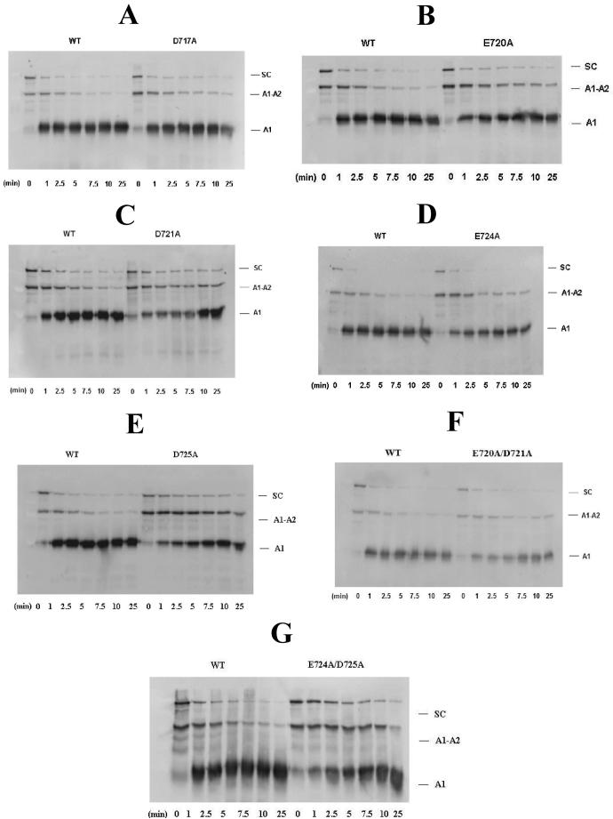
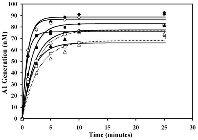
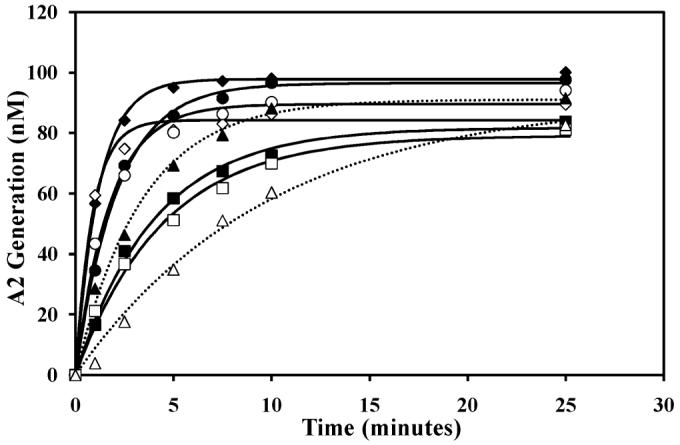
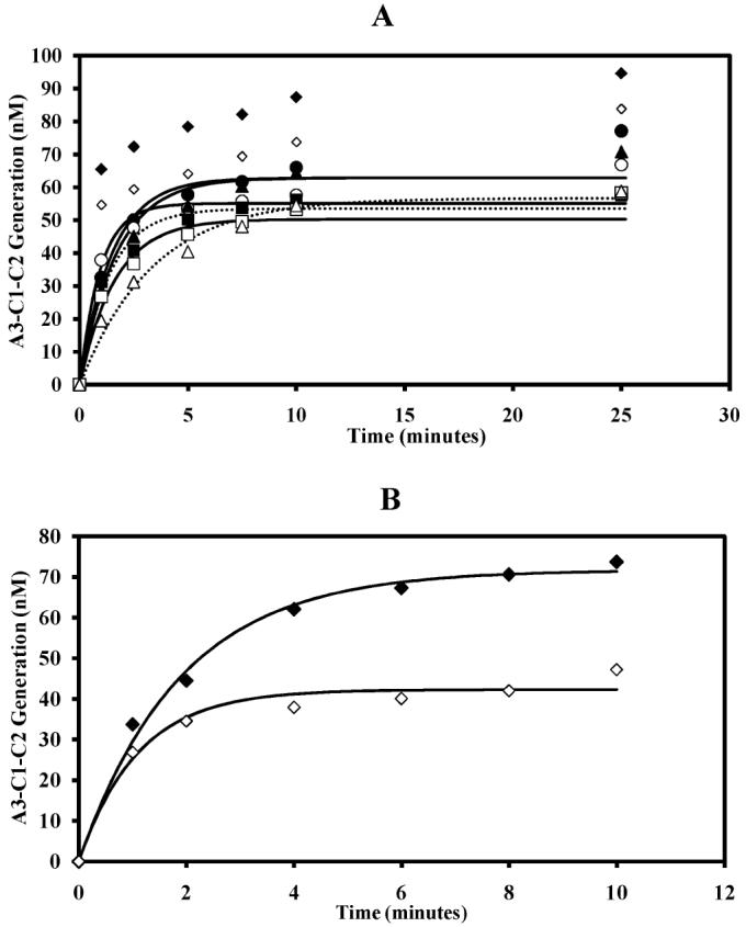
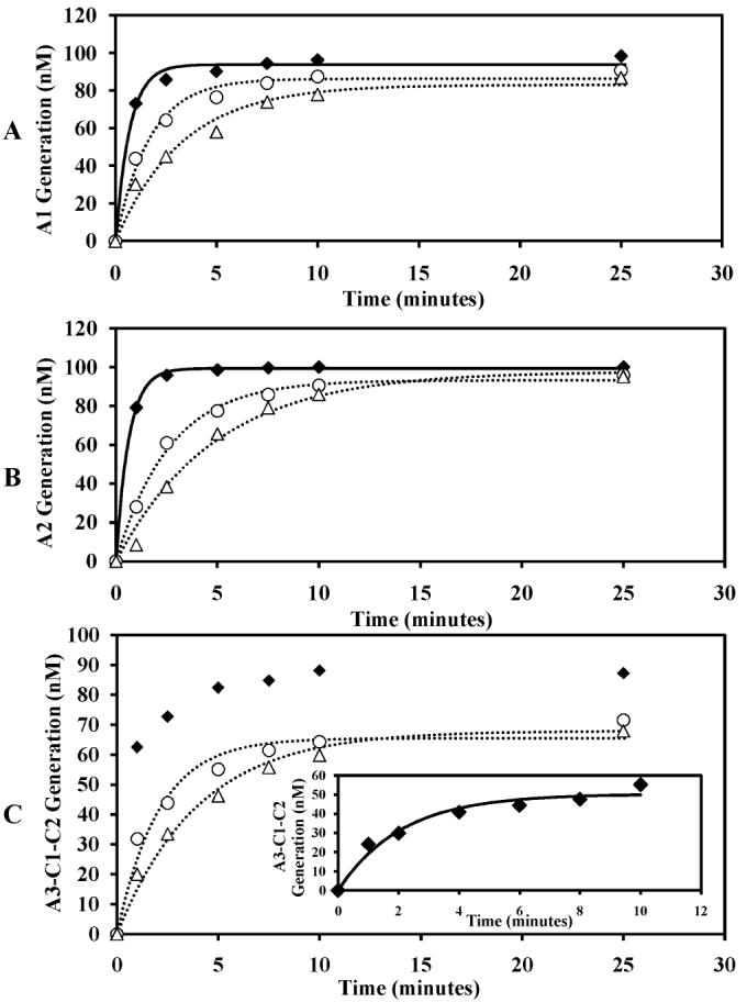
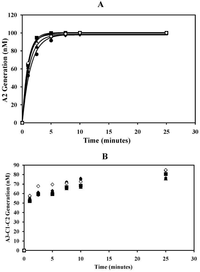
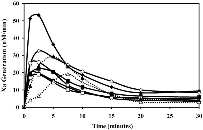
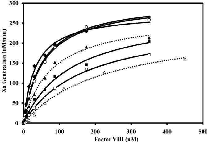
Factor VIII is activated by thrombin through proteolysis at Arg740, Arg372, and Arg1689. One region implicated in this exosite-dependent interaction is the factor VIII a2 segment (residues 711-740) separating the A2 and B domains. Residues 717-725 (DYYEDSYED) within this region consist of five acidic residues and three sulfo-Tyr residues, thus representing a high density of negative charge potential. The contributions of these residues to thrombin-catalyzed activation of factor VIII were assessed following mutagenesis of acidic residues to Ala or Tyr residues to Phe and expression and purification of the B-domainless proteins from stable-expressing cell lines. All mutations showed reduced specific activity from approximately 30% to approximately 70% of the wild-type value. While replacement of the Tyr residues showed little, if any, effect on rates of thrombin-catalyzed proteolysis of factor VIII and consequent activation, the acidic to Ala mutations Glu720Ala, Asp721Ala, Glu724Ala, and Asp725Ala showed decreased rates of proteolysis at each of the three P1 residues. Mutations at residues Glu724 and Asp725 were most affected with double mutations at these sites showing approximately 10-fold and approximately 30-fold reduced rates of cleavage at Arg372 and Arg1689, respectively. Factor VIII activation profiles paralleled the results assessing rates of proteolysis. Kinetic analyses revealed these mutations minimally affected apparent V max for thrombin-catalyzed cleavage but variably increased the K m for procofactor up to 7-fold, suggesting the latter parameter was dominant in reducing catalytic efficiency. These results suggest that residues Glu720, Asp721, Glu724, and Asp725 likely constitute an exosite-interactive region in factor VIII facilitating cleavages for procofactor activation.


Figures

FIGURE 1
FIGURE 1
Generation of A1 subunit following thrombin-catalyzed cleavage of recombinant factor VIII mutants. Factor VIII (100 nM) was reacted with thrombin (2.5 nM) for the indicated times as described in Materials and Methods. Samples were run on 8% polyacrylamide gels followed by Western blotting using an anti-A1 antibody. Blotting data are as follows: (A) Asp717Ala, (B) Glu720Ala, (C) Asp721Ala, (D) Glu724Ala, (E) Asp725Ala, (F) Glu720Ala/Asp721Ala, and (G) Glu724Ala/Asp725Ala. The abbreviations WT, A1-A2, and SC represent wild type, heavy chain (contiguous A1-A2 domains), and single chain, respectively.
FIGURE 2
FIGURE 2
A1 subunit generated following thrombin cleavage of factor VIII. Wild-type and mutant recombinant factor VIII (100 nM) were reacted with thrombin (2.5 nM) for the indicated times as described in Materials and Methods. Samples were run on 8% polyacrylamide gels followed by Western blotting using an anti-A1 antibody. Data was derived from densitometric analysis of A1 generation (nM) from blotting data (Figure 1). Symbols: ◆, wild type; ◇, Asp717Ala; ●, Glu720Ala; ○, Asp721Ala; ■, Glu724Ala; ◻, Asp725Ala; ▴, Glu720Ala/Asp721Ala; Δ, Glu724Ala/Asp725Ala. The data were fit to the single exponential equation using nonlinear least squares regression. Experiments were performed at least three separate times, and average values are shown. Continuous lines represent the single mutants while the double mutants are represented by the dotted lines.
FIGURE 3
FIGURE 3
Generation of A2 subunit by thrombin-catalyzed proteolysis of recombinant factor VIII. Wild-type and mutant recombinant factor VIII (100 nM) were reacted with thrombin (2.5 nM) for the indicated times as described in Materials and Methods. Samples were run on 8% polyacrylamide gels followed by Western blotting using the anti-A2 antibody R8B12. Data was derived from quantitative densitometry of A2 generation (nM) from blotting data (see Supporting Information for blots). Symbols: ◆, wild type; ◇, Asp717Ala; ●, Glu720Ala; ○, Asp721Ala; ■, Glu724Ala; ◻, Asp725Ala; ▴, Glu720Ala/Asp721Ala; Δ, Glu724Ala/Asp725Ala. The data were fitted to the single exponential equation using nonlinear least squares regression. Experiments were performed at least three separate times, and average values are shown. Continuous lines represent the single mutants while the double mutants are represented by the dotted lines.
FIGURE 4
FIGURE 4
Generation of A3-C1-C2 subunit following thrombin cleavage of factor VIII variants. Panel A: Recombinant wild-type and mutant factor VIII (100 nM) were reacted with thrombin (2.5 nM) for the indicated times and subjected to SDS-PAGE and blotting using the 2D2 antibody (see Supporting Information for blots). Densitometry data from blots are plotted as described in the legend to Figure 2. Panel B shows results obtained using wild-type and D717A factor VIII (100 nM) reacted with a lower thrombin concentration (0.125 nM). Symbols: ◆, wild type; ◇, Asp717Ala; ●, Glu720Ala; ○, Asp721Ala; ■, Glu724Ala; ◻, Asp725Ala; ▴, Glu720Ala/Asp721Ala; Δ, Glu724Ala/Asp725Ala. The data were fitted to the single exponential equation using nonlinear least squares regression. Experiments were performed at least three separate times, and average values are shown. Continuous lines represent the single mutants while the double mutants are represented by the dotted lines.
FIGURE 5
FIGURE 5
Generation of A1, A2, and A3-C1-C2 subunits at 37 °C and 140 mM NaCl following thrombin-catalyzed cleavage of recombinant wild-type and double mutant factor VIII. Wild-type and mutant recombinant factor VIII (100 nM) were reacted with thrombin (2.5 nM) at 37 °C in buffer containing 140 mM NaCl for the indicated times as described in Materials and Methods. Samples were run on 8% polyacrylamide gels and subjected to Western blotting with the following antibodies: (A) C5 (anti-A1), (B) R8B12 (anti-A2), and (C) 2D2 (anti-A3). Data were derived from densitometric analysis of A1, A2, and A3-C1-C2 subunit generation (nM) from blotting data (data not shown). Symbols: ◆, wild type; ○, Glu720Ala/Asp721Ala; Δ, Glu724Ala/Asp725Ala. The insert in panel C shows results obtained using wild-type factor VIII (100 nM) reacted with a lower thrombin concentration (0.125 nM). The data were fit to the single exponential equation using nonlinear least squares regression. Experiments were performed at least three separate times, and average values are shown. The continuous line represents wild-type factor VIII while the double mutants are represented by the dotted lines.
FIGURE 6
FIGURE 6
Western blot analysis of tyrosine sulfation mutants. Panel A: Recombinant wild-type and mutant factor VIII (100 nM) were reacted with thrombin (2.5 nM) for the indicated times and subjected to SDS-PAGE and blotting using the anti-A2 antibody (see Supporting Information for blots). Symbols: ◆, wild type; ●, Tyr718Phe; ■,Tyr719Phe; ▴, Tyr723Phe;◇, Tyr718Phe/Tyr719Phe/Tyr723Phe. Panel B: The wild-type and mutant factor VII reactions were repeated as described above except blotting with the anti-A3 antibody (see Supporting Information for blots). Symbols: ◆, wild type; ●, Tyr718Phe; ■, Tyr719Phe; ▴, Tyr723Phe; ◇, Tyr718Phe/Tyr719Phe/Tyr723Phe.
FIGURE 7
FIGURE 7
Activation of wild-type and mutant factor VIII by thrombin. Recombinant factor VIII WT and mutants (1 nM) were reacted with thrombin (0.05 nM) for the indicated times. Thrombin was inactivated by addition of hirudin (0.1 unit/mL), and factor VIIIa was reacted with factor IXa (20 nM) and phospholipid vesicles (10 µM). Factor Xa generation was initiated by addition of factor X (300 nM) as described in Materials and Methods. Symbols: ◆, wild type; ◇, Asp717Ala; ●, Glu720Ala; ○, Asp721Ala; ■, Glu724Ala; ◻, Asp725Ala; ▴, Glu720Ala/Asp721Ala Δ, Glu724Ala/Asp725Ala. Experiments were performed at least three separate times, and average values are shown. Continuous lines represent the single mutants while the double mutants are represented by the dotted lines.
FIGURE 8
FIGURE 8
Kinetics of thrombin activation of wild-type and double mutant factor VIII. Various concentrations of recombinant wild-type and mutant factor VIII were reacted with thrombin (0.05 nM) for 15 s. Thrombin was inactivated by addition of hirudin (0.1 unit/mL) in the presence of phospholipid vesicles (10 µM), and factor Xa generation was initiated by addition of factor X (300 nM) and factor IXa (20 nM) as described in Materials and Methods. Symbols: ◆, wild type; ●, Glu720Ala; ○, Asp721Ala; ■, Glu724Ala; ◻, Asp725Ala; ▴, Glu720Ala/Asp721Ala; Δ, Glu724Ala/Asp725Ala. Initial rates of factor Xa generation are plotted as a function of factor VIII concentration and fitted to the Michaelis-Menten equation by nonlinear least squares regression. Experiments were performed at least three separate times, and average values are shown. Continuous lines represent the single mutants while the double mutants are represented by the dotted lines.

Similar articles

Cited by

References

    1. Wood WI, Capon DJ, Simonsen CC, Eaton DL, Gitschier J, Keyt B, Seeburg PH, Smith DH, Hollingshead P, Wion KL. Expression of Active Human Factor VIII from Recombinant DNA Clones. Nature. 1984;312:330–337. - PubMed
    1. Vehar GA, Keyt B, Eaton D, Rodriguez H, O'Brien DP, Rotblat F, Oppermann H, Keck R, Wood WI, Harkins RN. Structure of Human Factor VIII. Nature. 1984;312:337–342. - PubMed
    1. Fay PJ. Activation of Factor VIII and Mechanisms of Cofactor Action. Blood Rev. 2004;18:1–15. - PubMed
    1. Mann KG, Nesheim ME, Church WR, Haley P, Krishnaswamy S. Surface-Dependent Reactions of the Vitamin K-Dependent Enzyme Complexes. Blood. 1990;76:1–16. - PubMed
    1. Eaton D, Rodriguez H, Vehar GA. Proteolytic Processing of Human Factor VIII. Correlation of Specific Cleavages by Thrombin, Factor Xa, and Activated Protein C with Activation and Inactivation of Factor VIII Coagulant Activity. Biochemistry. 1986;25:505–512. - PubMed

Publication types