. 2008 Sep;69(5):1304-15.
doi: 10.1111/j.1365-2958.2008.06363.x. Epub 2008 Jul 10.

Staphylococcus aureus haem oxygenases are differentially regulated by iron and haem

Affiliations

Staphylococcus aureus haem oxygenases are differentially regulated by iron and haem

Michelle L Reniere et al. Mol Microbiol. 2008 Sep.

Abstract

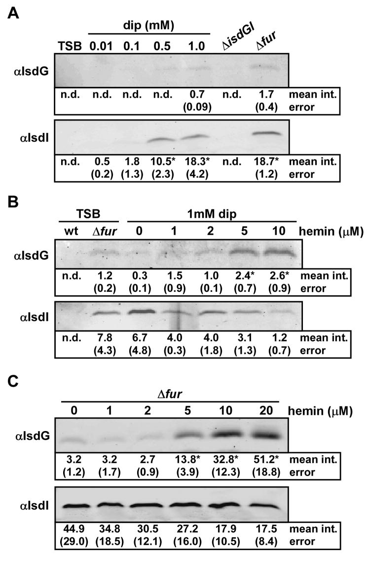
Iron acquisition is a vital process for most pathogenic bacteria, as iron is a limiting nutrient during infection. Staphylococcus aureus, an increasingly important pathogen, acquires iron from host haem via elaboration of the iron-regulated surface determinant system (Isd). IsdG and IsdI are haem oxygenases that have been proposed to degrade exogenous haem in the bacterial cytoplasm as a mechanism to liberate free iron for use as a nutrient source. Herein, we report that IsdG and IsdI are both important for S. aureus growth on haemin as a sole iron source and are necessary for full S. aureus pathogenesis. Investigations into the regulation of these enzymes revealed that IsdG and IsdI are differentially regulated by iron and haem through both transcriptional and post-transcriptional mechanisms. Additionally, IsdI was found to be expressed in infected tissues at the sites of abscess formation, suggesting that abscesses are iron-starved microenvironments inside the host. These findings suggest that S. aureus differentially regulates IsdG and IsdI in response to alterations in iron and haem availability during infection.


Figures

Figure 1
Figure 1. Growth of S. aureus heme oxygenase mutants on hemin as a sole iron source
(A) The colony diameter of S. aureus wild type, ΔisdG, ΔisdI, and ΔisdGI strains was measured after growth on solid media containing an iron chelator and various concentrations of hemin. Asterisks denote statistical significance between wild type and mutant strains, as measured by Student’s t test (p < 0.0001). (B and C) Strains listed in A were transformed with an empty vector (pOS1) or a vector expressing either isdG or isdI from a constitutive promoter (pisdG, pisdI) and colony diameter was measured as in A. A single time point at 48 hours is shown, however these trends were observed at all time points tested. Asterisks denote statistical significance between mutant and complemented strains, as measured by Student’s t test (p < 0.0001).
Figure 2
Figure 2. Regulation of IsdG and IsdI by iron and hemin
S. aureus strains were grown overnight in the indicated media and protoplast fractions were harvested and normalized by total protein concentration. The mean intensity of each band, as determined by densitometric analyses, is presented under the blots in arbitrary units, with corresponding standard error. Bands that were not detected for quantification are reported as not determined (n.d). Statistical significance was determined using the Student’s t test. Results are representative of at least four independent experiments. (A) The effect of iron chelation by 2,2’dipyridyl (dip) on expression of IsdG and IsdI was analyzed by immunoblot. Asterisks denote statistically significant increases, as compared to 0.01 mM dip (p < 0.0001). (B) The effect of exogenous hemin on expression of IsdG and IsdI in iron-deplete media (dip) was analyzed by immunoblot. Asterisks denote statistically significant increases, as compared to 0 μM hemin (p < 0.03). The slight decrease in IsdI abundance in 10 μM hemin compared to 0 μM hemin is not statistically significant (p > 0.06). (C) The effect of exogenous hemin on expression of IsdG and IsdI in an isogenic Δfur strain was analyzed by immunoblot. Asterisks denote statistically significant increases, as compared to 0 μM hemin (p < 0.0002). The slight decrease in IsdI abundance in 20 μM hemin compared to 0 μM hemin is not statistically significant (p > 0.1).
Figure 3
Figure 3. Hemin-dependent regulation of Isd protein expression
(A) Schematic representation of the isd locus. The genes are indicated by open arrows and the putative Fur-binding sites are denoted by black boxes upstream of putative transcription start sites (black arrows). orfX is an uncharacterized open reading frame upstream of isdI, and these genes are co-transcribed. (B) Expression of IsdG and IsdI was analyzed by immunoblot. Wild type S. aureus and ΔisdC were grown in iron-replete or iron-deplete (dip) media in the presence or absence of hemin (10 μM) and protoplast fractions were harvested and normalized by total protein concentration. (C) Immunoblot analysis of Isd proteins in S. aureus wild type grown in iron-deplete medium containing hemin (wt dip+hemin) compared to isogenic strains lacking individual genes of the isd locus in identical growth media. The isogenic Δfur strain was grown in TSB. All results are representative of at least three independent experiments.
Figure 4
Figure 4. Role of transcription in the hemin-dependent regulation of IsdG
(A) Transcript levels of isdC, isdD, and isdG were analyzed by real time RT-PCR and normalized to 16S rRNA. S. aureus wild type was grown in iron-deplete media (dip, gray bars) and iron-deplete media containing 5 μM hemin (dip + hemin, black bars) and transcript levels were compared to those grown in TSB. Error bars represent the average range of triplicate experiments. (B) IsdG abundance in S. aureus ΔisdG pisdG grown in iron-deplete media and various concentrations of hemin was analyzed by immunoblotting. Results depict normalized IsdG protein levels in arbitrary units (a.u.). Asterisk denotes statistical significance compared to iron-deplete media alone, as measured by Student’s t test (p < 0.015).
Figure 5
Figure 5. Role of hemin in the stability of IsdG and IsdI
(A and B) Phosphorimages of radiolabeled IsdG and IsdI immunoprecipitated from cells pulsed with 35S-methionine, followed by a chase with unlabeled methionine for the time indicated. Pulse-chase analyses were carried out in media treated with iron sulfate (+Fe) or 2,2’dipyridyl (-Fe), and 10 μM hemin (+H). Dash indicates control cells in which primary antibody was omitted during immunoprecipitation. (C and D) Quantification of IsdG and IsdI from quadruplicate experiments as described in A and B. Error bars represent standard deviation of data from at least three independent experiments. Asterisks denote statistical significance between conditions with and without hemin irrespective of iron status, as measured by Student’s t test (p < 0.003).
Figure 6
Figure 6. Contribution of IsdG and IsdI to S. aureus pathogenesis
S. aureus colonization of the hearts and kidneys of female Balb/c mice was analyzed four days post retroorbital infection. (A and C) Colonization was compared between wild type, ΔisdG, and ΔisdI strains. (B and D) In a separate experiment, colonization of wild type S. aureus was compared to the ΔisdGI strain. Each data point represents the colony forming units (CFU) per organ in a single animal. The horizontal lines represent the mean CFU/organ. Asterisks denote statistical significance between wild type and the representative mutant strain as measured by Student’s t test (p < 0.04).
Figure 7
Figure 7. In vivo bioluminescence imaging of IsdI expression
(A) S. aureus harboring pXen1 or pisdI.Xen1 was grown overnight in TSB or iron-deplete media (+dip). Serial dilutions were performed and luminescence was measured as described in the Experimental Procedures. Luminescence scale is in p/s/cm2/sr. (B) Quantification of luminescence depicted in A. Results are shown as fold change in luminescence in iron-deplete medium compared to TSB. Asterisks denote statistical significance between pXen1 (white bars) and pisdI.XenI (black bars), as measured by Student’s t test (p < 0.004). (C) At four days post-infection kidneys were removed from mice imaged ex vivo. Pictured are kidneys from mice infected with S. aureus carrying pXen1 or pisdI.Xen1 (left panels) and bioluminescent images of the same kidneys (right panels). Arrows indicate representative abscesses. Luminescence scale is as in A. Pictures and bioluminescent images are representative of at least five mice in each group. Bacterial load and number of abscesses were consistent between strains.

References

    1. Bae T, Schneewind O. Allelic replacement in Staphylococcus aureus with inducible counter-selection. Plasmid. 2006;55:58–63. - PubMed
    1. Bibb LA, Kunkle CA, Schmitt MP. The ChrA-ChrS and HrrA-HrrS Signal Transduction Systems Are Required for Activation of the hmuO Promoter and Repression of the hemA Promoter in Corynebacterium diphtheriae. Infect Immun. 2007;75:2421–2431. - PMC - PubMed
    1. Braun V, Gunter K, Hantke K. Transport of iron across the outer membrane. Biol Met. 1991;4:14–22. - PubMed
    1. Bubeck Wardenburg J, Williams WA, Missiakas D. Host defenses against Staphylococcus aureus infection require recognition of bacterial lipoproteins. Proc Natl Acad Sci U S A. 2006;103:13831–13836. - PMC - PubMed
    1. Bullen JJ, Griffiths E. Iron and Infection: Molecular, Physiological and Clinical Aspects. New York: John Wiley and Sons; 1999.

Publication types

MeSH terms