Review
. 2008 Jul 22;18(14):R620-31.
doi: 10.1016/j.cub.2008.05.039.

Ascidians and the plasticity of the chordate developmental program

Affiliations
Review

Ascidians and the plasticity of the chordate developmental program

Patrick Lemaire et al. Curr Biol. .

Abstract

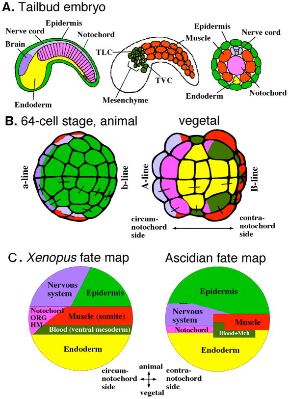
Little is known about the ancient chordates that gave rise to the first vertebrates, but the descendants of other invertebrate chordates extant at the time still flourish in the ocean. These invertebrates include the cephalochordates and tunicates, whose larvae share with vertebrate embryos a common body plan with a central notochord and a dorsal nerve cord. Tunicates are now thought to be the sister group of vertebrates. However, research based on several species of ascidians, a diverse and wide-spread class of tunicates, revealed that the molecular strategies underlying their development appear to diverge greatly from those found in vertebrates. Furthermore, the adult body plan of most tunicates, which arises following an extensive post-larval metamorphosis, shows little resemblance to the body plan of any other chordate. In this review, we compare the developmental strategies of ascidians and vertebrates and argue that the very divergence of these strategies reveals the surprising level of plasticity of the chordate developmental program and is a rich resource to identify core regulatory mechanisms that are evolutionarily conserved in chordates. Further, we propose that the comparative analysis of the architecture of ascidian and vertebrate gene regulatory networks may provide critical insight into the origin of the chordate body plan.


Figures

Figure 1
Figure 1. Ascidian Morphological Diversity
(A) Tadpole larvae of the solitary ascidian Ciona intestinalis (top), and the colonial ascidian Botrylloides violaceus (bottom). (B) Adult colony of the colonial ascidian Botryllus schlosseri. A single zooid (clone) is outlined. (C) Two adults (asexual clones) of the compound ascidian, Clavelina huntsmani. (D) Adult Ciona intestinalis. Both sperm and eggs are visible.
Figure 2
Figure 2. Fate Maps of the blastulae of ascidians and Xenopus
(A) Organization of ascidian tailbud embryos. Mid-sagittal plane, sagittal plane, and transverse section of the tail, showing the phylotypic features of chordates, including a dorsal nervous system and a tail with a notochord flanked by muscle. (B) Fate map of the 64-cell stage ascidian embryo. Animal and vegetal hemispheres are shown. Circum-notochord side is to the left. The color code is the same as in (A). Sister blastomeres generated during the previous cleavage are connected by bars. Coloring with a single color indicates that their fate is restricted to a single tissue type. (C) Schematic fate maps of ascidians and Xenopus blastulae. Lateral views. Circum-notochord side is to the left. Note the similarity of topography in presumptive tissue territories in the two fate maps. TLC, trunk lateral cells (precursors of blood and adult body wall muscle); TVC, trunk ventral cells (precursors of heart and adult body wall muscle); ORG, organizer; HM, head mesoderm; Mch, mesenchyme (precursors of adult tunic cells). (A, D) Modified from [25] and [21], respectively, with permission.
Figure 3
Figure 3. Comparison of early events on the embryonic-axis specification in ascidians and Xenopus eggs and embryos
(A) Specification of the animal-vegetal axis. In ascidians, yet unknown localized vegetal cytoplasmic determinants (shown in green) are initially distributed in a gradient throughout the egg. They become highly concentrated at the vegetal pole just after fertilization, before spreading to the entire vegetal hemisphere. During cleavage stages, these localized maternal determinants lead to the nuclear accumulation of maternal β-catenin (red dots) in the vegetal hemisphere (bottom). Nuclear β-catenin in turn activates vegetal-specific genes, and restricts the activity of the animal determinant GATA4/5/6 (violet dots) to the animal hemisphere. In Xenopus, specification of the animal-vegetal axis is mediated by vegetally localized maternal VegT mRNA, not by β-catenin. (B) Specification of the circum-/contra-notochord axis. In ascidians, muscle and contra-notochord side determinants (the postplasmic/PEM RNAs) distribute in gradient in the cortex of unfertilized eggs. They become highly concentrated at the vegetal pole by cortical and cytoplasmic movements collectively known as ooplasmic segregation just after fertilization. Then, they translocate to the future contra-notochord side before the first cleavage starts. In Xenopus, circum-notochord-side (dorsal) determinants are present at the vegetal pole in unfertilized eggs, then they move to the future circum-notochord side by a process called cortical rotation before the first cleavage starts. Note that cortically localized maternal determinants (blue) at the vegetal pole move towards the opposite sides along the circum-/contra-notochord axis in ascidians and Xenopus eggs. In contrast to ascidians, nuclear β-catenin appears in the notochord-side cells in Xenopus. This is promoted by aforementioned maternal circum-notochord-side determinants. (Fert: fertilization).
Figure 4
Figure 4. Inductive cell interactions in early ascidian embryos
(A) In the mesenchyme lineage, a common progenitor at the 32-cell stage cleaves to generate an induced mesenchyme daughter cell, which remains in contact with the endoderm, and a muscle daughter cell, in contact with the animal blastomeres at the 64-cell stage. Circum-notochord side is up. For the precise position of each blastomere and color code, see Figure 2. The FGF signal, coming from the endoderm, polarizes the mother cell, which divides asymmetrically to produce daughter cells with distinct identities. (B) During notochord induction, the situation is slightly more complex because FGF is expressed in the common notochord/posterior nervous system (p-NS) progenitor at the 32-cell stage as well as in the endoderm, and thus cannot itself polarize the mother cell. The polarizing signal originates in this case from the animal hemisphere, which expresses the Ephrin ligand. This ligand antagonizes the intracellular FGF signaling on the future posterior NS side of the mother cell, thus polarizing it. (C) During the induction of the animal neural precursors, a different system is used to select induced cells after the common precursors of epidermis (green) and neural tissue (blue) cleaved. The figure indicates the surface of contact established by the two sister cells with the inducing vegetal blastomere. Note the asymmetry of the contacts. All other animal cells establish surfaces of contact with inducing blastomeres smaller than that established by a6.5 and b6.5. Sister blastomeres are connected by bars. The color code is the same as in Figure 2. (a-NS: anterior nervous system; Ecto: cells of the animal hemisphere; En: endoderm; Mes: mesenchyme; Mus: muscle, Not: notochord; p-NS: posterior nervous system).
Figure 5
Figure 5. Larval precursors of primary adult organs
The major adult organs are color-coded on young adult Ciona (approximately 6 weeks post-fertilization). The approximate locations of the corresponding organ primordia are indicated on a late-larval stage Ciona (left).

References

    1. Haeckel E. Die Gastrea-Theorie, die phylogenetische Klassification des Tierreiches und Homologie der Keimblätter. Jena Z Naturwiss. 1874;8:1–55.
    1. Holland LZ, Laudet V, Schubert M. The chordate amphioxus: an emerging model organism for developmental biology. Cell Mol Life Sci. 2004;61:2290–2308. - PMC - PubMed
    1. Alldredge AaLPM. Pelagic tunicates: unique herbivores in the marine plankton. Bioscience. 1982;32:655–663.
    1. Satoh N. Developmental Biology of Ascidians. Cambridge: Cambridge University Press; 1994.
    1. Delsuc F, Brinkmann H, Chourrout D, Philippe H. Tunicates and not cephalochordates are the closest living relatives of vertebrates. Nature. 2006;439:965–968. - PubMed

LinkOut - more resources