. 2008 Oct;20(10):1830-8.
doi: 10.1016/j.cellsig.2008.06.010. Epub 2008 Jun 24.

EGFR activation confers protections against UV-induced apoptosis in cultured mouse skin dendritic cells

Affiliations

EGFR activation confers protections against UV-induced apoptosis in cultured mouse skin dendritic cells

Cong Cao et al. Cell Signal. 2008 Oct.

Erratum in

  • Cell Signal. 2009 Feb;21(2):365

Abstract

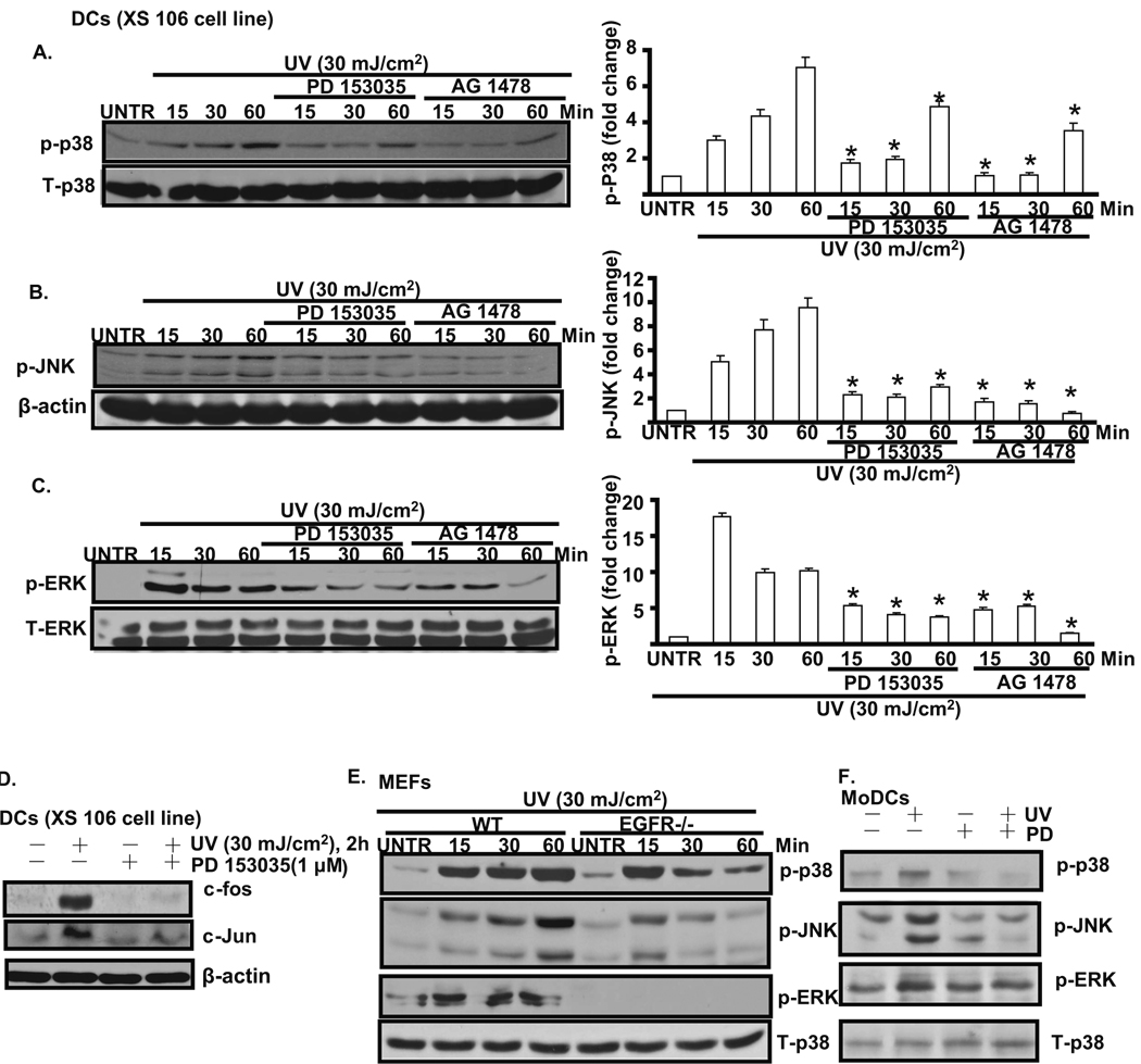
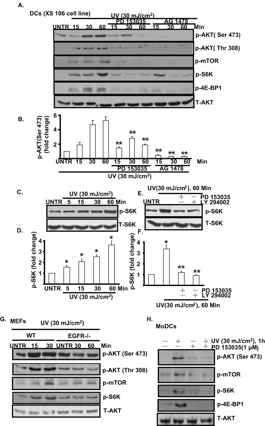
Ultraviolet radiation (UV) induces apoptosis and functional maturation in skin dendritic cells (DCs). However, the molecular mechanisms through which UV activates DCs have not been thoroughly investigated. In this study, we examined the mechanisms of activation and apoptosis of DCs after UV irradiation by focusing on epidermal growth factor receptor (EGFR). Our previous studies have demonstrated that in addition to cognate ligands, EGFR is also activated by UVB irradiation in cultured human skin keratinocytes in vitro and in human skin in vivo. We found for the first time in this study that UV also induces EGFR activation in cultured mouse skin DCs (XS 106 cell line) as well as mouse monocyte-derived dendritic cells (MoDCs). Pharmacological inhibition of EGFR tyrosine kinase significantly inhibits UV-induced ERK, p38, and JNK MAP kinases, and their effectors, transcription factors c-Fos and c-Jun. Inhibition of EGFR also suppresses UV-induced activation of PI3K/AKT/mTOR/S6K and NF-kappaB signal transduction pathways. Our data demonstrated that UV induces LKB1/AMPK pathway, also dependent on EGFR trans-activation. We further observed that MAPK, LKB1/AMPK, PI3K/AKT/mTOR/S6K as well as NF-kappaB activation are impaired in EGFR-/- cells compared to wide type MEF cells after UV radiation. Taken together, we conclude that UV induces multiple signaling pathways mediated by EGFR trans-activation leading to possible maturation, apoptosis and survival, and EGFR activation protects against UV-induced apoptosis in cultured mouse dendritic cells.


Figures

Fig. 1
Fig. 1. UV radiation transactivates EGFR in cultured mouse skin dendritic cells
Cultured mouse skin dendritic cells (DCs, XS 106 cells) were pre-treated with EGFR inhibitor PD 153035 (1 µM) for 1 hour, followed by UV radiation (30 mJ/cm2) (A) or EGF (100 ng/ml) (B) for indicated time points, p-EGFR (Tyr 1068, Tyr 1045) and T-EGFR were detected by Western blot. EGFR activation (Tyr 1068 and Tyr 1045) was detected in cultured human skin keratinocytes (HaCaT cell line) treated with UV or UV plus PD 153035 (C). Wild type and EGFR knockout MEFs (mouse embryonic fibroblasts) were treated with UV radiation (30 mJ/cm2) and cultured for indicated time points, p-EGFR (Tyr 1068), T-EGFR and β-actin were detected by Western blot (D). Primarily cultured mouse monocyte-derived dendritic cells (MoDCs) were treated with UV or UV plus PD 153035, p-EGFR and T-EGFR were detected by Western Blot (E). Data are presented as mean ± s.e.m for at least three independent experiments.
Fig. 2
Fig. 2. EGFR mediates UV-induced MAPK activation in cultured mouse skin dendritic cells
Cultured mouse skin dendritic cells (DCs, XS 106 cell) were treated with EGFR inhibitor PD 153035 (1 µM) or AG 1478 (1 µM) for 1 hour, followed by UV radiation (30 mJ/cm2) and cultured for 15, 30 and 60 min, p-p38 (A), p-JNK (B), p-ERK (C) were detected by Western blot and were quantified. c-fos and c-Jun level was detected in DCs treated with UV plus PD 153035 for 2 hours (D). Wild type and EGFR knockout MEFs (mouse embryonic fibroblasts) were treated with UV radiation (30 mJ/cm2) and cultured for 15, 30 and 60 min, p-p38, p-JNK, p-ERK were detected by Western blot (E). MAPK activation was detected in MoDCs treated with UV plus PD 153035 (F). *P<0.05 vs. UV treated group. Data are presented as mean ± s.e.m for at least three independent experiments.
Fig. 3
Fig. 3. EGFR mediates UV-induced AKT/mTOR/S6K/4E-BP1 activation in cultured mouse skin dendritic cells
Cultured mouse skin dendritic cells (DCs, XS 106 cell) were treated with EGFR inhibitor PD 153035 (1 µM) or AG 1478 (1 µM) for 1 hour, followed by UVB radiation (30 mJ/cm2) and cultured for 15, 30 and 60 min, p-AKT (Ser 473 and Thr 308), p-mTOR (Ser 2448), p-S6K (Thr 389), p-4E-BP1(Ser 65) and T- AKT were detected by Western blot (A) and AKT phosphorylation (Ser 473) was quantified (B). S6K phosphorylation was detected in DCs treated different time points after UV radiation (C and D). DCs were pre-treated with PD 153035 and LY 294002 for 1 hour, followed by UV radiation, S6K phosphorylation was detected by Western blot (E and F). UV-induced AKT/mTOR/S6K/4E-BP1 was detected in both wild type and EGFR knockout MEFs (G). AKT/mTOR/S6K/4E-BP1 activation was detected in MoDCs treated with UV plus PD 153035 (H). *P<0.05 vs. untreated group. **P<0.05 vs. UV treated group. Data are presented as mean ± s.e.m for at least three independent experiments.
Fig. 4
Fig. 4. EGFR mediates UV-induced NF-κB activation in cultured mouse skin dendritic cells
Cultured mouse skin dendritic cells (DCs, XS 106 cells) were treated with UV radiation (30 mJ/cm2) and cultured for 15, 30 60 and 120 min, p- IκBα (Ser 32/36), T-IκBα and β-actin were detected by Western blot (A and B). DCs (XS 106 cells) were treated with EGFR inhibitor PD 153035 (PD1, 1 μM) or AG 1478 (AG, 1 µM) for 1 hour, followed by UVB radiation (30 mJ/cm2) and cultured for 1 and 2 hours, p- IκBα (Ser 32/36), T-IκBα and β-actin were detected by Western blot (C and D). UV-induced NF-κB activation was detected in wild type but not in EGFR knockout MEFs (E and F). NF-κB activation was detected in MoDCs treated with UV plus PD 153035 (G and H). *P<0.05 vs. untreated group. #P<0.05 vs. UV treated group. Data are presented as mean ± s.e.m for at least three independent experiments.
Fig. 5
Fig. 5. EGFR mediates UV-induced AMPK activation in cultured mouse skin dendritic cells
Cultured mouse skin dendritic cells (DCs, XS 106 cells) were treated with EGFR inhibitor PD 153035 (1 µM) or AG 1478 (1 μM) for 1 hour, followed by UVB radiation (30 mJ/cm2) (A) or EGF (100 ng/ml) (B) and cultured for 15, 30 and 60 min, p-LKB1 (ser 428), p-AMPKα (Thr 172) and T-AMPK were detected by Western blot. Wild type and EGFR knockout MEFs (mouse embryonic fibroblasts) were treated with UV radiation (30 mJ/cm2) and cultured for indicated time points, p-LKB1 (Ser 428), p-AMPKα (Thr 172) and β-actin were detected by Western blot (C). LKB1 and AMPK phosphorylation were detected in MoDCs treated with UV with or without PD 153035 (D). *P<0.05 vs. untreated group. #P<0.05 vs. UV treated group. Data are presented as mean ± s.e.m for at least three independent experiments.
Fig. 6
Fig. 6. Trans-activation of EGFR protects dendritic cells from UV-induced cell death
Cultured mouse skin dendritic cells (DCs, XS 106 cells) were pre-treated with EGFR inhibitor PD153035 (PD1, 1 µM), AG1478 (AG, 1 µM), PI3K/AKT inhibitor LY294002 (LY, 1 µM), Wortmanin (WT, 1 µM), MEK/ERK inhibitor PD 98059 (PD9, 1 µM), U0126 (U, 1 µM), JNK inhibitor JNKi (JNKi, 1 µM) and p38 inhibitor SB 203580 (SB, 10 µM) for 1 hour, followed by UV radiation (30 mJ/cm2) for 24 hours, cell viability was detected by MTT assay (A). The effects of these inhibitors alone on cell viability were shown in (B). XS106 cells were pre-treated with or without EGF (100 ng/ml) for 1 hour, followed by UV radiation (30 mJ/cm2) for 24 hours, cell viability was detected by MTT assay (C). The apoptosis rate (Hoechst assay) of XS 106 cells treated with UV plus PD153035 (PD1, 1 µM) or AG 1478 (AG, 1 µM) for 18 hours was shown in (D). The cell viability of wild type and EGFR knockout MEFs treated with UV (30 mJ/cm2) with or without PD 153035 (1 µM) for 24 hours was shown in (E). Wild type, AKT1/2 knockout or AKT1 knockout MEFs were pre-treated with mTOR inhibitor rapamycin (20 nM) for 1 hour, followed by UV radiation (30 mJ/cm2), cell viability was detected by MTT assay (F). Cell viability and apoptosis rate (Hoechst assay) of MoDCs treated with UV (30 mJ/cm2) with or without PD1 (1 µM) were shown in (G) and (H) respectively. The data represent mean ± SE of triplicate experiments. *P<0.05 vs. untreated group. #P<0.05 vs. UV treated group. For the Hoechst experiment, a minimum of ten random fields and 500 cells were counted for apoptotic death rate.
Fig. 7
Fig. 7. Proposed model of UV-induced signal transduction pathways in mouse skin dendritic cells
(A) EGFR mediates UV induced AKT/mTOR activation in cultured skin dendritic cells, possibly leading to cell survival, (B) EGFR mediates UV-induced LKB1/AMPK activation, inhibiting mTOR, (C) EGFR is involved in UV- induced NF-κB activation, possibly leading to cell survival, activation or maturation of DCs, (D) EGFR is involved in UV induced-MAPK activation, possibly leading to cell survival or apoptosis.

Similar articles

Cited by

References

    1. Ruderman NB, Keller C, Richard AM, Saha AK, Luo Z, Xiang X, Giralt M, Ritov VB, Menshikova EV, Kelley DE, Hidalgo J, Pedersen BK, Kelly M. Diabetes. 2006;55 Suppl 2:S48–S54. - PubMed
    1. Vink AA, Strickland FM, Bucana C, Cox PA, Roza L, Yarosh DB, Kripke ML. J Exp Med. 1996;183(4):1491–1500. - PMC - PubMed
    1. Meunier L, Eur J. Dermatol. 1999;9(4):269–275. - PubMed
    1. Nakagawa S, Ohtani T, Mizuashi M, Mollah ZU, Ito Y, Tagami H, Aiba S. J Invest Dermatol. 2004;123(2):361–370. - PubMed
    1. Zhuang L, Wang B, Shinder GA, Shivji GM, Mak TW, Sauder DN. J Immunol. 1999;162(3):1440–1447. - PubMed

Publication types

MeSH terms