. 2008 Sep;36(16):e103.
doi: 10.1093/nar/gkn398. Epub 2008 Jul 21.

Transduction of artificial transcriptional regulatory proteins into human cells

Affiliations

Transduction of artificial transcriptional regulatory proteins into human cells

Chae-Ok Yun et al. Nucleic Acids Res. 2008 Sep.

Abstract

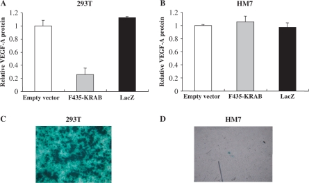
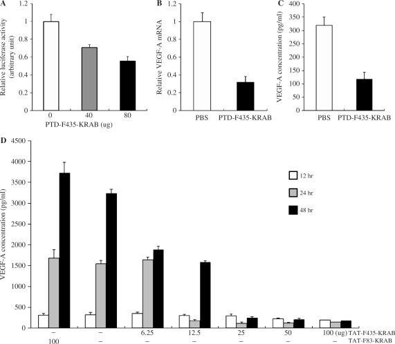
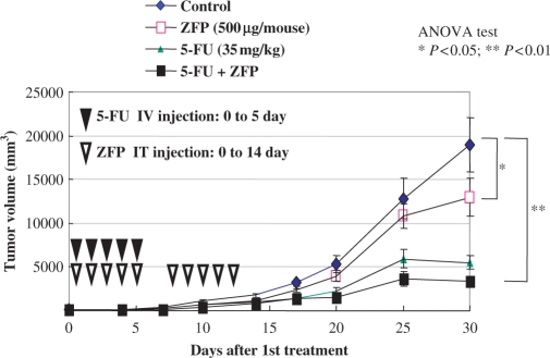
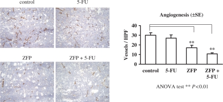
Protein transduction (PT) is a method for delivering proteins into mammalian cells. PT is accomplished by linking a small peptide tag--called a PT domain (PTD)--to a protein of interest, which generates a functional fusion protein that can penetrate efficiently into mammalian cells. In order to study the functions of a transcription factor (TF) of interest, expression plasmids that encode the TF often are transfected into mammalian cells. However, the efficiency of DNA transfection is highly variable among different cell types and is usually very low in primary cells, stem cells and tumor cells. Zinc-finger transcription factors (ZF-TFs) can be tailor-made to target almost any gene in the human genome. However, the extremely low efficiency of DNA transfection into cancer cells, both in vivo and in vitro, limits the utility of ZF-TFs. Here, we report on an artificial ZF-TF that has been fused to a well-characterized PTD from the human immunodeficiency virus-1 (HIV-1) transcriptional activator protein, Tat. This ZF-TF targeted the endogenous promoter of the human VEGF-A gene. The PTD-attached ZF-TF was delivered efficiently into human cells in vitro. In addition, the VEGF-A-specific transcriptional repressor retarded the growth rate of tumor cells in a mouse xenograft experiment.


Figures

Figure 1.
Figure 1.
Liposome-mediated transfection of plasmids that encode either a ZF-TF or LacZ into HEK 293 cells and HM7 cancer cells. HEK 293T cells (A) and HM7 human cancer cells (B) were transfected with a plasmid that expressed F435-KRAB or the LacZ gene product, β-galactosidase (pcDNA3.1/His/LacZ; Invitrogen) using Lipofectamine 2000 (Invitrogen) and the amounts of secreted VEGF-A were measured by ELISA at 2 days after transfection. The F435-KRAB-encoding plasmid efficiently suppressed the expression of VEGF-A in 293T cells, but not in HM7 cells. (C and D) HEK 293T cells and HM7 cells transfected with the LacZ plasmid were stained to visualize the enzyme activity. Greater than 90% of the transfected 293T cells were LacZ positive, but <1% of the HM7 cells were LacZ positive.
Figure 2.
Figure 2.
Regulation of luciferase reporter activity by PTDTAT–F435-KRAB. (A) HEK 293 cells were cotransfected with 15 ng of pGL-VEGF and 68.5 ng of the Renilla luciferase expression plasmid pRL-SV40. The cells were then treated with the indicated amount of purified PTDTAT–F435-KRAB 24 h after transfection. Luciferase activity was measured 24 h after protein transduction. (B) Repression of endogenous VEGF-A at the mRNA level. HEK 293 cells (105 cells/24-well plate) were treated with either PBS or PTDTAT–F435-KRAB (250 μg) for 2 h, then the culture medium was replaced with fresh growth medium (DMEM, 10% FBS). VEGF-A mRNA was analyzed by RT–PCR using specific primers. The amount of VEGF mRNA was normalized to the amount of GAPDH mRNA from the same RT product, then relative VEGF mRNA amounts from PTDTAT–F435-KRAB-treated cells were represented as values compared to normalized values from control PBS-treated cells. (C) From the same experiments described in (B), the culture medium at 24 h after transduction with either PBS (control) or effector (PTDTAT–F435-KRAB) was harvested. The amount of secreted VEGF-A protein was measured by ELISA with an antibody specific to human VEGF-A. (D) Dose-dependent repression of VEGF-A protein expression by PTDTAT–F435-KRAB. HEK293F cells were treated with various amounts of PTDTAT–F435-KRAB for 2 h, and then the culture medium was replaced with fresh growth medium. Culture supernatants were harvested at the indicated times (white bar, after 12 h; gray bar, after 24 and black bar, after 48 h). Secreted VEGF-A amounts were measured by ELISA. TAT-F83-KRAB was used as a negative control. All of the results in Figure 1 are the mean values and SEs of three independent experiments.
Figure 3.
Figure 3.
Tumor growth inhibition in xenograft mice. Human colorectal cancer cells (HM7 cell line) were xenografted into nude mice. The mice either received no treatment (control) or were treated by injection with PTDTAT–F435-KRAB (ZFP) alone; 5-FU in combination with PTDTAT–F435-KRAB (ZFP + 5-FU) or 5-FU alone at the indicated dose and injection time (see Materials and methods section). Tumor volumes were assessed for 30 days after the first injection of the various substances. All data are presented as means and SEMs. IV, intravenous; IT, intratumoral.
Figure 4.
Figure 4.
Immunohistochemistry of tumor blood vessels. Cryosections of tumors from xenografted mice described in Figure 2 were stained to visualize CD31 (PECAM-1). Angiogenesis indices were determined by calculating vessel densities from at least three cryosections prepared from mice of each group. The differences between the angiogenesis index of tumors from control (PBS-treated) mice and the angiogenesis indices of tumors from mice treated with PTDTAT–F435-KRAB (ZFP) or PTDTAT–F435-KRAB and 5-FU (ZFP + 5-FU) were statistically significant (++) (ANOVA test, P < 0.01). The angiogenesis indices shown are the mean values and SE of three independent experiments.

References

    1. Nagahara H, Vocero-Akbani AM, Snyder EL, Ho A, Latham DG, Lissy NA, Becker-Hapak M, Ezhevsky SA, Dowdy SF. Transduction of full-length TAT fusion proteins into mammalian cells: TAT-p27Kip1 induces cell migration. Nat. Med. 1998;4:1449–1452. - PubMed
    1. Schwarze SR, Ho A, Vocero-Akbani A, Dowdy SF. In vivo protein transduction: delivery of a biologically active protein into the mouse. Science. 1999;285:1569–1572. - PubMed
    1. Schwarze SR, Hruska KA, Dowdy SF. Protein transduction: unrestricted delivery into all cells? Trends Cell Biol. 2000;10:290–295. - PubMed
    1. Wadia JS, Dowdy SF. Protein transduction technology. Curr. Opin. Biotechnol. 2002;13:52–56. - PubMed
    1. Xia H, Mao Q, Davidson BL. The HIV Tat protein transduction domain improves the biodistribution of beta-glucuronidase expressed from recombinant viral vectors. Nat. Biotechnol. 2001;19:640–644. - PubMed

Publication types

MeSH terms

Substances