. 2008 Oct;28(19):5851-64.
doi: 10.1128/MCB.00405-08. Epub 2008 Jul 21.

Evidence for the bifunctional nature of mitochondrial phosphatidylserine decarboxylase: role in Pdr3-dependent retrograde regulation of PDR5 expression

Affiliations

Evidence for the bifunctional nature of mitochondrial phosphatidylserine decarboxylase: role in Pdr3-dependent retrograde regulation of PDR5 expression

Kailash Gulshan et al. Mol Cell Biol. 2008 Oct.

Abstract

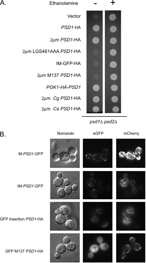
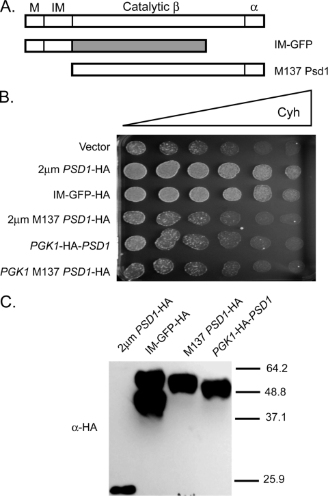
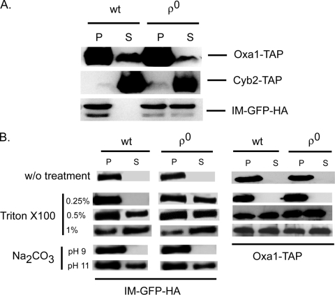
Multidrug resistance in the yeast Saccharomyces cerevisiae is sensitive to the mitochondrial genome status of cells. Cells that lose their organellar genome ([rho(0)] cells) dramatically induce transcription of multiple or pleiotropic drug resistance genes via increased expression of a zinc cluster-containing transcription factor designated Pdr3. A major Pdr3 target gene is the ATP-binding cassette transporter-encoding gene PDR5. Pdr5 has been demonstrated to act as a phospholipid floppase catalyzing the net outward movement of phosphatidylethanolamine (PE). Since the mitochondrially localized Psd1 enzyme provides a major route of PE biosynthesis, we evaluated the potential linkage between Psd1 function and PDR5 regulation. Overproduction of Psd1 in wild-type ([rho(+)]) cells was found to induce PDR5 transcription and drug resistance in a Pdr3-dependent manner. Loss of the PSD1 gene from [rho(0)] cells prevented the normal activation of PDR5 expression. Surprisingly, expression of a catalytically inactive form of Psd1 still supported PDR5 transcriptional activation, suggesting that PE levels were not the signal triggering PDR5 induction. Expression of green fluorescent protein fusions mapped the region required to induce PDR5 expression to the noncatalytic amino-terminal portion of Psd1. Psd1 is a novel bifunctional protein required both for PE biosynthesis and regulation of multidrug resistance.


Figures

FIG. 1.
FIG. 1.
Overproduction of Psd1 elevates cycloheximide resistance. (A) Biosynthetic pathway leading to production of the three major phospholipids in S. cerevisiae. Phosphatidylserine (PS) is decarboxylated by the enzymes Psd1 or Psd2 to produce PE. PE is then methylated by the Cho2 and Opi3 enzymes to form PC. (B) A wild-type strain was transformed with low-copy-number plasmids carrying transcriptional fusions between the GAL promoter and the indicated ORF. Transformants were grown to mid-log phase in selective medium with galactose as the carbon source and then placed on solid medium containing a gradient of cycloheximide (Cyh) (indicated by the bar of increasing width). Each spot contained 1,000 cells of each indicated transformant strain.
FIG. 2.
FIG. 2.
Processing and induction of drug resistance by Psd1. (A) A high-copy-number clone (2μm PSD1-HA) expressing epitope-tagged Psd1 was introduced into wild-type cells and tested for the ability to elevate resistance to cycloheximide (CYH) or oligomycin (OLI). Serial dilutions of cells transformed with the vector plasmid alone were used as negative controls. (B) A wild-type strain was transformed with an empty high-copy-number plasmid (vector), a low-copy-number (CEN) or high-copy-number (2μm) plasmid expressing a wild-type form of PSD1-HA, or a high-copy-number plasmid containing a mutant form of PSD1 (LGS461AAA PSD1-HA). Transformants were grown to mid-log phase and placed on rich medium containing a gradient of cycloheximide indicated as described for Fig. 1. (C) The transformants from panel B were analyzed by Western blotting using antibodies against the epitope tag (α-HA) or Vph1 (α-Vph1) as a loading control. Molecular mass standards in kilodaltons are denoted by the numbers at the right.
FIG. 3.
FIG. 3.
Induction of Pdr5-dependent transport and expression by overproduction of Psd1. (A) Wild-type (wt) or isogenic pdr5Δ cells were transformed by a high-copy-number vector plasmid or the same plasmid containing the PSD1 gene. These transformants were grown to mid-log phase and then analyzed for rhodamine G efflux activity as described previously (29). Efflux measurements were conducted after de-energizing the cells in the presence or absence of added 1 mM glucose to restore ATP levels. Rhodamine fluorescence in the supernatants of the cells was determined and plotted on the graph. These data are representative values that varied less than 20% over at least two independent trials. (B) The chromosomal PSD1 promoter was replaced by strong glycolytic glyceraldehyde-3-phosphate dehydrogenase (encoded by the TDH3 gene) promoter using a PCR-based recombination approach (22). This otherwise wild-type strain (TDH3-PSD1) was grown along with the isogenic wild-type and pdr5Δ cells to mid-log phase. Whole-cell protein extracts were prepared, resolved by SDS-PAGE, and analyzed by Western blotting. The antibodies used were directed against Pdr5 (α-Pdr5) or Vph1 (α-Vph1).
FIG. 4.
FIG. 4.
The Pdr pathway responds to elevated Psd1 levels. (A) Wild-type (wt) or isogenic TDH3-PSD1 cells were transformed with low-copy-number plasmids containing the indicated lacZ fusion genes. Transformants were grown to mid-log phase and assayed for β-galactosidase activities as described previously (16). (B) A collection of isogenic strains containing variable dosages of PDR1 and/or PDR3 were transformed with a high-copy-number plasmids containing PSD1 or the empty vector. Transformants were grown to mid-log phase and then placed on medium containing a gradient of cycloheximide (Cyh).
FIG. 5.
FIG. 5.
PSD1 is required for [rho0] signaling to the nucleus. (A) Isogenic [rho+] and [rho0] lacking the indicated nuclear genes were transformed with low-copy-number plasmids carrying either a PDR5- or PDR15-lacZ fusion gene. Transformants were grown and assayed for β-galactosidase levels as described for Fig. 4. (B) A low-copy-number plasmid containing a PDR3-lacZ fusion gene was introduced into the indicated strains. Transformants were assayed for β-galactosidase activity using the fluorescent substrate as described for Fig. 3. (C) Strains containing the relevant genotypes indicated at the left hand were tested for resistance to cycloheximide (Cyh) using a gradient plate assay as described for Fig. 1. wt, wild type.
FIG. 6.
FIG. 6.
Complementation of ethanolamine auxotrophy by and localization of Psd1 derivatives. (A) Cells lacking phosphatidylserine decarboxylase activity (psd1Δ psd2Δ) were transformed with plasmids expressing the indicated forms of Psd1. Transformants were spotted on medium containing (+) or lacking (−) ethanolamine and then incubated at 30°C. (B) Plasmids expressing the indicated forms of Psd1 were introduced into wild-type cells along with a control plasmid expressing mitochondrially targeted mCherry (3). Transformants were grown to mid-log phase and visualized by differential interference (Nomarski) or fluorescence microscopy for GFP or mCherry.
FIG. 7.
FIG. 7.
The amino terminus of Psd1 is sufficient to induce drug resistance. (A) The potential functional domains of Psd1 are represented along with two mutant constructs used to map the minimal signaling domain. Wild-type Psd1 is shown at the top. M indicates the mitochondrial targeting signal, while IM denotes the inner membrane localization motif. The two catalytic subunits are designated a and b. The small a subunit is generated by autoproteolysis between G462 and S463 in the C terminus of Psd1. The IM-GFP construct replaces the catalytic a and b domains with GFP. The M137 Psd1 derivative deletes the targeting information from the protein and initiates translation at methionine 137. (B) High-copy-number plasmids expressing the indicated forms of Psd1 were tested for the ability to elevate cycloheximide (Cyh) resistance as described for Fig. 1. (C) Whole-cell protein extracts were prepared from wild-type cells expressing the indicated forms of Psd1 and analyzed by Western blotting using an anti-HA antibody. Molecular mass standards are indicated on the right.
FIG. 8.
FIG. 8.
Overproduction of Candida Psd1 homologues elevate drug resistance. Wild-type cells were transformed with a low-copy-number plasmid expressing epitope-tagged Psd1 (PSD1-HA), an empty high-copy-number vector, or high-copy-number plasmids expressing S. cerevisiae (2μm PSD1-HA), C. glabrata (2μm Cg PSD1-HA), or C. albicans (2μm Ca PSD1-HA) epitope-tagged Psd1 homologues. Transformants were grown to mid-log phase and tested for cycloheximide (Cyh) resistance using a gradient plate assay (A) or Western blotting (B) with the indicated antisera. Molecular mass standards are indicated on the right.
FIG. 9.
FIG. 9.
Fractionation of Psd1 is altered in [rho0] cells. Isogenic [rho+] or [rho0] strains were transformed with cassettes expressing TAP fusion proteins (Oxa1-TAP or Cyb2-TAP) or a low-copy-number plasmid producing the IM-GFP-HA form of Psd1. Transformants were grown in selective minimal medium to mid-log phase. (A) Whole-cell protein extracts were generated by glass bead lysis using a sorbitol-containing buffer (54), and these extracts were separated into P10 (P) and S10 (S) fractions by centrifugation at 10,000 × g. Equal amounts of protein from each fraction were then resolved on SDS-PAGE and analyzed by Western blotting with anti-TAP or anti-HA antibodies. (B) The P10 fraction from panel A was resuspended in sorbitol buffer (without [w/o] treatment) or in sorbitol buffer containing the indicated concentrations of Triton X-100 or Na2CO3 added to raise the pH to the value noted. These samples were then mixed and centrifuged again at 10,000 × g. These P10 and S10 fractions were Western blotted as described for panel A. wt, wild type.

Similar articles

Cited by

References

    1. Balzi, E., W. Chen, S. Ulaszewski, E. Capieaux, and A. Goffeau. 1987. The multidrug resistance gene PDR1 from Saccharomyces cerevisiae. J. Biol. Chem. 26216871-16879. - PubMed
    1. Balzi, E., M. Wang, S. Leterme, L. Van Dyck, and A. Goffeau. 1994. PDR5: a novel yeast multidrug resistance transporter controlled by the transcription regulator PDR1. J. Biol. Chem. 2692206-2214. - PubMed
    1. Bevis, B. J., and B. S. Glick. 2002. Rapidly maturing variants of the Discosma red fluorescent protein (DsRed). Nat. Biotechnol. 2083-87. - PubMed
    1. Birner, R., M. Burgermeister, R. Schneiter, and G. Daum. 2001. Roles of phosphatidylethanolamine and of its several biosynthetic pathways in Saccharomyces cerevisiae. Mol. Biol. Cell 12997-1007. - PMC - PubMed
    1. Bissinger, P. H., and K. Kuchler. 1994. Molecular cloning and expression of the S. cerevisiae STS1 gene product. J. Biol. Chem. 2694180-4186. - PubMed

Publication types

MeSH terms