. 2008 Oct;28(19):5874-85.
doi: 10.1128/MCB.00821-08. Epub 2008 Jul 21.

Regulation of Chk2 ubiquitination and signaling through autophosphorylation of serine 379

Affiliations

Regulation of Chk2 ubiquitination and signaling through autophosphorylation of serine 379

Christine M Lovly et al. Mol Cell Biol. 2008 Oct.

Abstract

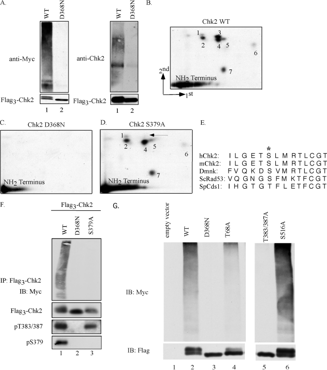
The Chk2 protein kinase protects genome integrity by promoting cell cycle arrest or apoptosis in response to DNA double-strand breaks, and Chk2 mutations are found in both familial and sporadic cancers. Exposure of cells to ionizing radiation (IR) or radiomimetic drugs induces Chk2 phosphorylation by ATM, followed by Chk2 oligomerization, auto-/transphosphorylation, and activation. Here we demonstrate that Chk2 is ubiquitinated upon activation and that this requires Chk2 kinase activity. Serine 379 (S379) was identified as a novel IR-inducible autophosphorylation site required for ubiquitination of Chk2 by a Cullin 1-containing E3 ligase complex. Importantly, S379 was required for Chk2 to induce apoptosis in cells with DNA double-strand breaks. Thus, auto-/transphosphorylation of S379 is required for Chk2 ubiquitination and effector function.


Figures

FIG. 1.
FIG. 1.
Chk2 autophosphorylation on S379 regulates ubiquitination. (A) HEK-293 cells were transfected with plasmids encoding Flag3-Chk2 (WT and D368N) and Myc-tagged ubiquitin. At 42 h posttransfection, cells were treated with MG132 for 2 h, and Chk2 proteins were immunoprecipitated using an antibody specific for the Flag epitope. Ubiquitinated proteins were visualized by immunoblotting with antibodies specific for either Myc (top left panel) or Chk2 (top right panel). Precipitated Flag3-Chk2 was detected with antibodies specific for Flag (bottom left panel) or Chk2 (bottom right panel). (B to D) Asynchronously growing HEK-293 cells expressing Flag3-Chk2-WT (B), Flag3-Chk2-D368N (C), or Flag3-Chk2-S379A (D) were incubated with 32P-labeled inorganic phosphate in the presence of 25 μM VP16. Radiolabeled Chk2 proteins were digested with trypsin, and peptides were subjected to two-dimensional phosphopeptide mapping. The arrow in panel D denotes the absence of a phosphopeptide that is present in the map of wild-type Chk2. (E) Alignment of sequences inclusive of and surrounding S379 in the indicated Chk2 orthologs. The asterisk denotes S379 in human Chk2 and conserved residues in other orthologs. (F) HEK-293 cells were transfected with plasmids encoding Flag3-Chk2 (WT, D368N, and S379A) and Myc-tagged ubiquitin. At 48 h posttransfection, cells were lysed and Chk2 proteins were immunoprecipitated (IP) using an antibody specific for the Flag epitope. Ubiquitinated proteins were visualized by immunoblotting (IB) with an antibody to the Myc tag. The Myc blots were then stripped and probed with an antibody specific for the Flag tag. Phosphorylation of Chk2 on T383/387 and S379 was examined on total lysates using antibodies specific for these sites. (G) HEK-293 cells were transfected with expression plasmids encoding Flag3-Chk2 (WT, D368N, T68A, T383/387A, or S516A) and Myc-tagged ubiquitin (Myc-Ub). Chk2 proteins were immunoprecipitated using an antibody specific for the Flag epitope. Ubiquitinated proteins were visualized by immunoblotting with an antibody to the Myc tag. The Myc blots were then stripped and probed with an antibody specific for the Flag tag.
FIG. 2.
FIG. 2.
Phosphorylation and kinase activity of Chk2 S379A. (A) Lysates of HEK-293 cells expressing Flag3-Chk2-WT or Flag3-S379A were resolved by SDS-PAGE and blotted with an antibody specific for Chk2 phosphorylated on S379. The blot was then stripped and reprobed with an antibody specific for the Flag epitope. (B) Lysates from HEK-293 cells that had been mock irradiated (lane 1) or exposed to 10 Gy IR (lane 2) were resolved by SDS-PAGE and blotted with an antibody specific for Chk2 phosphorylated on S379 and an antibody specific for Chk2. (C) His6-Chk2-WT, -D368N, and -S379A were purified from bacteria. Matched amounts of protein were resolved by SDS-PAGE and blotted with the indicated antibodies. (D) Lysates from Chk2 null MEFs expressing Flag3Chk2-WT, -D368N, or -S379A were incubated with an antibody specific for the Flag epitope. Precipitates were resolved by SDS-PAGE and blotted with the indicated phospho-specific antibodies. The phospho-specific blots were then stripped and reprobed with an antibody specific for the Flag tag. (E) Myc3-tagged Chk2 proteins (WT and S379A) were precipitated from U2OS/FRT stable lines 30 min after mock irradiation (−) or exposure to 10 Gy IR (+). The precipitated Chk2 proteins were tested for their ability to phosphorylate GST-Cdc25C(200-256) in vitro. 32P-labeled GST-Cdc25C(200-256) was quantified using a Storm Imager. Error bars indicate the standard errors of the means for triplicate reactions performed for each Chk2 protein.
FIG. 3.
FIG. 3.
Chk2 ubiquitination by Cul 1 E3 ligase complex. (A) HEK-293 cells were transfected with plasmids encoding Flag3-Chk2-WT and the indicated Myc-tagged Cullin proteins for 48 h. Lysates were prepared and either resolved directly by SDS-PAGE (bottom panel) or first incubated with an antibody specific for the Flag epitope (top panel), and precipitates were resolved by SDS-PAGE. Western blotting was performed with Myc antibody to detect tagged Cullin proteins and with Flag antibody to detect precipitated Flag3-Chk2. (B) HEK-293 cells were transfected with plasmids encoding hemagglutinin-tagged ubiquitin (HA-Ub), Myc3-Chk2, and dominant-negative forms of Cul 1, 3, and 4B. Myc3-Chk2 was immunoprecipitated (IP), and ubiquitinated Chk2 was examined by Western blotting (IB) with HA-specific antibody. The blot was then stripped and blotted with Myc antibody. Total lysates were examined for Cullin levels by Western blotting with a Flag-specific antibody.
FIG. 4.
FIG. 4.
Chk2 is ubiquitinated in vitro by a Cul 1-containing E3 ligase. (A) HEK-293 cells were transfected with plasmids encoding Flag3-Chk2 (WT, D368N, or S379A) and Myc-Cul 1. Flag3-tagged proteins were isolated using Flag-agarose, and precipitates were examined for the presence of Myc-Cul 1 by Western blotting with antibodies specific for the Myc and Flag tags (top panel). Levels of Myc-Cul 1 were determined by Western blotting total cell lysates (bottom panel). (B) Chk2 proteins were purified as Flag-tagged proteins as described in Materials and Methods and incubated in vitro with purified ubiquitin, E1 (Ube1), and E2 (UbcH5c) in ubiquitin reaction buffer at 37°C for 1 h. Reactions were resolved by SDS-PAGE and subjected to Western blotting (IB) with antibodies specific for ubiquitin (top panel) or Flag (bottom panel).
FIG. 5.
FIG. 5.
Ubiquitination of Chk2 does not affect Chk2 stability. (A) HEK-293 cells were treated with cycloheximide and then either mock or gamma irradiated and collected at the indicated times post-IR. Lysates were incubated in the absence or presence of λ phosphatase. Chk2 protein levels were monitored by Western blotting using a Chk2 monoclonal antibody. An antibody specific for actin was used to verify similar loading in each lane. Densitometry analysis was performed using Image J software and normalized to the level of actin. Chk2 protein levels at each time point are shown relative to those in mock-irradiated samples (time zero is set at 1.0). (B) Flag3-Chk2 proteins (WT, D368N, and S379A) were expressed in HEK-293 cells. Cells were cultured in the presence of cycloheximide and collected at the indicated time points. Flag3-Chk2 protein levels were examined using an anti-Flag antibody. Densitometry analysis was performed as for panel A.
FIG. 6.
FIG. 6.
Phosphorylation of S379 in activation of Chk2 and apoptosis induction. (A) U2OS/FRT parental cells were stably transfected with plasmids encoding Myc3-Chk2-WT, Myc3-Chk2-D368N, or Myc3-Chk2-S379A as described in Materials and Methods. Lysates were blotted with an antibody against Chk2, and the relative expression level of Myc3-Chk2 versus endogenous Chk2 for each cell line was quantified by chemiluminescence using a Storm Imager; the ratio of ectopic to endogenous Chk2 expression is indicated. (B) U2OS/FRT parental and Chk2-expressing cells were treated with etoposide for the indicated times. The percentage of cells containing sub-2N DNA content was assessed by propidium iodide staining and fluorescence-activated cell sorter analysis. Error bars denote standard errors of the means (n = 4). (C and D) U2OS/FRT parental and Chk2-expressing cells were exposed to IR. Lysates were prepared and Western blotting was performed with the indicated antibodies.

References

    1. Abramoff, M. D., P. J. Magelhaes, and S. J. Ram. 2004. Image processing with Image J. Biophotonics Int. 1136-42.
    1. Ahn, J.-Y., X. Li, H. L. Davis, and C. E. Canman. 2002. Phosphorylation of threonine 68 promotes oligomerization and autophosphorylation of the Chk2 protein kinase via the Forkhead-associated (FHA) domain. J. Biol. Chem. 27719389-19395. - PubMed
    1. Ahn, J.-Y., J. K. Schwarz, H. Piwnica-Worms, and C. E. Canman. 2000. Threonine 68 phosphorylation by ataxia telangiectasia mutated is required for efficient activation of Chk2 in response to ionizing radiation. Cancer Res. 605934-5936. - PubMed
    1. Bartek, J., and J. Lukas. 2001. Pathways governing G1/S transition and their response to DNA damage. FEBS Lett. 490117-122. - PubMed
    1. Bartkova, J., J. Falck, E. Rajpert-De Meyts, N. E. Skakkebaek, J. Lukas, and J. Bartek. 2001. Chk2 tumour suppressor protein in human spermatogenesis and testicular germ-cell tumours. Oncogene 205897-5902. - PubMed

Publication types