. 2008 Oct;28(19):5886-98.
doi: 10.1128/MCB.01265-07. Epub 2008 Jul 21.

The forkhead transcription factor FOXO3a increases phosphoinositide-3 kinase/Akt activity in drug-resistant leukemic cells through induction of PIK3CA expression

Affiliations

The forkhead transcription factor FOXO3a increases phosphoinositide-3 kinase/Akt activity in drug-resistant leukemic cells through induction of PIK3CA expression

Rosaline C-Y Hui et al. Mol Cell Biol. 2008 Oct.

Abstract

The phosphoinositide-3 kinase (PI3K)/Akt signal pathway plays a key role in the tumorigenesis of many cancers and in the subsequent development of drug resistance. Using the K562 chronic myelogenous leukemia (CML) cell line and the doxorubicin-resistant derivative lines KD30 and KD225 as models, we observed that enhanced PI3K/Akt activity and the acquisition of chemoresistance correlated unexpectedly with the increased expression and nuclear accumulation of FOXO3a. Moreover, we found that the induction of FOXO3a activity in naïve K562 cells was sufficient to enhance PI3K/Akt activity and to confer resistance to the cytotoxic effects of doxorubicin. Conversely, the knockdown of endogenous FOXO3a expression reduced PI3K/Akt activity and sensitized these cells to doxorubicin. Further chromatin immunoprecipitation and promoter mutation analyses demonstrated that FOXO3a regulates the expression of the PI3K catalytic subunit p110alpha through the activation of a promoter region proximal to a novel untranslated exon upstream from the reported transcription start site of the p110alpha gene PIK3CA. As was the case for FOXO3a, the expression or knockdown of p110alpha was sufficient to amplify or reduce PI3K/Akt activity, respectively. Thus, our results suggest that the chronic activation of FOXO3a by doxorubicin in CML cells can enhance survival through a feedback mechanism that involves enhanced p110alpha expression and hyperactivation of the PI3K/Akt pathway.


Figures

FIG. 1.
FIG. 1.
Doxorubicin resistance correlates with FOXO3a expression and Akt phosphorylation in K562 cells. (A) Growth curves of K562, KD30, and KD225 cells in the presence or absence of 1 μM doxorubicin. (B) The naïve and drug-resistant K562 cell lines, as indicated, were analyzed by Western blotting using specific antibodies against P-Akt(Ser-473), P-Akt(Thr-308), Akt, P-FOXO3a(Thr-32), FOXO3a, and β-tubulin. (C) Cytoplasmic and nuclear extracts isolated from K562 cell lines were Western blotted with antibodies against FOXO3a(Thr-32), FOXO3a, actin, and lamin B1. (D) K562, KD30, and KD225 cells were cultured for 24 h in 0.5% FCS and then restimulated with 10% FCS for 24 h. Cell lysates from these K562 clones before and after serum stimulation were Western blotted for P-Akt(Ser-473), Akt, P-FOXO3a(Thr-32), FOXO3a, and β-tubulin.
FIG. 2.
FIG. 2.
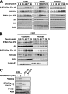
Doxorubicin treatment causes an induction of FOXO3a expression and activity and Akt phosphorylation. Naïve and drug-resistant K562 cells were treated with 1 μM doxorubicin. (A) Protein lysates were prepared at the times indicated, and protein expression levels were analyzed by Western blotting using specific antibodies against P-Akt(Ser-473), Akt, P-FOXO3a(Thr-32), FOXO3a, ID1, and actin. The lower exposure blots for P-FOXO3a and P-Akt are shown in order to demonstrate the kinetics of induction upon doxorubicin treatment. (B) Cytoplasmic and nuclear extracts isolated from K562 cells treated with 1 μM doxorubicin were Western blotted with antibodies against P-Akt(Ser-473), Akt, P-FOXO3a(Thr-32), FOXO3a, actin, and lamin B1. (C) Lysates were prepared from K562 cells with or without 1 μM doxorubicin for 24 h and HEC1B and NIH 3T3 cells and immunoblotted for FOXO3a, FOXO1, and FOXO4 expression.
FIG. 3.
FIG. 3.
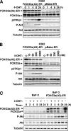
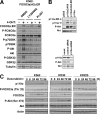
Effect of FOXO3a induction on Akt phosphorylation and activity in K562 and BaF3 cells. K562-FOXO3a(A3):ER and K562-ER cells were treated with 4-OHT for the indicated times. (A) Cell lysates were prepared at the times indicated, separated on polyacrylamide gels, and subjected to immunoblotting with specific antibodies. The expression levels of FOXO3a, P-FOXO3a, p27Kip1, P-Akt(473), Akt, and β-tubulin were analyzed by Western blotting. (B) Protein lysates were prepared from K562 cells treated with indicated doses of 4-OHT for 24 h, and protein expression levels were analyzed by Western blotting using specific antibodies against FOXO3a, P-FOXO3a, p27Kip1, P-Akt(473), Akt, and β-tubulin. (C) BaF3-FOXO3a(A3):ER and BaF3-ER cells were treated with 4-OHT for the indicated times. Protein lysates were prepared from BaF3 cells treated with 4-OHT for the indicated times, and protein expression levels were analyzed by Western blotting using specific antibodies against FOXO3a, P-FOXO3a, P-Akt(473), Akt, and β-tubulin.
FIG. 4.
FIG. 4.
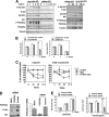
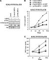
Effects of Akt induction and FOXO3a silencing on K562 proliferation. (A) K562 cells harboring (myr)Akt:ER and K562-ER cells were treated with 4-OHT for the indicated times. Cell lysates were prepared at the times indicated, separated on polyacrylamide gels, and subjected to immunoblotting with specific antibodies. The expression levels of FOXO3a, P-FOXO3a, p27Kip1, P-Akt(473), Akt, and β-tubulin were analyzed by Western blotting. (B) (myr)Akt:ER and K562-ER cells were treated with or without 4-OHT for 2 h and then treated with 1 mM doxorubicin. Cells were counts at the times indicated following doxorubicin treatment. (C) (myr)Akt:ER and K562-ER cells were treated with or without 4-OHT for 2 h and then treated with 1 mM doxorubicin. MTT assays were performed at the times indicated following doxorubicin treatment. (D) KD562 cells were transiently transfected using Nucleofect solution V (Amaxa) with either no siRNA, FOXO3a Smartpool siRNA, or control Smartpool siRNA (Dharmacon). Cell lysates and mRNA were prepared from these transfectants after 48 h and were subjected to Western blotting with FOXO3a, P-Akt(473), and Akt antibodies and RTQ-PCR analysis for FOXO3a and L19 RNA expression, respectively. (E) The siRNA-transfected K562 cells were then treated with 1 mM doxorubicin and counted at the times indicated following doxorubicin treatment. All data shown represent the averages of data from three independent experiments performed together, and the error bars show the standard deviations.
FIG. 5.
FIG. 5.
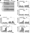
FOXO3a induced p110α gene expression directly to mediate Akt phosphorylation. K562-FOXO3a(A3):ER and K562-ER cells were treated with 4-OHT for the indicated times. (A) Cell lysates were prepared at the times indicated, separated on polyacrylamide gels, and subjected to immunoblotting with specific antibodies. The expression levels of FOXO3a, P-FOXO3a, p27Kip1, P-Akt(473), Akt, and β-tubulin were analyzed by Western blotting. (B) Total RNA was extracted and analyzed for p85α, p110α, PDK1, and IGFR1 mRNA expression using RTQ-PCR as described in the text and normalized to the level of L19 RNA. (C) K562-FOXO3a(A3):ER and K562-ER cells were pretreated with 100 μM cycloheximide for 30 min before stimulation with 4-OHT for the indicated times. Total RNA was extracted and analyzed for p110α and IGFR1 mRNA expression using RTQ-PCR as described in the text and normalized to the level of L19 RNA. All data shown represent the averages of data from three experiments, and the error bars show the standard deviations.
FIG. 6.
FIG. 6.
FOXO3a induces Akt phosphorylation and activation via p110α in K562 cells. (A) K562-FOXO3a(A3):ER cells were cultured for 24 h with or without 4-OHT in the presence of vehicle (dimethyl sulfoxide [DMSO]), 30 μM LY294002, or 15 μM PI-387. Cell lysates prepared from these cells were separated on polyacrylamide gels and subjected to immunoblotting with specific antibodies. The expression of levels of Akt, P-Akt(473), FOXO3a, P-FOXO3a(Thr-32), p70S6K, P-p70S6K, GSK3β, and P-GSK3β were analyzed by Western blotting. (B) K562 cells were either mock transfected or transfected with p110α, control Smartpool siRNA (Dharmacon), pSG5, pSG5-p110α, pSG5-p110αCAAX-ER, or pSG5-p110α. Cell lysates were prepared from these transfectants after 48 h and Western blotted for p110α, Akt, and P-Akt(473). (C) The naïve K562 and drug-resistant KD30 and KD225 cell lines were treated with 1 μM doxorubicin. Protein lysates were prepared at the times indicated, and protein expression levels were analyzed by Western blotting using specific antibodies against p110α, P-Akt(Ser-473), P-Akt(Thr-308), Akt, P-FOXO3a(Thr-32), FOXO3a, and β-tubulin.
FIG. 7.
FIG. 7.
Effects of p110α silencing on Akt activity and K562 proliferation. (A) K562 cells harboring FOXO3a:ER cells were transfected with control siRNA and siRNA and then treated with 4-OHT for 24 h. Cell lysates were prepared, separated on polyacrylamide gels, and subjected to immunoblotting with specific antibodies. The expression levels of FOXO3a:ER, P-Akt(473), Akt, and p110α were analyzed by Western blotting. These results indicate that p110α silencing significantly decreases P-Akt expression. (B) Cell counts were performed on these cells, showing that the knockdown of p110α decreases the cell proliferation rate but has no effect on cell proliferation in response to FOXO3a induction. (C) MTT assays were performed on these cells, again indicating that the knockdown of p110α decreases the cell proliferation rate but has no effect on cell proliferation in response to FOXO3a induction.
FIG. 8.
FIG. 8.
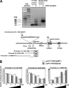
FOXO3a induces the expression of the human PIK3CA gene through a promoter region upstream of a novel 5′ exon. (A) Mapping of the human p110α transcription start site using 5′-RACE. The total RNA prepared from K562-FOXO3a(A3):ER cells after 24 h of 4-OHT induction was reverse transcribed, and the cDNA was subjected to two sequential PCRs using primers from the Ambion FirstChoice RLM-RACE kit and nested PCR primers specific to exon 2 of the human p110α gene. The nested PCR products were analyzed by agarose gel electrophoresis, and the major PCR product was clearly visible as a band of about 300 bp. Molecular size markers (base pairs) are indicated on the left of the promoter sequence relative to the major transcription start site. (Bottom) Schematic representation of the 5′ region of the human PIK3CA genes. The exons (boxes), their sizes in base pairs, and their positions relative to the major transcription start site are indicated. Introns are represented by thick lines. The sequence and genome location of the novel exon 1A are shown. Boxes underneath show the relative positions of the three putative human PIK3CA promoter constructs A, B, and C. (B) K562 cells were transiently transfected with 1 μg of each of the three putative human p110α promoter/reporter constructs, together with increasing amounts (0, 1, 5, and 10 μg) of pLPCFOXO3a-WT or -A3. Cells were harvested 24 h after transfection and assayed for luciferase activity. All relative luciferase activity values are corrected for cotransfected Renilla activity. All data shown represent the averages of data from three independent experiments, and the error bars show the standard deviations. (C) K562 cells were transiently transfected with 1 μg of each of the human PIK3CA promoter/reporter constructs, together with 0 or 5 μg of pLPCFOXO3a-A3, and processed as described above. The induction of the PIK3CA promoter by FOXO3a is shown on the right. (D) ChIP analysis of the human PIK3CA promoter. Protein-DNA complexes from K562 FOXO3a(A3):ER cells cultured in the presence or absence of 4-OHT for 4 h were subjected to immunoprecipitation with antibodies against immunoglobulin G (IgG) (nonspecific) and FOXO3a, as indicated. After cross-link reversal, the coimmunoprecipitated DNA was amplified by PCR using the indicated primers and resolved in 2% agarose gels. The primer pairs used were 5′-GCTTTTTCTGCTATGACACACAACTTC-3′ and 5′-GCACCTGGCCTATTTGTGATTTTTA-3′ (positions −2000 to −1741), 5′-GTGAAACTACGACCACAAAGAGGA-3′ and 5′-AGCATCGGTTCCTCCTTGAA-3′ (positions −1127 to −946) 5′-CGCCTTCGGGATGGTATACAA-3′ and 5′-GAGGGTGTTGTGTCATCCTAGGAC-3′ (positions −548 to −251), and 5′-GACACAACACCCTCACTACTGCA-3′ and 5′-GGCAGAGCCTACAATCCCC-3′ (positions −264 to −55).
FIG. 8.
FIG. 8.
FOXO3a induces the expression of the human PIK3CA gene through a promoter region upstream of a novel 5′ exon. (A) Mapping of the human p110α transcription start site using 5′-RACE. The total RNA prepared from K562-FOXO3a(A3):ER cells after 24 h of 4-OHT induction was reverse transcribed, and the cDNA was subjected to two sequential PCRs using primers from the Ambion FirstChoice RLM-RACE kit and nested PCR primers specific to exon 2 of the human p110α gene. The nested PCR products were analyzed by agarose gel electrophoresis, and the major PCR product was clearly visible as a band of about 300 bp. Molecular size markers (base pairs) are indicated on the left of the promoter sequence relative to the major transcription start site. (Bottom) Schematic representation of the 5′ region of the human PIK3CA genes. The exons (boxes), their sizes in base pairs, and their positions relative to the major transcription start site are indicated. Introns are represented by thick lines. The sequence and genome location of the novel exon 1A are shown. Boxes underneath show the relative positions of the three putative human PIK3CA promoter constructs A, B, and C. (B) K562 cells were transiently transfected with 1 μg of each of the three putative human p110α promoter/reporter constructs, together with increasing amounts (0, 1, 5, and 10 μg) of pLPCFOXO3a-WT or -A3. Cells were harvested 24 h after transfection and assayed for luciferase activity. All relative luciferase activity values are corrected for cotransfected Renilla activity. All data shown represent the averages of data from three independent experiments, and the error bars show the standard deviations. (C) K562 cells were transiently transfected with 1 μg of each of the human PIK3CA promoter/reporter constructs, together with 0 or 5 μg of pLPCFOXO3a-A3, and processed as described above. The induction of the PIK3CA promoter by FOXO3a is shown on the right. (D) ChIP analysis of the human PIK3CA promoter. Protein-DNA complexes from K562 FOXO3a(A3):ER cells cultured in the presence or absence of 4-OHT for 4 h were subjected to immunoprecipitation with antibodies against immunoglobulin G (IgG) (nonspecific) and FOXO3a, as indicated. After cross-link reversal, the coimmunoprecipitated DNA was amplified by PCR using the indicated primers and resolved in 2% agarose gels. The primer pairs used were 5′-GCTTTTTCTGCTATGACACACAACTTC-3′ and 5′-GCACCTGGCCTATTTGTGATTTTTA-3′ (positions −2000 to −1741), 5′-GTGAAACTACGACCACAAAGAGGA-3′ and 5′-AGCATCGGTTCCTCCTTGAA-3′ (positions −1127 to −946) 5′-CGCCTTCGGGATGGTATACAA-3′ and 5′-GAGGGTGTTGTGTCATCCTAGGAC-3′ (positions −548 to −251), and 5′-GACACAACACCCTCACTACTGCA-3′ and 5′-GGCAGAGCCTACAATCCCC-3′ (positions −264 to −55).

Similar articles

Cited by

References

    1. Birkenkamp, K. U., A. Essafi, K. E. van der Vos, M. da Costa, R. C. Hui, F. Holstege, L. Koenderman, E. W. Lam, and P. J. Coffer. 2007. FOXO3a induces differentiation of Bcr-Abl-transformed cells through transcriptional down-regulation of Id1. J. Biol. Chem. 2822211-2220. - PubMed
    1. Brunet, A., L. B. Sweeney, J. F. Sturgill, K. F. Chua, P. L. Greer, Y. Lin, H. Tran, S. E. Ross, R. Mostoslavsky, H. Y. Cohen, L. S. Hu, H. L. Cheng, M. P. Jedrychowski, S. P. Gygi, D. A. Sinclair, F. W. Alt, and M. E. Greenberg. 2004. Stress-dependent regulation of FOXO transcription factors by the SIRT1 deacetylase. Science 3032011-2015. - PubMed
    1. Charvet, C., I. Alberti, F. Luciano, A. Jacquel, A. Bernard, P. Auberger, and M. Deckert. 2003. Proteolytic regulation of Forkhead transcription factor FOXO3a by caspase-3-like proteases. Oncogene 224557-4568. - PubMed
    1. Daitoku, H., M. Hatta, H. Matsuzaki, S. Aratani, T. Ohshima, M. Miyagishi, T. Nakajima, and A. Fukamizu. 2004. Silent information regulator 2 potentiates Foxo1-mediated transcription through its deacetylase activity. Proc. Natl. Acad. Sci. USA 10110042-10047. - PMC - PubMed
    1. Delpuech, O., B. Griffiths, P. East, A. Essafi, E. W. Lam, B. Burgering, J. Downward, and A. Schulze. 2007. Induction of Mxi1-SRα by FOXO3a contributes to repression of Myc-dependent gene expression. Mol. Cell. Biol. 274917-4930. - PMC - PubMed

Publication types