. 2008 Sep;28(18):5747-63.
doi: 10.1128/MCB.02070-07. Epub 2008 Jul 21.

The chaperone-mediated autophagy receptor organizes in dynamic protein complexes at the lysosomal membrane

Affiliations

The chaperone-mediated autophagy receptor organizes in dynamic protein complexes at the lysosomal membrane

Urmi Bandyopadhyay et al. Mol Cell Biol. 2008 Sep.

Abstract

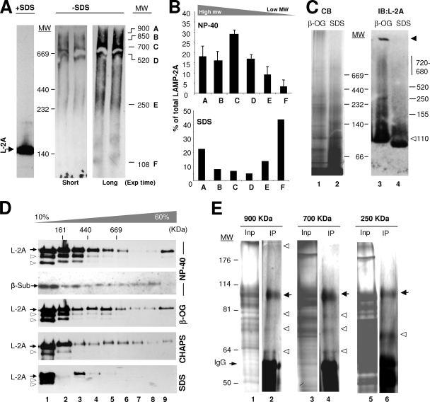
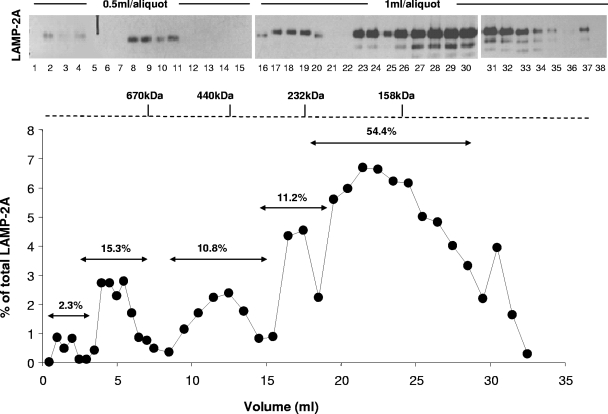
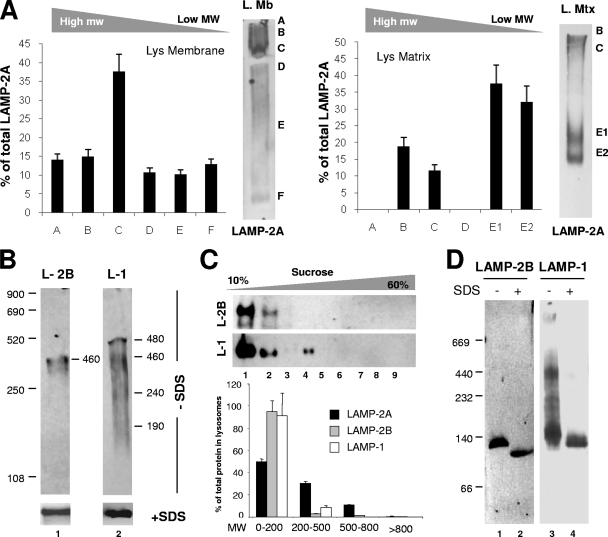
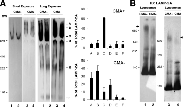
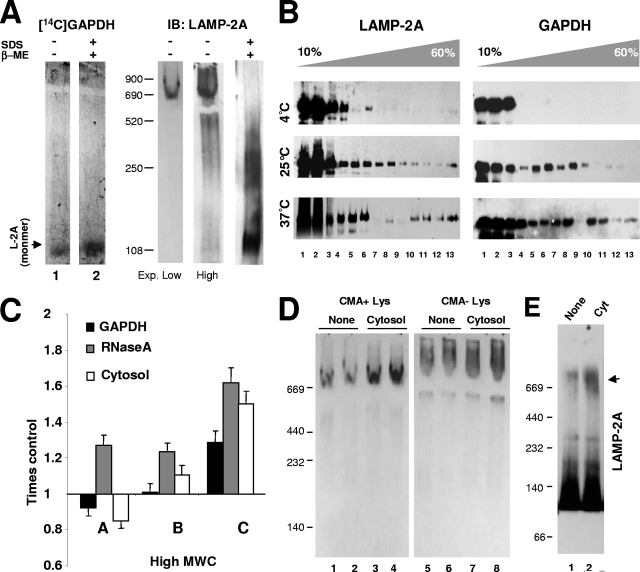
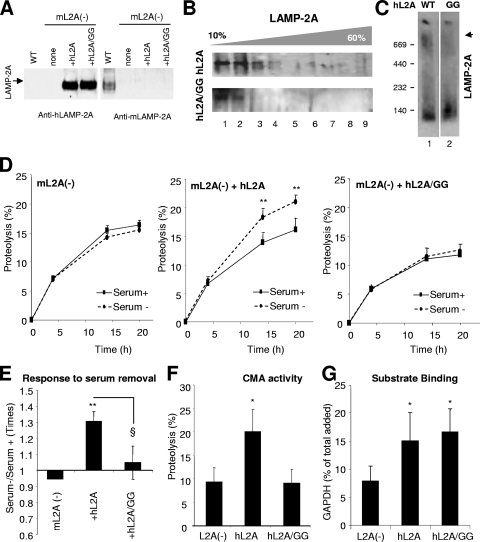
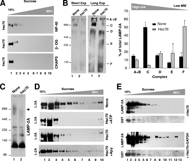
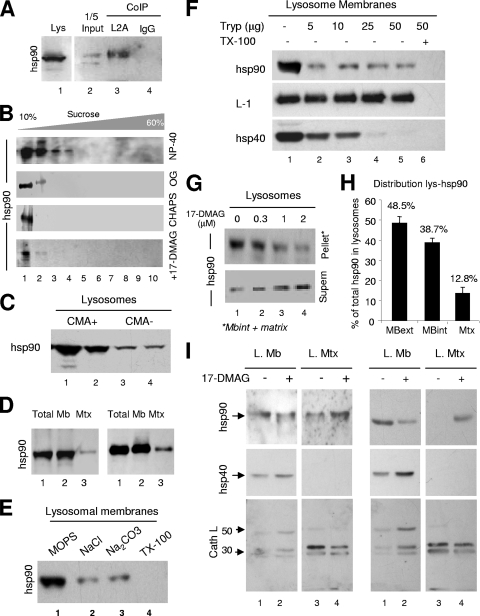
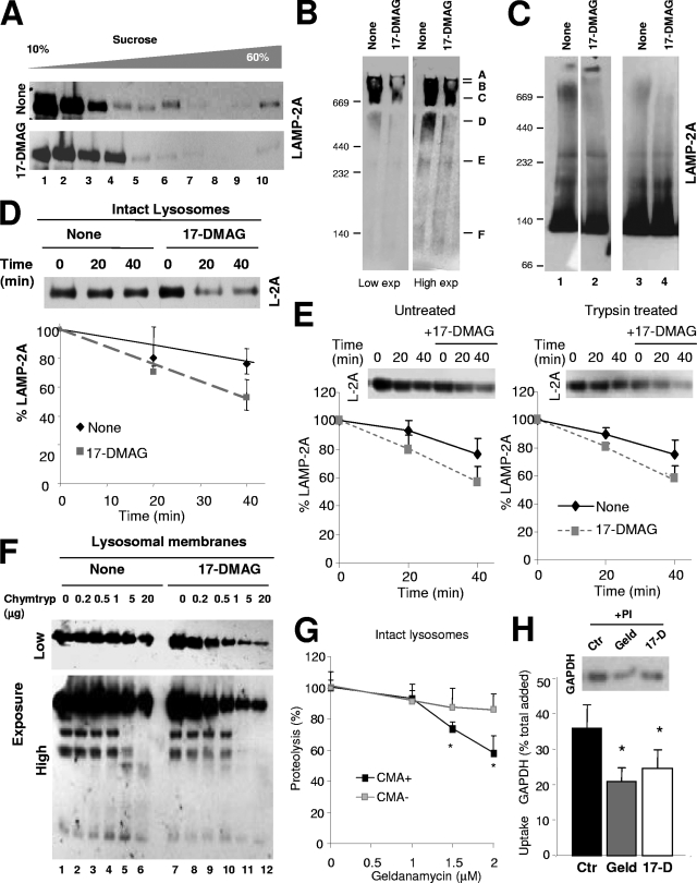
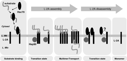
Chaperone-mediated autophagy (CMA) is a selective type of autophagy by which specific cytosolic proteins are sent to lysosomes for degradation. Substrate proteins bind to the lysosomal membrane through the lysosome-associated membrane protein type 2A (LAMP-2A), one of the three splice variants of the lamp2 gene, and this binding is limiting for their degradation via CMA. However, the mechanisms of substrate binding and uptake remain unknown. We report here that LAMP-2A organizes at the lysosomal membrane into protein complexes of different sizes. The assembly and disassembly of these complexes are a very dynamic process directly related to CMA activity. Substrate proteins only bind to monomeric LAMP-2A, while the efficient translocation of substrates requires the formation of a particular high-molecular-weight LAMP-2A complex. The two major chaperones related to CMA, hsc70 and hsp90, play critical roles in the functional dynamics of the LAMP-2A complexes at the lysosomal membrane. Thus, we have identified a novel function for hsc70 in the disassembly of LAMP-2A from these complexes, whereas the presence of lysosome-associated hsp90 is essential to preserve the stability of LAMP-2A at the lysosomal membrane.


Figures

FIG. 1.
FIG. 1.
LAMP-2A organizes into defined protein complexes at the lysosomal membrane. (A) Lysosomes isolated from the livers of 48-h-starved rats solubilized in 0.5% NP-40 were subjected to electrophoresis in the presence or absence of SDS and without β-mercaptoethanol and immunoblotted for LAMP-2A (L-2A). The LAMP-2A complexes are named alphabetically from A to F, and their estimated molecular weights (MW) are indicated in thousands. Exp, exposure. (B) Effect of the solubilization of lysosomes with NP-40 (top) or SDS (bottom) on the distribution of LAMP-2A among different molecular complexes resolved by native gel electrophoresis. Values are expressed as percentages of the total LAMP-2A in the membrane present in each complex and are the means ± SE of the densitometric quantification of two to six immunoblots as the one shown in panel A. (C) Isolated lysosomal membranes solubilized with the indicated detergents were subjected to BNE and stained with Coomassie blue (CB) (left) or subjected to immunoblotting (IB) for LAMP-2A (L-2A) (right). The black arrowhead indicates protein retained in the well, and the white arrowhead indicates full-size monomeric LAMP-2A. β-OG, β-octyl-glycoside. (D) Lysosomal membranes isolated and solubilized with the indicated detergents were subjected to continuous sucrose density gradient centrifugation. Immunoblots for LAMP-2A (L-2A) of aliquots collected from the top to the bottom of the gradients are shown. The estimated molecular masses were calculated with molecular markers run through the gradients. The distribution of the 20S proteasome β subunits (β-Sub) after subjecting cytosol to the same gradients is shown as a control of the >700-kDa complexes. The arrowheads point to partially deglycosylated forms of LAMP-2A. (E) Protein composition of three of the LAMP-2A-containing complexes. Solubilized lysosomal membranes were resolved by gel filtration, and the fractions in which the 900-, 700-, and 250-kDa LAMP-2A-containing complexes were detected were subjected to immunoprecipitation with an antibody specific against LAMP-2A. The inputs (Inp; 1/10 of total) and immunoprecipitates (IP) were subjected to SDS-PAGE, and the gels were stained with a periodic acid-modified silver staining procedure to stain heavily glycosylated proteins. The bands corresponding to IgG are indicated. Black arrows indicate LAMP-2A, and white arrowheads indicate proteins reproducibly coimmunoprecipitated with LAMP-2A in each fraction.
FIG. 2.
FIG. 2.
Resolution of different LAMP-2A-containing protein complexes at the lysosomal membrane by gel filtration. Distribution of LAMP-2A through the different fractions collected after subjecting lysosomes isolated from 48-h-starved rats and solubilized in 0.5% NP-40 to gel filtration chromatography. The graph (bottom) shows the percentages of the total LAMP-2A at the lysosomal membrane present in each of the fractions monitored by immunoblotting (top). The relative percentage of LAMP-2A in the resolved peaks was calculated after the addition of the LAMP-2A present in the different aliquots included in each peak.
FIG. 3.
FIG. 3.
Organization into protein complexes of different membrane proteins in lysosomes. (A) Lysosomal membranes (L. Mb) and matrices (L. Mtx) solubilized with 0.5% NP-40 were subjected to native gel electrophoresis and immunoblotting for LAMP-2A. Graphs show the means + SE of the densitometric quantification of three different experiments as the ones shown. Values are expressed as percentages of the total LAMP-2A present in each complex. Two bands (E1 and E2) with different molecular weights (MW) were detected in the region that corresponds to complex E in total lysosomes (Fig. 1A). (B) Lysosomal membranes solubilized as described for panel A were subjected to native gel electrophoresis (−SDS; top) or SDS-PAGE (+SDS; bottom) and immunoblotted for LAMP-2B (L-2B) and LAMP-1 (L-1). The estimated molecular weights of the immunoreactive bands are indicated. (C) Lysosomal membranes solubilized as described for panel A were subjected to continuous sucrose density gradient centrifugation. The content of LAMP-2A, LAMP-2B (L-2B), and LAMP-1 (L-1) in each of the aliquots collected from the gradients was determined by immunoblotting with specific antibodies. Values are expressed as percentages of the total protein in lysosomes and correspond to the densitometric quantification of five immunoblots as the ones shown here. Aliquots were grouped by MW (in thousands) regions as shown in panel D. (D) Lysosomal membranes solubilized as described for panel C were subjected to BNE and immunoblotting for LAMP-2B or LAMP-1.
FIG. 4.
FIG. 4.
The organization of LAMP-2A into protein complexes is different in lysosomes with different CMA activity. (A) Lysosomes with high (CMA+) and low (CMA−) CMA activity isolated from the livers of 48-h-starved rats were subjected to native gel electrophoresis and immunoblotted for LAMP-2A. Left, two different exposures of a representative immunoblot. Right, mean values + SE of the distribution of LAMP-2A in the different complexes were calculated by the densitometric quantification of three to four immunoblots as the ones shown. Values are expressed as percentages of the total amount of LAMP-2A at the lysosomal membrane. (B) The same groups of lysosomes were subjected to BNE and immunoblotting (IB) of LAMP-2A. The immunoblot on the right shows a gel run for a longer time to increase the resolution of the intermediate complexes. Arrowheads indicate the approximately 700-kDa complex.
FIG. 5.
FIG. 5.
Functional differences of the LAMP-2A complexes at the lysosomal membrane. (A) Lysosomal membranes isolated from the livers of 48-h-starved rats were subjected to native and reducing electrophoresis as labeled and electrotransferred to nitrocellulose membranes which were then incubated in renaturalization buffer with [14C]GAPDH and exposed to a phosphorimager screen (left) or subjected to immunoblotting (IB) for LAMP-2A (L-2A) (right). Two different time exposures (Exp.) are shown for the immunoblot. β-ME, β-mercaptoethanol. (B) Lysosomes isolated as described for panel A were incubated with GAPDH at the indicated temperatures, cross-linked, and then subjected to centrifugation through sucrose density gradients. Aliquots from the top to the bottom of the gradient were subjected to SDS-PAGE and immunoblotting for LAMP-2A (left) or GAPDH (right). (C) Lysosomes incubated with the indicated CMA substrates were solubilized and subjected to native gel electrophoresis and immunoblotted for LAMP-2A. Changes in the percentage of LAMP-2A in the indicated complexes (as defined in Fig. 1A) relative to the amount in lysosomes incubated without additions (control) are shown and are the means + SE of the results from four to five different experiments. (D) The effect of the incubation of lysosomes (Lys) with high (+) and low (−) CMA activity with cytosolic substrates on the organization of LAMP-2A at their membranes was analyzed by native gel electrophoresis and immunoblotting for LAMP-2A. Samples from two different animals are shown. (E) Lysosomes incubated alone or with a pool of cytosolic proteins (Cyt) were solubilized and subjected to BNE and immunoblotting for LAMP-2A. Incubation with substrates increased the intensity of the LAMP-2A-containing complex of approximately 700 kDa (indicated by the black arrowhead) by 4.2- ± 0.8- fold.
FIG. 6.
FIG. 6.
Identification of a LAMP-2A-containing protein complex required for substrate uptake by lysosomes via CMA. NIH 3T3 mouse fibroblasts subjected to stable LAMP-2A RNAi [mL2A(−)] (27) were transfected with cDNA encoding for the wild type (+hL2A) or a GG/AA mutant (+hL2A/GG) of human LAMP-2A. (A) Immunoblot with an antibody against human LAMP-2A (left) or against mouse LAMP-2A (right) of total cellular extracts of untransfected mouse fibroblasts subjected to LAMP-2A RNAi (none) or the same fibroblasts transfected with the indicated constructs. A cellular extract of mouse fibroblasts not subjected to RNAi (WT) is shown in lane 1. (B and C) Lysosomes from the LAMP-2A RNAi-treated cells transfected with wild-type (hL2A) or mutant (hL2A/GG) human LAMP-2A were subjected to centrifugation through sucrose density gradients (B) or BNE (C). The SDS-PAGE of the aliquots from the top to the bottom of the gradient and the BNE were subjected to immunoblotting for human LAMP-2A. (D) Rates of long-lived-protein degradation via CMA (sensitive to ammonium chloride and insensitive to 3-methyl adenine) were calculated in the cells maintained in the presence (Serum+) or absence (Serum−) of serum, as described in Materials and Methods. Values are expressed as percentages of the total acid-precipitable radioactivity (proteins) transformed into acid-soluble radioactivity (amino acids and small peptides) at the indicated time points and are means + SE of the results from three different experiments with triplicate wells. *, P < 0.01; **, P < 0.001. (E) Changes in protein degradation in response to serum removal were analyzed in the cells described for panel D. Values are expressed as the fold increases in protein degradation in untransfected wild-type cells and are the means + SE of the results from two different experiments with triplicate wells. * (P < 0.001) and § (P < 0.01) are differences with respect to the control and the cells transfected with the wild-type protein, respectively. (F) Proteolysis of a pool of labeled cytosolic proteins by intact lysosomes isolated as described for panel B. Values are shown as percentages of proteolysis and are the means + SE of the results from two different experiments with triplicate samples. *, P < 0.01. (G) Binding of GAPDH to intact lysosomes isolated as described for panel B. Lysosomes were incubated at 4°C to eliminate differences due to impaired uptake. Values are shown as percentages of the total GAPDH associated with lysosomes collected by centrifugation after correcting for the total amount of lysosomal protein in each sample and are the means + SE of the results from three individual experiments. *, P < 0.05.
FIG. 7.
FIG. 7.
Hsc70 induces disassembly of LAMP-2A from the high-molecular-weight protein complexes at the lysosomal membrane. (A) Lysosomes isolated from the livers of 48-h-starved rats were solubilized with the indicated detergents and subjected to sucrose density gradient centrifugation and immunoblotting for hsc70. (B to D) Lysosomes isolated as described for panel A were incubated with GST-hsc70 and/or ATP and apyrase (Apy), as labeled, solubilized, and subjected to native gel electrophoresis (two different exposures [Exp] are shown) (B), BNE (C), or sucrose density gradient centrifugation (D) and immunoblotted for LAMP-2A (L-2A). Changes in the distribution of LAMP-2A throughout the native gel were calculated by the densitometric quantification of the immunoblots from three different experiments. Values are expressed as times the values in lysosomes incubated without additions. The black arrowhead in panel C indicates the approximately 700-kDa complex. (E) Lysosomes incubated with the indicated additions were subjected to sucrose density gradient centrifugation and immunoblotted for LAMP-2A or GST.
FIG. 8.
FIG. 8.
Characterization of lysosome-associated hsp90. (A) Lysosomes from the livers of 48-h-starved rats were solubilized and subjected to immunoprecipitation (CoIP) with an antibody against LAMP-2A (L2A) or an irrelevant immunoglobulin (IgG). Precipitates were subjected to SDS-PAGE and immunoblotting for hsp90. Lane 1 shows total lysosomes (Lys), and lane 2 contains 1/5 of the starting solubilized membranes. (B) Lysosomes isolated from the livers of 48-h-starved rats were solubilized with the indicated detergents and subjected to sucrose density gradient centrifugation and immunoblotting for hsp90. Bottom panel, lysosomes were incubated with the geldanamycin derivative 17-DMAG before solubilization with NP-40. (C) Intact lysosomes with high (CMA+) and low (CMA−) CMA activity isolated from the livers of 48-h-starved rats were subjected to SDS-PAGE and immunoblotted for hsp90 (duplicate samples for each type of lysosome are shown). (D) Intact lysosomes (total) with high CMA activity were subjected to hypotonic shock and centrifugation, and membranes (Mb) and matrices (Mtx) collected by centrifugation were processed as described for panel C. Duplicate samples are shown. (E) Intact lysosomes were subjected to washes with MOPS buffer, 1 M NaCl, 0.1 M Na2CO3, or 1% Triton X-100 as labeled. The amount of hsp90 associated with the membrane was detected after isolation of the membranes as above and immunoblotting for hsp90. (F) Intact lysosomes were incubated with increasing concentrations of trypsin (Tryp) supplemented or not with 1% Triton X-100, as labeled. Samples were subjected to SDS-PAGE and immunoblotting for the indicated proteins. (G) Intact lysosomes with high CMA activity were incubated with increasing concentrations of 17-DMAG, collected by centrifugation and supernatant (Supern) and pellet were subjected to SDS-PAGE and immunoblotting for hsp90. Mbint, luminal part of the membrane. (H) The percentage of lysosomal hsp90 present on the cytosolic side of the lysosomal membrane (MBext), the luminal part of the membrane (MBint), and the lysosomal lumen (Mtx) was calculated by combining the results obtained after membrane/matrix separation (D), membrane washing (E) and trypsinization (F), and treatment with hsp90 inhibitors (G). Values are the means ± SE of the results from five to eight different experiments. (I) Membranes (L. Mb) and matrices (L. Mtx) from lysosomes incubated alone (−) or with (+) 17-DMAG were subjected to immunoblotting for hsp90, hsp40, or cathepsin L (Cath L). Two representative immunoblots are shown. Arrows indicate the precursor (50 kDa) and active subunits (20 and 30 kDa) of cathepsin L.
FIG. 9.
FIG. 9.
Novel role for hsp90 as a stabilizer of LAMP-2A at the lysosomal membrane. (A) Intact rat liver lysosomes with high CMA activity were incubated alone or with the geldanamycin derivative 17-DMAG. At the end of the incubation, lysosomes were subjected to sucrose density gradient centrifugation (A), native gel electrophoresis (B), or BNE (C) and immunoblotted for LAMP-2A. Two exposures (Exp) of the same immunoblot are shown in panel B to better appreciate the region corresponding to the monomeric form of LAMP-2A. Two independent experiments are shown in C (lanes on the left were run in the same gel but were cut as they were separated by irrelevant samples). (D) Resealed membranes from lysosomes preincubated or not with 17-DMAG were incubated at 37°C in an isotonic buffer, and the remaining levels of LAMP-2A at the indicated times were detected by immunoblotting. Values are expressed as percentages of the LAMP-2A at time zero and are the means ± SE of the densitometric quantification of the immunoblots from three different experiments. A representative immunoblot is shown. (E) Intact lysosomes untreated or previously preincubated with 25 μg of trypsin and with 17-DMAG as indicated were incubated (after neutralization of the protease with a trypsin inhibitor) at 37°C in an isotonic buffer, and the remaining levels of LAMP-2A at the indicated times were detected by immunoblotting. Values are expressed as described for panel D and are the means ± SE of the densitometric quantification of the immunoblots from two different experiments. (F) Proteolytic cleavage pattern of LAMP-2A in lysosomal membranes isolated from lysosomes incubated as described for panel B and treated with increasing concentrations of chymotrypsin (Chymtryp). Images of a low (top) and a high (bottom) exposure time of the immunoblot for LAMP-2A are shown. (G to H) Effect of geldanamycin (Geld) and 17-DMAG (17-D) on CMA of GAPDH by isolated lysosomes. Panel G shows the effect of increasing concentrations of geldanamycin on the degradation of GAPDH by intact lysosomes with high (+) or low (−) CMA activity. Values are expressed as percentages of the degradation in untreated lysosomes to be able to compare the samples despite the low rates of degradation observed in the CMA lysosomes and are the means + SE of the results from two different experiments with triplicate samples. In panel H, untreated lysosomes (control; Ctr) or lysosomes treated with the indicated hsp90 inhibitors and preincubated or not with protease inhibitors (PI) were incubated with GAPDH. Lysosomes were collected by centrifugation, and the amount of GAPDH associated with each sample was determined by SDS-PAGE and immunoblotting. Uptake was calculated after densitometric quantification as the difference between association and binding. Values are expressed as percentages of total GAPDH added and are the means + SE of six different experiments. *, P < 0.01.
FIG. 10.
FIG. 10.
Hypothetical model for the dynamic assembly and disassembly of LAMP-2A (L-2A) into multimeric complexes at the lysosomal membrane (L. Mb). Based on the results presented in this work, we hypothesize that CMA substrates delivered by cytosolic hsc70 bind preferentially to monomers of LAMP-2A at the lysosomal membrane and that the binding of substrates promotes the multistep organization of LAMP-2A into higher-order multimeric complexes. The translocation of substrates requires the formation of an approximately 700-kDa LAMP-2A complex that is disassembled into smaller complexes in an hsc70-dependent manner when substrates are no longer present. The interaction of hsp90 with LAMP-2A at the luminal side of the lysosomal membrane stabilizes this receptor while transitioning between the multimeric membrane complexes. L. Mtx, lysosomal matrix.

References

    1. Agarraberes, F., and J. F. Dice. 2001. A molecular chaperone complex at the lysosomal membrane is required for protein translocation. J. Cell Sci. 1142491-2499. - PubMed
    1. Agarraberes, F., S. Terlecky, and J. Dice. 1997. An intralysosomal hsp70 is required for a selective pathway of lysosomal protein degradation. J. Cell Biol. 137825-834. - PMC - PubMed
    1. Aniento, F., E. Roche, A. M. Cuervo, and E. Knecht. 1993. Uptake and degradation of glyceraldehyde-3-phosphate dehydrogenase by rat liver lysosomes. J. Biol. Chem. 26810463-10470. - PubMed
    1. Auteri, J. S., A. Okada, V. Bochaki, and J. F. Dice. 1983. Regulation of intracellular protein degradation in IMR-90 human diploid fibroblasts. J. Cell. Physiol. 115167-174. - PubMed
    1. Bandhyopadhyay, U., and A. M. Cuervo. 2006. Chaperone-mediated autophagy in aging and neurodegeneration: lessons from alpha-synuclein. Exp. Gerontol. 42120-128. - PubMed

Publication types

MeSH terms

Substances