. 2008 Jul 28;182(2):327-40.
doi: 10.1083/jcb.200712125. Epub 2008 Jul 21.

Hormone-dependent nuclear export of estradiol receptor and DNA synthesis in breast cancer cells

Affiliations

Hormone-dependent nuclear export of estradiol receptor and DNA synthesis in breast cancer cells

Maria Lombardi et al. J Cell Biol. .

Abstract

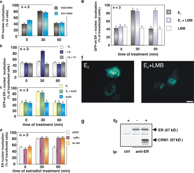
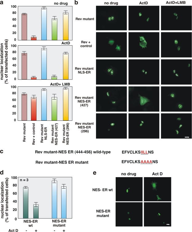
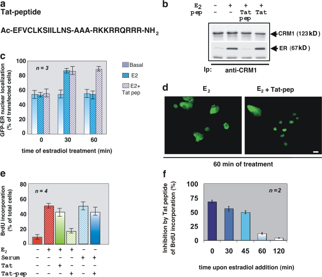
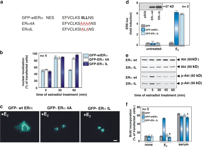
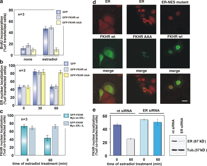
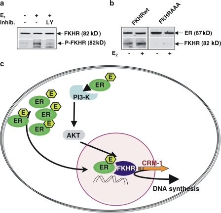
In breast cancer cells, cytoplasmic localization of the estradiol receptor alpha (ERalpha) regulates estradiol-dependent S phase entry. We identified a nuclear export sequence (NES) in ERalpha and show that its export is dependent on both estradiol-mediated phosphatidylinositol-3-kinase (PI3K)/AKT activation and chromosome region maintenance 1 (CRM1). A Tat peptide containing the ERalpha NES disrupts ERalpha-CRM1 interaction and prevents nuclear export of ERalpha- and estradiol-induced DNA synthesis. NES-ERalpha mutants do not exit the nucleus and inhibit estradiol-induced S phase entry; ERalpha-dependent transcription is normal. ERalpha is associated with Forkhead proteins in the nucleus, and estradiol stimulates nuclear exit of both proteins. ERalpha knockdown or ERalpha NES mutations prevent ERalpha and Forkhead nuclear export. A mutant of forkhead in rhabdomyosarcoma (FKHR), which cannot be phosphorylated by estradiol-activated AKT, does not associate with ERalpha and is trapped in the nucleus, blocking S phase entry. In conclusion, estradiol-induced AKT-dependent phosphorylation of FKHR drives its association with ERalpha, thereby triggering complex export from the nucleus necessary for initiation of DNA synthesis and S phase entry.


Figures

Figure 1.
Figure 1.
Estradiol induces nuclear export of ERα, which is regulated by the PI3K–AKT pathway and depends on CRM1. Quiescent MCF-7 cells were used. (a) Cells were untreated or treated with 10 nM estradiol (E2) for the indicated times (min). ERα localization was analyzed by immunofluorescence using the indicated antibodies. (b and c) Cells were transfected with GFP-wtERα then left untreated or treated with the indicated compounds. OH-tamoxifen (Tx; AstraZeneca) was used at 0.1 μM; actinomycin D (Act D) was added at 5 μg/ml, 1 h before estradiol stimulation. GFP-wtERα localization was determined by fluorescence. (d) Cells were transfected with the indicated plasmids and left untreated or treated with 10 nM estradiol for the indicated times (min). The Myc-tagged pSG5, Δp85α, or Myc-His–tagged dominant-negative AKT ectopically expressed in MCF-7 cells were visualized by immunofluorescence, as described in Materials and methods. ERα localization was analyzed by immunofluorescence using the rat H222 anti-ERα mAb. (e) Cells were transfected with GFP-wtERα and then left untreated or treated with 10 nM estradiol in the absence or presence of LMB (at 5 ng/ml). LMB was added 30 min before the hormone. Cells were also treated with LMB in the absence of hormone. GFP-wtERα localization was determined by fluorescence. (a, b, c, d, and e) Cells that fell into the category of exclusively nuclear fluorescence were scored, and data was expressed as a percentage of total cells (in a) or transfected cells (in b, c, d, and e). Data were derived from at least 1,000 scored cells. The results of several independent experiments were averaged; means and SEM are shown. n represents the number of experiments. (f) Images from one experiment in panel e were captured. Panels show GFP-wtERα localization in MCF-7 cells stimulated for 60 min with estradiol (E2) in the absence or presence of LMB. Bar, 5 μm. (g) 35S-labeled HA-CRM1 was incubated with recombinant ERα in the absence or presence of 10 nM estradiol. The purified recombinant RanQ69L (at 1 μM) was included in the incubation mixture of each sample. Proteins were immunoprecipitated with rabbit polyclonal anti-ERα antibody. Eluted proteins were immunoblotted with anti-ERα antibody (top) or revealed by fluorography (bottom).
Figure 2.
Figure 2.
The ERα 444–456 sequence restores export activity of the NES-deficient REV1.4-GFP. Growing MCF-7 cells were used. (a and b) Cells were transfected with the indicated constructs. After transfection, the cells were left untreated (no drug) or treated with actinomycin D (ActD) at 5 μg/ml, alone or together with 5 ng/ml LMB. The subcellular distribution of GFP proteins was determined by fluorescence microscopy, and cells that fell into the category of exclusively nuclear fluorescence were scored. Data are expressed as a percentage of transfected cells, with mean values taken from at least three experiments. For each experiment, at least 600 cells were scored. (b) Images of one experiment in panel a. (c) The wt ERα 444–456 sequence (NES ERα wt) as well as its mutated version (NES ERα mutant). The putative NES-ERα sequence is indicated by the underlined amino acids, which were substituted with alanine residues in the mutant sequence. The NES-ERα wt as well as the NES-ERα mutant subcloned into the Rev mutant were transfected (d and e) in growing MCF-7 cells. After transfection, the cells were left untreated (−) or treated with 5 μg/ml Act D (+). The percentage of cells with nuclear GFP protein was determined by fluorescence microscopy and graphically shown in panel d. Data were derived from at least 600 scored cells. The results of several independent experiments were averaged. (a and d) Means and SEM are shown. n represents the number of experiments. (e) Images of one experiment in panel d. Bars, 5 μm
Figure 3.
Figure 3.
A peptide mimicking the ERα 444–456 sequence displaces the CRM1–ERα interaction, sequesters the receptor into nuclei, and inhibits S phase entry in estradiol-treated MCF-7 cells. (a) The amino acidic sequence of the Tat-conjugated ERα (444–456) peptide. (b) 35S-labeled HA-CRM1 was incubated with recombinant ERα from baculovirus in the absence or presence of 10 nM estradiol alone or together with a 200-fold excess of the Tat-conjugated peptide (Tat-pep). A 200-fold excess of a nonspecific peptide (Tat) was used as a control. CRM1 was immunoprecipitated with the anti-HA mAb, and proteins in immunocomplexes were revealed by fluorography (top) and immunoblotting with the anti-ERα antibody (bottom). (c) Quiescent MCF-7 cells transfected with GFP-HEG0 were incubated for 1 h with the Tat-conjugated peptide (Tat pep). Thereafter, the cells were left untreated or treated for the indicated times with 10 nM estradiol. The proportion of cells with nuclear GFP-wtERα protein was determined by fluorescence microscopy and graphically shown. For each experiment, at least 200 cells were scored. The results of several independent experiments were averaged. n represents the number of experiments. (d) Images of one experiment in panel c. This shows the localization of GFP-wtERα in MCF-7 cells treated for 60 min with estradiol in the absence or presence of Tat-pep. Bar, 5 μm. (e) MCF-7 cells on coverslips were made quiescent or serum starved (maintained for 24 h in 0.5% FCS). The cells were incubated for 24 h with 10 nM estradiol or 20% serum in the absence or presence of the Tat-conjugated ERα (444–456) peptide (Tat-pep) or Tat alone. After in vivo pulse with BrdU, DNA synthesis was analyzed and BrdU incorporation was expressed as a percentage of total nuclei. For each experiment, at least 200 cells were scored. The results of several independent experiments were averaged. n represents the number of experiments. (f) Quiescent MCF-7 cells were stimulated with 10 nM estradiol for 24 h, and the Tat peptide was added after the addition of the hormone at the time points indicated in the figure. After an in vivo pulse with BrdU, DNA synthesis was analyzed and expressed as a percentage of BrdU incorporation inhibition. For each experiment, at least 300 cells were scored. The results of two independent experiments were averaged. (c, e, and f) Means and SEM are shown.
Figure 4.
Figure 4.
Mutations of NES-ERα impair nuclear export of full-length ERα and prevent S phase entry in MCF-7 cells stimulated by estradiol. (a) The wild-type NES-ERα endowed in the amino acid 444–456 sequence is shown in bold. Mutated amino acids are underlined. The NES mutants, GFP-HEG4A and GFP-HEGIL, were prepared as described in Materials and methods. (b) Quiescent MCF-7 cells were transfected with the indicated plasmids and then left unstimulated or stimulated with 10 nM estradiol for the indicated times. The percentage of cells with nuclear GFP protein was determined by fluorescence microscopy and shown graphically. For each experiment, at least 150 cells were scored. The results of different independent experiments were averaged; n represents the number of experiments. (c) Images of one experiment in panel b that shows the intracellular distribution of GFP-wtERα, GFP-ERα 4A, or GFP-ERα IL in MCF-7 cells treated for 1 h with estradiol. Bar, 5 μm. (d–f) Growing NIH3T3 fibroblasts were used. The Western blot in panel d shows that NIH3T3 fibroblasts are ERα negative. In the graph in panel d, cells were transfected with an ERE-Luc construct along with the indicated plasmids. After transfection, the cells were made quiescent and then left unstimulated or stimulated with 10 nM estradiol. Luciferase activity was assayed, normalized using β-gal as an internal control, and expressed as fold induction. (e) Cells were transfected with the indicated plasmids and made quiescent. The cells were left unstimulated or stimulated for 5 min with 10 nM estradiol. Lysates were analyzed for AKT activation using the anti–P-Ser-473 antibody (bottom). The nitrocellulose filters were then reprobed with anti-AKT antibody (top). (f) Cells on coverslips were transfected with the indicated plasmids and made quiescent. The cells were left unstimulated or stimulated for 18 h with 10 nM estradiol or 20% serum. In cells transfected with GFP-wtERα, the inhibitory action of Tat peptide on estradiol-induced BrdU incorporation was analyzed by including this compound (at 1 μM) to the cell medium (bars with asterisks). After in vivo pulse with BrdU, DNA synthesis was analyzed by immunofluorescence. In transfected cells, BrdU incorporation was calculated by the formula (percentage of BrdU-positive cells = [number of transfected-positive cells/number of transfected cells] × 100) and compared with BrdU incorporation of untransfected cells from the same coverslips. For each plasmid, data were derived from at least 500 scored cells. The results of several independent experiments were averaged; n represents the number of experiments. (b, d, and f) Means and SEM are shown.
Figure 5.
Figure 5.
FKHR nuclear export: regulation by estradiol and a role in hormone-induced DNA synthesis in MCF-7 cells. Quiescent MCF-7 cells on coverslips were used. (a) Cells were transfected with the indicated plasmids then left unstimulated or stimulated for 24 h with 10 nM estradiol. After in vivo pulse with BrdU, DNA synthesis was analyzed by immunofluorescence and BrdU incorporation was calculated as in Fig. 4. (b) Cells were transfected with the indicated plasmids then left unstimulated or stimulated with 10 nM estradiol for the indicated times. Endogenous ERα localization as well as expression of GFP, GFP-FKHR wt, or GFP-FKHR AAA mutant was monitored by confocal microscopy. Cells that fell into the category of exclusively ERα nuclear fluorescence were scored, and data were expressed as a percentage of transfected cells. (c) Cells were cotransfected with the indicated plasmids then left unstimulated or stimulated with 10 nM estradiol for 60 min. Localization of GFP-FKHR wt, Myc-HEG0, or Myc-HEGIL mutant was monitored by confocal microscopy. Cells that fell into the category of exclusively FKHR nuclear fluorescence were scored and the data were expressed as a percentage of cotransfected cells. For each experiment in panels a–c, data were derived from at least 500 scored cells. The results of several independent experiments were averaged; n represents the number of experiments. (d) Images from one experiment in b or c are shown. They represent the staining of endogenous ERα (red) in MCF-7 cells expressing GFP-FKHR wt (left, green) or the mutant, GFP-FKHR AAA (middle, green), and treated for 60 min with estradiol. (right) The staining of Myc-tagged NES-ERα mutant (red) in MCF-7 cells coexpressing GFP-FKHR wt (green) and treated for 60 min with estradiol. Merged images are also shown on the bottom. Bar, 5 μm. (e) The cells were cotransfected with ERα siRNA (ERα siRNA) or nontargeting siRNA (nt siRNA) and GFP-FKHR wt. The cells were then left unstimulated or stimulated with 10 nM estradiol for the indicated times. GFP-FKHR wt localization was monitored by confocal microscopy. Cells that fell into the category of exclusively GFP-FKHR wt nuclear fluorescence were scored and data were expressed as a percentage of transfected cells. Data were derived from at least 200 scored cells. The results of two independent experiments were averaged. The blot in panel e confirms the silencing of ERα in MCF-7 cells transfected with ERα siRNA (top). The bottom shows the blot of loading proteins revealed using the anti-tubulin antibody. (a, b, c, and e) Means and SEM are shown.
Figure 6.
Figure 6.
Estradiol-induced FKHR activation and Erα–FKHR complex assembly in MCF-7 cells. Model of ERα nuclear export. Quiescent MCF-7 cells were used. (a) Cells were left unstimulated or stimulated for 5 min with 10 nM estradiol in the absence or presence of LY294002 (at 5 μM; Qbiogene). Lysates were analyzed for FKHR phosphorylation using the anti–P-Ser-256–FKHR antibody (bottom). The nitrocellulose filter was then reprobed with anti-FKHR antibody (top). (b) The cells were transfected with the indicated plasmids (FKHR wt or FKHR AAA) then left unstimulated or stimulated for 30 min with 10 nM estradiol. Lysates were immunoprecipitated with anti-ERα antibody. Immunocomplexes were immunoblotted using the antibodies against the indicated proteins. (c) The model of ERα nuclear export is depicted. Estradiol stimulates the PI3K–AKT pathway, leading to FKHR phosphorylation and triggering the functionally associated export of FKHR–ERα. This export results in stimulation of DNA synthesis.

Similar articles

Cited by

References

    1. Abbondanza, C., V. Rossi, A. Roscigno, L. Gallo, A. Belsito, G. Piluso, N. Medici, V. Nigro, A.M. Molinari, B. Moncharmont, and G.A. Puca. 1998. Interaction of vault particles with estrogen receptor in the MCF-7 breast cancer cell. J. Cell Biol. 141:1301–1310. - PMC - PubMed
    1. Alt, J.R., J.L. Cleveland, M. Hannink, and J.A. Diehl. 2000. Phosphorylation-dependent regulation of cyclin D1 nuclear export and cyclin D1-dependent cellular transformation. Genes. Dev. 14:3102–3114. - PMC - PubMed
    1. Askjaer, P., A. Bachi, M. Wilm, F.R. Bischoff, D.L. Weeks, V. Ogniewski, M. Ohno, C. Niehrs, J. Kjems, I.W. Mattaj, and M. Fornerod. 1999. Ran-regulated interactions of CRM1 with nucleoporins and a shuttling DEAD-Box helicase. Mol. Cell. Biol. 19:6276–6285. - PMC - PubMed
    1. Biggs, W.H. III, J. Meisenhelder, T. Hunter, W.K. Cavenee, and K.C. Arden. 1999. Protein kinase B/Akt-mediated phosphorylation promotes nuclear exclusion of the winged helix transcription factor FKHR1. Proc. Natl. Acad. Sci. USA. 96:7421–7426. - PMC - PubMed
    1. Birkenkamp, K.U., and P.J. Coffer. 2003. Regulation of cell survival and proliferation by the FOXO (Forkhead box, class O) subfamily of Forkhead transcription factors. Biochem. Soc. Trans. 31:292–297. - PubMed

Publication types

MeSH terms