. 2008 Jul 28;182(2):355-66.
doi: 10.1083/jcb.200801056. Epub 2008 Jul 21.

Phosphatidylinositol 4,5-bisphosphate regulates SNARE-dependent membrane fusion

Affiliations

Phosphatidylinositol 4,5-bisphosphate regulates SNARE-dependent membrane fusion

Declan J James et al. J Cell Biol. .

Abstract

Phosphatidylinositol 4,5-bisphosphate (PI 4,5-P(2)) on the plasma membrane is essential for vesicle exocytosis but its role in membrane fusion has not been determined. Here, we quantify the concentration of PI 4,5-P(2) as approximately 6 mol% in the cytoplasmic leaflet of plasma membrane microdomains at sites of docked vesicles. At this concentration of PI 4,5-P(2) soluble NSF attachment protein receptor (SNARE)-dependent liposome fusion is inhibited. Inhibition by PI 4,5-P(2) likely results from its intrinsic positive curvature-promoting properties that inhibit formation of high negative curvature membrane fusion intermediates. Mutation of juxtamembrane basic residues in the plasma membrane SNARE syntaxin-1 increase inhibition by PI 4,5-P(2), suggesting that syntaxin sequesters PI 4,5-P(2) to alleviate inhibition. To define an essential rather than inhibitory role for PI 4,5-P(2), we test a PI 4,5-P(2)-binding priming factor required for vesicle exocytosis. Ca(2+)-dependent activator protein for secretion promotes increased rates of SNARE-dependent fusion that are PI 4,5-P(2) dependent. These results indicate that PI 4,5-P(2) regulates fusion both as a fusion restraint that syntaxin-1 alleviates and as an essential cofactor that recruits protein priming factors to facilitate SNARE-dependent fusion.


Figures

Figure 1.
Figure 1.
Microdomains of PI 4,5-P2 on the plasma membrane. (A–F) PC12 cell membrane sheets were incubated with 2 μM PLCδ1–PH-GFP fusion protein and 100 μM FM4-64 to image PI 4,5-P2 (A and B) or membrane area (C and D), respectively. (E and F) Membrane sheets were incubated with PLCδ1–PH-GFP fusion protein after incubations with or without 2 mM MgATP, respectively. (G–I) Quantification of membrane PI 4,5-P2. (G) Supported planar bilayers containing indicated mole percentage of PI 4,5-P2 and 0.1 mol% rhodamine-PE were incubated with 2 μM PLCδ1–PH-GFP fusion protein and imaged in red (top) and green (bottom) channels. (H) Fluorescence intensity per pixel was plotted for each supported bilayer to generate a calibration curve. (I) From calibration curves prepared in parallel, pixel intensity on PC12 cell membrane sheets (incubated with or without MgATP) was used to infer PI 4,5-P2 concentrations in microdomains, between microdomains, and averaged across the membrane. Mean ± SEM values are shown for +MgATP (n = 13) and −MgATP (n = 5). (J and K) PI 4,5-P2 microdomains overlap with clusters of CAPS and docked vesicles. (J) PI 4,5-P2 microdomains in PC12 cell membrane sheets were localized by incubation with PLCδ1–PH-GFP and fixed for immunolocalization with CAPS polyclonal or chromogranin B monoclonal antibodies. (K) Triple channel colocalizations were quantified as described in Materials and methods. Mean ± SEM (n = 9) values are shown.
Figure 2.
Figure 2.
PI 4,5-P2 inhibits SNARE-dependent liposome fusion. (A) Acceptor liposomes with 40 copies of syntaxin-1/SNAP-25 (t-SNARE) in 90:10 mol% PC/PI 4,5-P2 or 85:15 mol% PC/PS were mixed with fluorescent PC/PS donor liposomes with 100 copies of VAMP-2. Parallel reactions with Pf acceptor liposomes at 90:10 mol% PC/PI 4,5-P2 or 85:15 mol% PC/PS were conducted. The kinetics of fusion are shown as a percentage of total NBD fluorescence after correction for Pf liposomes (see Materials and methods). Points represent mean values ± SEM for five independent experiments. (B) Acceptor liposomes reconstituted with syntaxin-1/SNAP-25 and the indicated mole percentage PI 4,5-P2 were mixed with donor liposomes at a 9:1 ratio (∼2 mM lipid) and NBD fluorescence was monitored. The extent of fusion at 120 min is expressed as a percentage of fusion relative to that observed using acceptor liposomes with 85:15 mol% PC/PS. Bars indicate mean values ± SEM for three independent experiments (*, P < 0.01). (C) Liposome fusion reactions were conducted with VAMP-2 donor liposomes incubated with syntaxin-1/SNAP-25 acceptor liposomes containing 5 mol% PI 4-P, PI 4,5-P2, or PI 3,4-P2 as indicated. The extent of fusion at 120 min is expressed as in B (**, P < 0.001 compared with PC/PS donor–acceptor liposomes). (D) Inhibition of fusion by inverted cone lipid LPC. Acceptor liposomes with 40 copies of syntaxin-1/SNAP-25 in 85:15 mol% PC/PS or 90:5 mol% PC/PI 4,5-P2 as indicated were incubated 5 min at 30°C without or with 0.2 mM LPC and mixed with donor liposomes to monitor fusion kinetics. Values (mean ± SEM; n = 6) of percentage of maximal NBD fluorescence at 120-min incubation times are shown (**, P < 0.001 compared with untreated PC/PS liposomes). (E) The extent of liposome fusion at 120 min is shown for reactions containing donor liposomes mixed with acceptor liposomes containing wild-type or mutant (K252,253A, K264,265A, and K252,253,264,265A) syntaxins in 85:15 mol% PC/PS or 90:10 mol% PC/PI 4,5-P2 as indicated. Extent of fusion is shown as a percentage relative to wild-type syntaxin in PC/PS liposomes (**, P < 0.001 compared with WT syntaxin containing PC/PS liposomes).
Figure 3.
Figure 3.
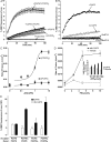
CAPS accelerates liposome fusion in a PI 4,5-P2– and SNARE-dependent manner. (A) Acceptor liposomes with 40 copies of syntaxin-1/SNAP-25 (indicated by t for t-SNAREs) in 90:10 mol% PC/PI 4,5-P2 were incubated with donor liposomes with 100 copies of VAMP-2 in 85:15 mol% PC/PS in the absence or presence of 1 μM CAPS. Similar incubations with donor and acceptor liposomes in 85:15 mol% PC/PS in the absence or presence of 1 μM CAPS were conducted. NBD fluorescence in parallel reactions with Pf liposomes was used to correct all data. Mean values ± SEM for five independent experiments are shown. (B) Acceptor liposomes with 40 copies of syntaxin-1/SNAP-25 in 90:10 mol% PC/PI 4,5-P2 were incubated with VAMP-2–containing donor liposomes in the absence or presence of 1 μM CAPS. Parallel incubations used botulinum neurotoxin B–treated donor liposomes incubated without or with 1 μM CAPS. (C) Syntaxin-1/SNAP-25 acceptor liposomes (indicated by t for t-SNAREs) with 10 mol% PI 4,5-P2 or with 15% PS were incubated with VAMP-2 donor liposomes in the presence of indicated CAPS concentrations. Exponential fits of time courses were used to derive rate constants (k). Mean ± SEM values are shown for three independent experiments. (D) Rate constants were determined for fusion reactions without or with 1 μM CAPS in incubations that contained syntaxin-1/SNAP-25 acceptor liposomes with the indicated mole percentage PI 4,5-P2. Data in inset show the percentage of maximal NBD fluorescence at 120 min in incubations without or with CAPS. Mean ± SEM values are shown for four independent experiments. (E) Extent of fusion in 120 min was determined in the absence or presence of 1 μM CAPS with donor VAMP-2 or acceptor syntaxin-1/SNAP-25 liposomes that contained 85:15 mol% PC/PS or 90:10 mol% PC/PI 4,5-P2 as indicated. Mean ± SEM values are shown for three independent experiments (*, P < 0.01 for PC/PIP2 compared with PC/PS; †, P = 0.05 compared with parallel incubations without CAPS).
Figure 4.
Figure 4.
CAPS acceleration of fusion requires a functional PH domain. (A) CAPS PH domain fails to accelerate fusion but inhibits CAPS stimulation. Fusion reactions with 5 mol% PI 4,5-P2–containing acceptor liposomes were conducted in the absence (PH −CAPS) or presence (PH +CAPS) of 1 μM CAPS, and the indicated concentrations of a CAPS GST-PH domain fusion protein. For clarity, points were selected and plotted every 7.5 min. Exponential fits of time courses are shown. (B) Phosphoinositide binding characteristics of CAPS PH domain. Indicated amounts of CAPS GST-PH domain fusion protein (either wild-type PH or mutant PHDEE) immobilized on glutathione agarose beads were incubated with liposomes containing PI 4-P, PI 4,5-P2, or PI 3,4-P2 as described in Materials and methods. (C) Liposome fusion reactions were conducted with VAMP-2 donor liposomes incubated with syntaxin-1/SNAP-25 acceptor liposomes containing 5 mol% PI 4-P, PI 4,5-P2, or PI 3,4-P2 as indicated. Rate constants were determined by exponential fit to time courses in the absence or presence of 1 μM CAPS. Mean ± SEM values are shown for three independent experiments. (D) The CAPS PH domain mutant R558D/K560E/K561E with impaired phosphoinositide binding fails to accelerate fusion. Fusion reactions with 10 mol% PI 4,5-P2–containing acceptor liposomes were conducted in the absence or presence of 1 μM wild-type CAPS or mutant CAPS (CAPS PHDEE).

Similar articles

Cited by

References

    1. Aikawa, Y., and T.F. Martin. 2003. ARF6 regulates a plasma membrane pool of phosphatidylinositol(4,5)bisphosphate required for regulated exocytosis. J. Cell Biol. 162:647–659. - PMC - PubMed
    1. Aikawa, Y., X. Xia, and T.F. Martin. 2006. SNAP25, but not syntaxin 1A, recycles via an ARF6-regulated pathway in neuroendocrine cells. Mol. Biol. Cell. 17:711–722. - PMC - PubMed
    1. Aoyagi, K., T. Sugaya, M. Umeda, S. Yamamoto, S. Terakawa, and M. Takahashi. 2005. The activation of exocytotic sites by the formation of phosphatidylinositol 4,5-bisphosphate microdomains at syntaxin clusters. J. Biol. Chem. 280:17346–17352. - PubMed
    1. Avery, J., D.J. Ellis, T. Lang, P. Holroyd, D. Riedel, R.M. Henderson, J.M. Edwardson, and R. Jahn. 2000. A cell-free system for regulated exocytosis in PC12 cells. J. Cell Biol. 148:317–324. - PMC - PubMed
    1. Bai, J., W.C. Tucker, and E.R. Chapman. 2004. PIP2 increases the speed of response of synaptotagmin and steers its membrane-penetration activity toward the plasma membrane. Nat. Struct. Mol. Biol. 11:36–44. - PubMed

Publication types

MeSH terms