

Oncogenic Ras and transforming growth factor-beta synergistically regulate AU-rich element-containing mRNAs during epithelial to mesenchymal transition
- PMID: 18644977
- PMCID: PMC2572152
- DOI: 10.1158/1541-7786.MCR-07-2095
Oncogenic Ras and transforming growth factor-beta synergistically regulate AU-rich element-containing mRNAs during epithelial to mesenchymal transition
Abstract
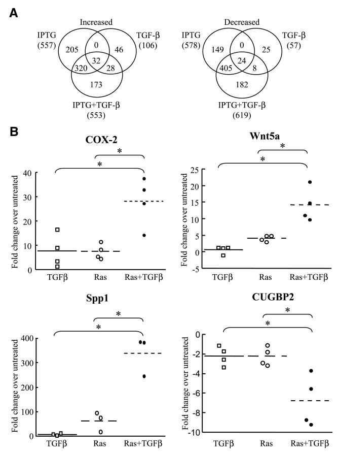
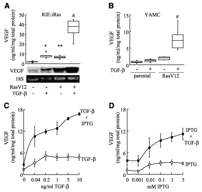
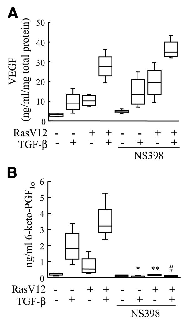
Colon cancer progression is characterized by activating mutations in Ras and by the emergence of the tumor-promoting effects of transforming growth factor-beta (TGF-beta) signaling. Ras-inducible rat intestinal epithelial cells (RIE:iRas) undergo a well-described epithelial to mesenchymal transition and invasive phenotype in response to H-RasV12 expression and TGF-beta treatment, modeling tumor progression. We characterized global gene expression profiles accompanying Ras-induced and TGF-beta-induced epithelial to mesenchymal transition in RIE:iRas cells by microarray analysis and found that the regulation of gene expression by the combined activation of Ras and TGF-beta signaling was associated with enrichment of a class of mRNAs containing 3' AU-rich element (ARE) motifs known to regulate mRNA stability. Regulation of ARE-containing mRNA transcripts was validated at the mRNA level, including genes important for tumor progression. Ras and TGF-beta synergistically increased the expression and mRNA stability of vascular endothelial growth factor (VEGF), a key regulator of tumor angiogenesis, in both RIE:iRas cells and an independent cell culture model (young adult mouse colonocyte). Expression profiling of human colorectal cancers (CRC) further revealed that many of these genes, including VEGF and PAI-1, were differentially expressed in stage IV human colon adenocarcinomas compared with adenomas. Furthermore, genes differentially expressed in CRC are also significantly enriched with ARE-containing transcripts. These studies show that oncogenic Ras and TGF-beta synergistically regulate genes containing AREs in cultured rodent intestinal epithelial cells and suggest that posttranscriptional regulation of gene expression is an important mechanism involved in cellular transformation and CRC tumor progression.
Conflict of interest statement
Figures


Similar articles
-
Transforming growth factor-beta1 promotes invasiveness after cellular transformation with activated Ras in intestinal epithelial cells.Exp Cell Res. 2001 Jun 10;266(2):239-49. doi: 10.1006/excr.2000.5229. Exp Cell Res. 2001. PMID: 11399052
-
Transformation by oncogenic Ras expands the early genomic response to transforming growth factor beta in intestinal epithelial cells.Neoplasia. 2008 Oct;10(10):1073-82. doi: 10.1593/neo.07739. Neoplasia. 2008. PMID: 18813357 Free PMC article.
-
Transforming growth factor-beta and Ras regulate the VEGF/VEGF-receptor system during tumor angiogenesis.Int J Cancer. 2002 Jan 10;97(2):142-8. doi: 10.1002/ijc.1599. Int J Cancer. 2002. PMID: 11774256
-
The epithelial-mesenchymal transition (EMT) and colorectal cancer progression.Cancer Biol Ther. 2005 Apr;4(4):365-70. doi: 10.4161/cbt.4.4.1655. Epub 2005 Apr 4. Cancer Biol Ther. 2005. PMID: 15846061 Review.
-
Mechanisms of the epithelial-mesenchymal transition by TGF-beta.Future Oncol. 2009 Oct;5(8):1145-68. doi: 10.2217/fon.09.90. Future Oncol. 2009. PMID: 19852727 Free PMC article. Review.
Cited by
-
The mRNA binding proteins HuR and tristetraprolin regulate cyclooxygenase 2 expression during colon carcinogenesis.Gastroenterology. 2009 May;136(5):1669-79. doi: 10.1053/j.gastro.2009.01.010. Epub 2009 Jan 15. Gastroenterology. 2009. PMID: 19208339 Free PMC article.
-
Caveolin-1 promotes autoregulatory, Akt-mediated induction of cancer-promoting growth factors in prostate cancer cells.Mol Cancer Res. 2009 Nov;7(11):1781-91. doi: 10.1158/1541-7786.MCR-09-0255. Epub 2009 Nov 10. Mol Cancer Res. 2009. PMID: 19903767 Free PMC article.
-
The novel tumor suppressor NOL7 post-transcriptionally regulates thrombospondin-1 expression.Oncogene. 2013 Sep 12;32(37):4377-86. doi: 10.1038/onc.2012.464. Epub 2012 Oct 22. Oncogene. 2013. PMID: 23085760 Free PMC article.
-
Mechanistic aspects of COX-2 expression in colorectal neoplasia.Recent Results Cancer Res. 2013;191:7-37. doi: 10.1007/978-3-642-30331-9_2. Recent Results Cancer Res. 2013. PMID: 22893198 Free PMC article. Review.
-
Post-transcriptional control during chronic inflammation and cancer: a focus on AU-rich elements.Cell Mol Life Sci. 2010 Sep;67(17):2937-55. doi: 10.1007/s00018-010-0383-x. Epub 2010 May 22. Cell Mol Life Sci. 2010. PMID: 20495997 Free PMC article. Review.
References
-
- Bos J, Fearon E, Hamilton S, et al. Prevalence of ras gene mutations in human colorectal cancers. Nature. 1987;327:293–7. - PubMed
-
- Lee S, Lee J, Soung Y, et al. Colorectal tumors frequently express phosphorylated mitogen-activated protein kinase. APMIS. 2004;112:233–8. - PubMed
-
- Kurokowa M, Lynch K, Podolsky D. Effects of growth factors on an intestinal epithelial cell line: transforming growth factor β inhibits proliferation and stimulates differentiation. Biochem Biophys Res Commun. 1987;142:775–82. - PubMed
-
- Filmus J, Zhao J, Buick R. Overexpression of H-ras oncogene induces resistance to the growth-inhibitory action of transforming growth factor β-1 (TGF-β1) and alters the number and type of TGF-β1 receptors in rat intestinal epithelial cell clones. Oncogene. 1992;7:521–6. - PubMed
-
- Shi Y, Massague J. Mechanisms of TGF-β signaling from cell membrane to the nucleus. Cell. 2003;113:685–700. - PubMed
Publication types
MeSH terms
Substances
Grants and funding
LinkOut - more resources
Full Text Sources
Miscellaneous
