. 2008 Jul;6(7):1215-24.
doi: 10.1158/1541-7786.MCR-08-0087.

The pak4 protein kinase plays a key role in cell survival and tumorigenesis in athymic mice

Affiliations

The pak4 protein kinase plays a key role in cell survival and tumorigenesis in athymic mice

Yingying Liu et al. Mol Cancer Res. 2008 Jul.

Abstract

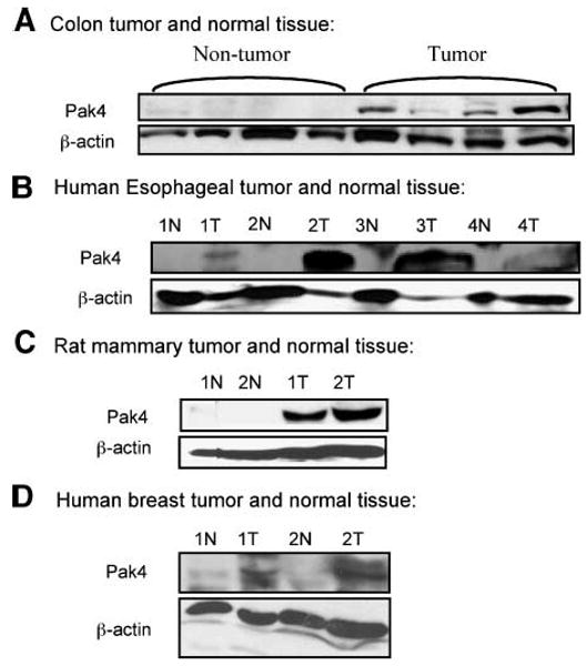
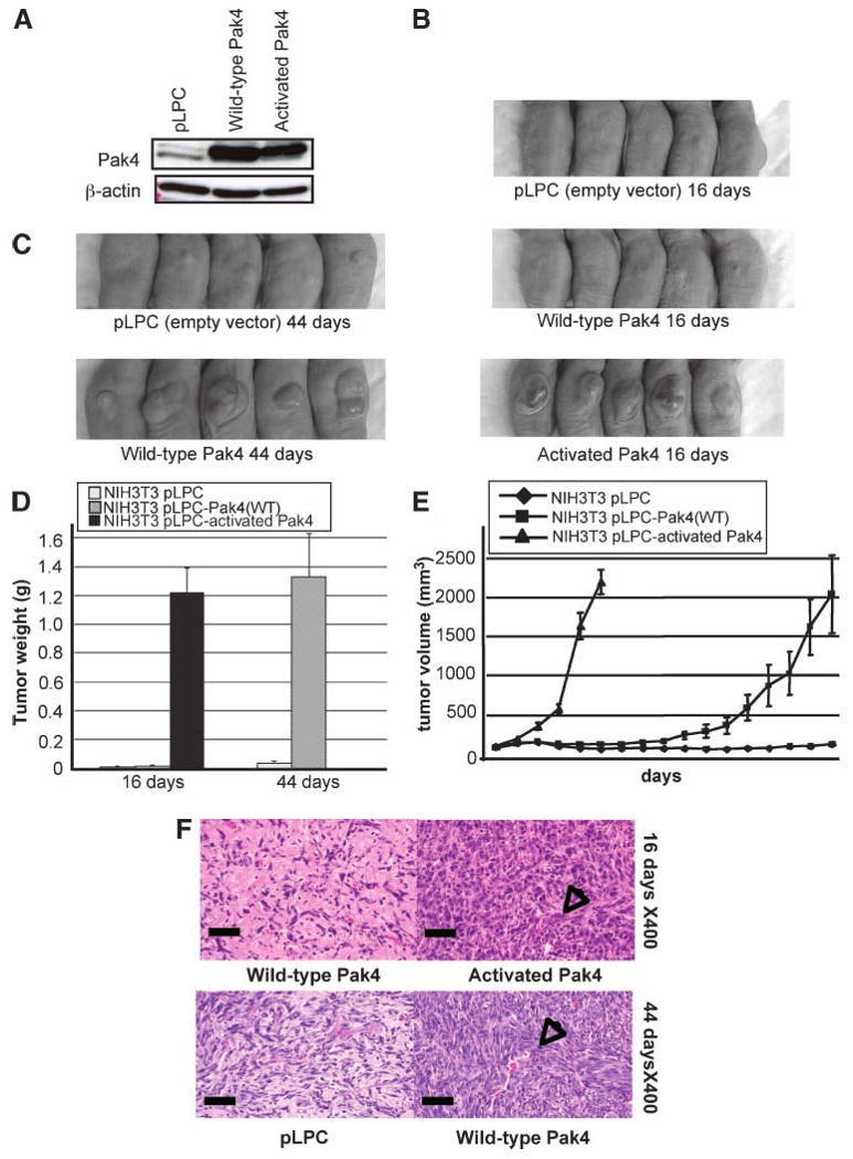
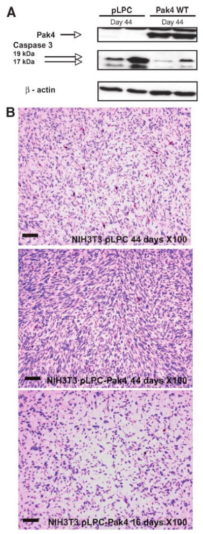
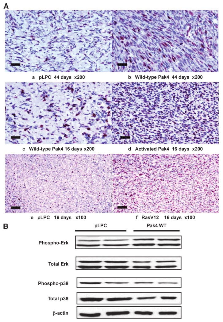
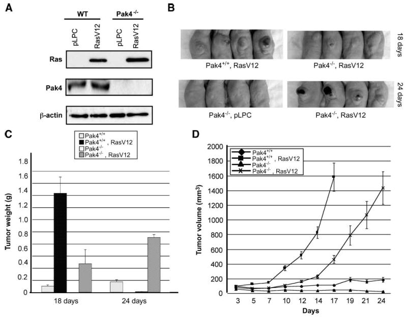
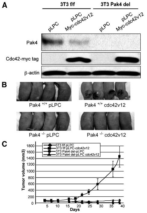
Pak4 is a member of the B group of p21-activated (Pak) kinases, originally identified as an effector protein for Cdc42. Although Pak4 is expressed at low levels in most adult tissues, it is highly overexpressed in tumor cell lines. Here, we show that Pak4 is also overexpressed in primary tumors, including colon, esophageal, and mammary tumors. Overexpression of Pak4 also leads to tumor formation in athymic mice, whereas deletion of Pak4 inhibits tumorigenesis. Although a constitutively active Pak4 mutant was previously shown to promote oncogenic transformation in cultured cells, our results are the first to show that Pak4 also promotes tumorigenesis in experimental animals. Furthermore, these results show for the first time that not only constitutively active Pak4, but also wild-type Pak4, is transforming, when experimental animals are used. These results are highly significant because wild-type Pak4, rather than activated Pak4, is overexpressed in tumor cells. Our results suggest that overexpression or activation of Pak4 is a key step in oncogenic transformation, due to its ability to promote cell survival and subsequent uncontrolled proliferation. The finding that Pak4 is up-regulated in so many types of cancers indicates that Pak4 may play a vital role in a wide range of different types of cancer. This makes it an attractive candidate for drug therapy for different types of cancer.


Conflict of interest statement

Disclosure of Potential Conflicts of Interest: No potential conflicts of interest were disclosed.

Figures

FIGURE 1
FIGURE 1
Pak4 is overexpressed in primary tumors. Western blot analysis illustrates Pak4 expression in tumor and normal tissues. The following tumor tissues were analyzed: A. Mouse colon tumor isolated from azoxymethane-treated CF-1 female mice; nontumor tissue was adjacent to normal tissue. B. Human esophageal squamous cell carcinoma (T) and normal tissues (N). Paired normal tissue was pathologically normal and from the same patient. C. Rat mammary tissue was originally isolated from N-methylnitrosourea treated Sprague-Dawley female rats; normal tissue was isolated from nontreated rats. D. Human breast tumor and normal tissues. Paired normal tissue was pathologically normal and from the same patient. In all cases, 100 μg of protein extract were used; β-actin served as a loading control.
FIGURE 2
FIGURE 2
Ectopic overexpression of Pak4 leads to tumor formation. A. NIH3T3 cells were transfected with empty vector (pLPC), wild-type Pak4, or activated Pak4–containing pLPC-puro retroviral vectors. Cell extracts were prepared for Western blotting with the indicated antibodies. In each case, 20 μg of protein extract were used and β-actin served as a loading control. B. Cells containing activated Pak4 lead to tumor formation in athymic mice by 16 d postinjection. C. By 44 d, cells containing wild-type Pak4 also form tumors in athymic mice. D and E. Tumor weights and volumes from mice injected with NIH3T3 cells containing empty vector, wild-type Pak4, or activated Pak4 were assessed (activated Pak4-injected mice were sacrificed after day 16). F. Histology of tumor tissue isolated from mice injected with cells overexpressing wild-type or activated Pak4 (top), or empty vector or wild-type Pak4 (bottom), 16 or 44 d after injection. Arrows, areas with large amounts of spindle-shaped cells typical of sarcoma tissue. Scale bars, 50 μm.
FIGURE 3
FIGURE 3
Activated caspase-3 levels are abrogated in tumors derived from Pak4-overexpressing cells. A. Activated caspase-3 (cleaved into 19 and 17 kDa fragments) was assessed by Western blot analysis of tumor or normal tissue that was isolated from mice injected with cells overexpressing empty vector (pLPC) or wild-type Pak4, 44 d after injection. The antibody recognizes only cleaved (activated) caspase-3. Pak4 expression and β-actin expression were also assessed. B. Activated caspase-3 (cleaved into 19 and 17 kDa fragments) was assessed by immunostaining of sections from tumors that were isolated from mice injected with cells overexpressing wild-type Pak4. Tissue sections from mice injected with cells containing empty vector (pLPC) were analyzed as a control. Tumor tissues were examined 16 and 44 d after injection. Areas containing active caspase-3 appear as red spots on the H&E-stained sections. Cells in individual sections were counted and at day 44; an average of 1.32% of the cells in control tissue stained positively for active caspases-3, whereas only 0.25% of the cells in the tumor tissue formed by wild-type Pak4 stained positively. Even at 16 d postinjection, as seen above, only 0.26% of the cells from the injection sites of mice injected with wild-type Pak4 stained positively for caspase-3, although tumors had not begun to form at that time point. In both cases, the difference between Pak4-containing cells and empty vector cells is statistically significant. Scale bars, 100 μm.
FIGURE 4
FIGURE 4
Increased proliferation is in response to Pak4 expression. Mice were injected with NIH3T3 cells containing empty vector (pLPC), wild-type Pak4, activated Pak4, or oncogenic RasV12, as a positive control. A. Tissue sections were taken from the injection sites of all of the mice 16 or 44 d after injection. Sections were stained with H&E and with antibody against Ki67, which recognizes proliferating cells (dark purple). The results indicate that wild-type and activated Pak4 lead to an increased number of proliferating cells in the tissue sections. Cells in individual sections were counted and at day 44; an average of 20.4% of the cells from control tissue stained positively for Ki67, whereas 36.4% of cells from tumors derived from wild-type Pak4 stained positively. At 16 d, 31.7% of cells from the injection sites of mice injected with wild-type Pak4 stained positively for Ki67. In both cases, the difference between Pak4-containing cells and empty vector cells was statistically significant. This was seen even at 16 d, at which point wild-type Pak4 had not yet caused tumors to form. Scale bars, 50 μm. B. Phospho-ERK and total ERK levels were assessed by Western blot analysis of tumor or normal tissue that was isolated from mice injected with cells overexpressing empty vector (pLPC) or wild-type Pak4, 44 d after injection. Top band, ERK2; bottom band, ERK1. As a negative control, phosphorylation of p38 was also examined by Western blot. The results indicated that wild-type Pak4 can lead to a slight increase in phosphorylation of the ERK mitogen-activated protein kinase pathway, which is often associated with increased proliferation.
FIGURE 5
FIGURE 5
Tumor formation is attenuated in Pak4 null cells when transfected with Ras. A. Immortalized wild-type and Pak4−/− fibroblasts were infected with either empty vector (pLPC) or RasV12-containing pLPC retroviral vectors. Two days after infection, cells were selected with puromycin for 2 wk, after which cell extracts were prepared for Western blotting with the indicated antibodies. In each case, 20 μg of protein extract were used. β-Actin served as a loading control. B. Top, mice were injected with Pak4+/+ cells (wild-type) or Pak4−/− cells that were infected with oncogenic Ras. Tumors were examined 18 d after injection (for both cell types, cells infected with empty vector did not form tumors; not shown). Bottom, mice were injected with Pak4−/− cells containing empty vector (pLPC; left), or with RasV12 (right). Tumors were examined 24 d after injection (the Pak4+/+, RasV12 condition is not shown at 24 d because the tumors became too large and the mice had to be sacrificed). C and D. Tumor weight and tumor volume change in mice injected with Pak4+/+ and Pak4−/− cells with or without RasV12 were assessed (tumor weight in mice injected RasV12; Pak4+/+ cells were only analyzed at day 18 because the mice had to be sacrificed due to the large size of the tumors).
FIGURE 6
FIGURE 6
Tumor formation is completely abrogated in Pak4 null cells when transfected with Cdc42V12. A. Immortalized wild-type and Pak4 conditional knockout fibroblasts were infected with either empty vector (pLPC) or Cdc42V12-containing pLPC retroviral vectors. Two days after infection, cells were selected with puromycin for 2 wk, after which cell extracts were prepared for Western blotting with the indicated antibodies. In each case, 20 μg of protein extract were used. β-Actin served as a loading control. B. Top, mice were injected with Pak4+/+ cells (wild-type) that were infected with empty vector (pLPC; left) or with Cdc42V12 (right). Bottom, mice were injected with Pak4−/− cells containing empty vector (pLPC; left) or with Cdc42V12 (right). Tumors were examined 38 d after injection. For both cell types, cells infected with empty vector did not form tumors. Wild-type cells infected with Cdc42V12 formed tumors; however, tumor formation were completely abrogated in Pak4 null cells when transfected with Cdc42V12. C. Tumor volumes in mice injected with Pak4+/+ and Pak4 conditional knockout cells with or without Cdc42V12 were assessed at the indicated time points.

Similar articles

Cited by

References

    1. Daniels RH, Bokoch GM. p21-Activated protein kinase: a crucial component of morphological signaling? Trends Biochem Sci. 1999;24:350–5. - PubMed
    1. Knaus UG, Bokoch GM. The p21Rac/Cdc42-activated kinases (PAKs) Int J Biochem Cell Biol. 1998;30:857–62. - PubMed
    1. Sells MA, Chernoff J. Emerging from the Pak: the p21-activated protein kinase family. Trends Cell Biol. 1997;7:162–7. - PubMed
    1. Jaffer ZM, Chernoff J. p21-Activated kinases: three more join the Pak. Int J Biochem Cell Biol. 2002;34:713–7. - PubMed
    1. Abo A, Qu J, Cammarano MS, et al. PAK4, a novel effector for Cdc42Hs, is implicated in the reorganization of the actin cytoskeleton and in the formation of filopodia. EMBO J. 1998;17:6527–40. - PMC - PubMed

Publication types

MeSH terms