. 2008 Jul;7(7):2003-11.
doi: 10.1158/1535-7163.MCT-08-0095.

Inhibition of centromere dynamics by eribulin (E7389) during mitotic metaphase

Affiliations

Inhibition of centromere dynamics by eribulin (E7389) during mitotic metaphase

Tatiana Okouneva et al. Mol Cancer Ther. 2008 Jul.

Abstract

Eribulin (E7389), a synthetic analogue of halichondrin B in phase III clinical trials for breast cancer, binds to tubulin and microtubules. At low concentrations, it suppresses the growth phase of microtubule dynamic instability in interphase cells, arrests mitosis, and induces apoptosis, suggesting that suppression of spindle microtubule dynamics induces mitotic arrest. To further test this hypothesis, we measured the effects of eribulin on dynamics of centromeres and their attached kinetochore microtubules by time-lapse confocal microscopy in living mitotic U-2 OS human osteosarcoma cells. Green fluorescent protein-labeled centromere-binding protein B marked centromeres and kinetochore-microtubule plus-ends. In control cells, sister chromatid centromere pairs alternated under tension between increasing and decreasing separation (stretching and relaxing). Eribulin suppressed centromere dynamics at concentrations that arrest mitosis. At 60 nmol/L eribulin (2 x mitotic IC(50)), the relaxation rate was suppressed 21%, the time spent paused increased 67%, and dynamicity decreased 35% (but without reduction in mean centromere separation), indicating that eribulin decreased normal microtubule-dependent spindle tension at the kinetochores, preventing the signal for mitotic checkpoint passage. We also examined a more potent, but in tumors less efficacious antiproliferative halichondrin derivative, ER-076349. At 2 x IC(50) (4 nmol/L), mitotic arrest also occurred in concert with suppressed centromere dynamics. Although media IC(50) values differed 15-fold between the two compounds, the intracellular concentrations were similar, indicating more extensive relative uptake of ER-076349 into cells compared with eribulin. The strong correlation between suppression of kinetochore-microtubule dynamics and mitotic arrest indicates that the primary mechanism by which eribulin blocks mitosis is suppression of spindle microtubule dynamics.


Conflict of interest statement

Conflicts of Interest: Drs. Okouneva, Azarenko, Wilson, and Jordan received or were supported in part by a research grant from Eisai Research Institute. Dr. Littlefield is an employee of Eisai Research Institute.

Figures
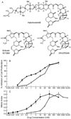
Fig. 1
Fig. 1
A) The structures of halichondrin B, eribulin, and ER-076349. B) Inhibition of proliferation of U-2 OS human osteosarcoma cells by eribulin (circles) and ER-076349 (squares). Cell proliferation was determined by counting live cells at the time of drug addition and 28 h later. IC50, eribulin, 30 nmol/L; ER-076349, 3 nmol/L. C) Accumulation of cells in mitosis after incubation with eribulin (circles) and ER-076349 (squares). Mitotic accumulation was determined by counting cells by microscopy following fixation and staining of microtubules and chromatin. Half-maximal mitotic arrest occurred at 30 nmol/L for eribulin and at 2 nmol/L for ER-076349. Values are means and SEM of 5 independent experiments.
Fig. 2
Fig. 2
(A–D) Centromeres, microtubules, and chromosomes in U-2 OS cells in the absence (A, B) or presence (C, D) of eribulin. Cells expressing GFP-CENP-B (green) were incubated with or without eribulin (28 h), fixed, and stained with antibody to α-tubulin (red) and with DAPI for chromosomes. In control cells in metaphase (A), pairs of sister centromeres are present on sister chromatids and are separated by variable distance. In anaphase (B), sister chromatids and centromeres have separated. At half-maximal mitotic arrest (30 nmol/L eribulin (C, D) and 2 nmol/L ER-076349, data not shown), spindles were abnormal bipolar with uncongressed chromosomes (arrow) (C) or multipolar (D). At 60 nmol/L eribulin or 4 nmol/L ER-076349 most spindles were multipolar. (E–H) Dynamic behavior of centromeres in living U-2 OS cells in the absence of drug. Images of GFP-centromeres were collected from a single plane by confocal microscopy at 5-s intervals. Arrowheads indicate the positions of sister centromeres at time 50s (separated by 0.93 µm) (E); in the second (F), third (G) and fourth (H) panels (120, 275, and 310 s) they are separated by 0.70, 1.2, 0.59 µm.
Fig. 3
Fig. 3
Time course of changes in the center-to-center separation distance between sister centromeres of a pair in the absence (upper trace) and presence of 60 nmol/L eribulin (lower trace). Images of GFP-centromeres were collected from a single plane by confocal microscopy at 5-s intervals. Separation distances between the 2 members of pairs that remained well focused for the 7 min recording period were measured as described in Materials and Methods. For the centromere pair in the absence of drug, the centromeres were separated by 0.5 µm at zero time. The pair then underwent multiple episodes of stretching and relaxing. For example, at 45 s, the pair was separated by 0.79 µm; at 55 s, the separation distance decreased to 0.41 µm, the minimum separation observed for this pair; and at 275 s, it increased to 1.0 µm, the maximum separation observed for this pair. In the presence of 60 nmol/L eribulin, the separation distance was reduced overall, and ranged from 0.73 - 0.28 µm for the centromere pair shown in the lower panel.
Fig. 4
Fig. 4
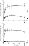
Time course of uptake of A) [3H]eribulin (squares, 60 nmol/L; circles, 30 nmol/L) and B) [3H]ER-076349 (squares, 4 nmol/L; circles, 2 nmol/L) into U-2 OS cells. [3H]eribulin or [3H]ER-076349, were added to U-2 OS cells growing exponentially (Materials and Methods). At time intervals from 30 min to 20 h, intracellular radioactivity was determined. After 20 h, cells were washed with 2 ml PBS and fresh media was added (arrow) for 1 h (last data point) and for 24 h (data not shown) to determine radioactivity retained after washout. Values are means and SEM of 5 independent experiments.
Fig. 5
Fig. 5
Histogram comparing the percent suppression of the major parameters of centromere dynamics in U-2 OS cells by eribulin (60 nmol/L, left-hand dark hatched bars) with those previously found to be induced by vinblastine (50 nmol/L, interlaced hatched bars), vinorelbine (50 nmol/L, dotted bars), vinflunine (50 nmol/L, light hatched bars) (13), and paclitaxel (100 nmol/L, solid.bars)(12). The drug concentrations were all ≥ 2× the concentration that induced half-maximal mitotic arrest in order to ensure that the observed suppression was associated with significant mitotic arrest. Specifically, the concentrations were 2 × IC50, 8 × IC50, 7 × IC50, 3 × IC50 and 2 × IC50, respectively.

References

    1. Jordan MA, Kamath K, Manna T, et al. The primary antimitotic mechanism of action of the synthetic halichondrin E7389 is suppression of microtubule growth. Mol Cancer Ther. 2005;4:1086–1095. - PubMed
    1. Dabydeen D, Burnett J, Bai R, et al. Comparison of the activities of the truncated halichondrin B analog NSC 707389 (E7389) with those of the parent compound and a proposed binding site on tubulin. Mol Pharmacol. 2006;70:1866–1875. - PubMed
    1. Kuznetsov G, Towle MJ, Cheng H, et al. Induction of morphological and biochemical apoptosis following prolonged mitotic blockage by halichondrin B macrocyclic ketone analog E7389. Cancer Res. 2004;64:5760–5766. - PubMed
    1. Towle MJ, Salvato KA, Budrow J, et al. In vitro and in vivo anticancer activities of synthetic macrocyclic ketone analogs of halichondrin B. Cancer Res. 2001;61:1013–1021. - PubMed
    1. Shelby RD, Hahn KM, Sullivan KF. Dynamic elastic behavior of alpha-satellite DNA domains visualized in situ in living human cells. J Cell Biol. 1996;135:545–557. - PMC - PubMed

Publication types

MeSH terms