Controlled Clinical Trial
. 2008 Jul;144(7):851-8.
doi: 10.1001/archderm.144.7.851.

Effect of increased pigmentation on the antifibrotic response of human skin to UV-A1 phototherapy

Affiliations
Controlled Clinical Trial

Effect of increased pigmentation on the antifibrotic response of human skin to UV-A1 phototherapy

Frank Wang et al. Arch Dermatol. 2008 Jul.

Abstract

Objective: To investigate the efficacy, potential limitations, and biological mechanisms of UV-A1 phototherapy for skin sclerosis due to collagen deposition disorders.

Design: Before-and-after trial of UV-A1 irradiation of sclerotic skin; in vivo biochemical analyses after UV-A1 irradiation of normal skin.

Setting: Academic referral center.

Participants: Patients with morphea/scleroderma or sclerodermoid graft-vs-host disease and volunteers without skin disease. Intervention Sclerotic skin was treated with high-dose (130 J/cm(2); n = 12) or medium-dose (65 J/cm(2); n = 6) UV-A1 phototherapy 3 times per week for 14 weeks; normal skin was treated with UV-A1 irradiation at various doses and frequencies, with biopsies performed afterwards.

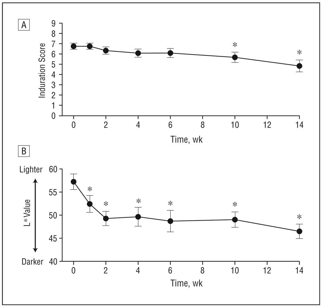
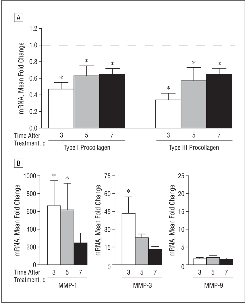
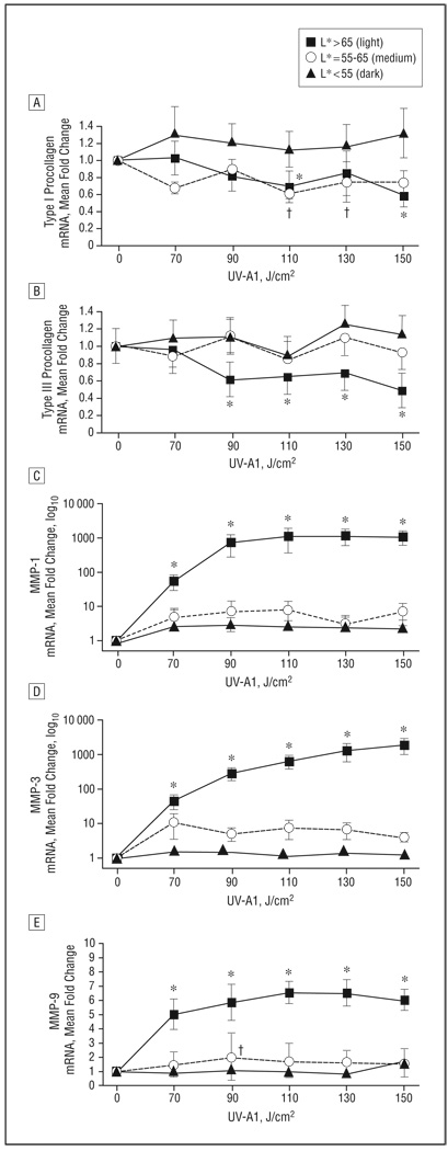
Main outcome measures: In sclerotic skin, induration was clinically assessed using a scoring scale. In normal skin, quantitative polymerase chain reaction was used to assess antifibrotic responses, defined as decreased type I and type III procollagen and increased matrix metalloproteinase levels.

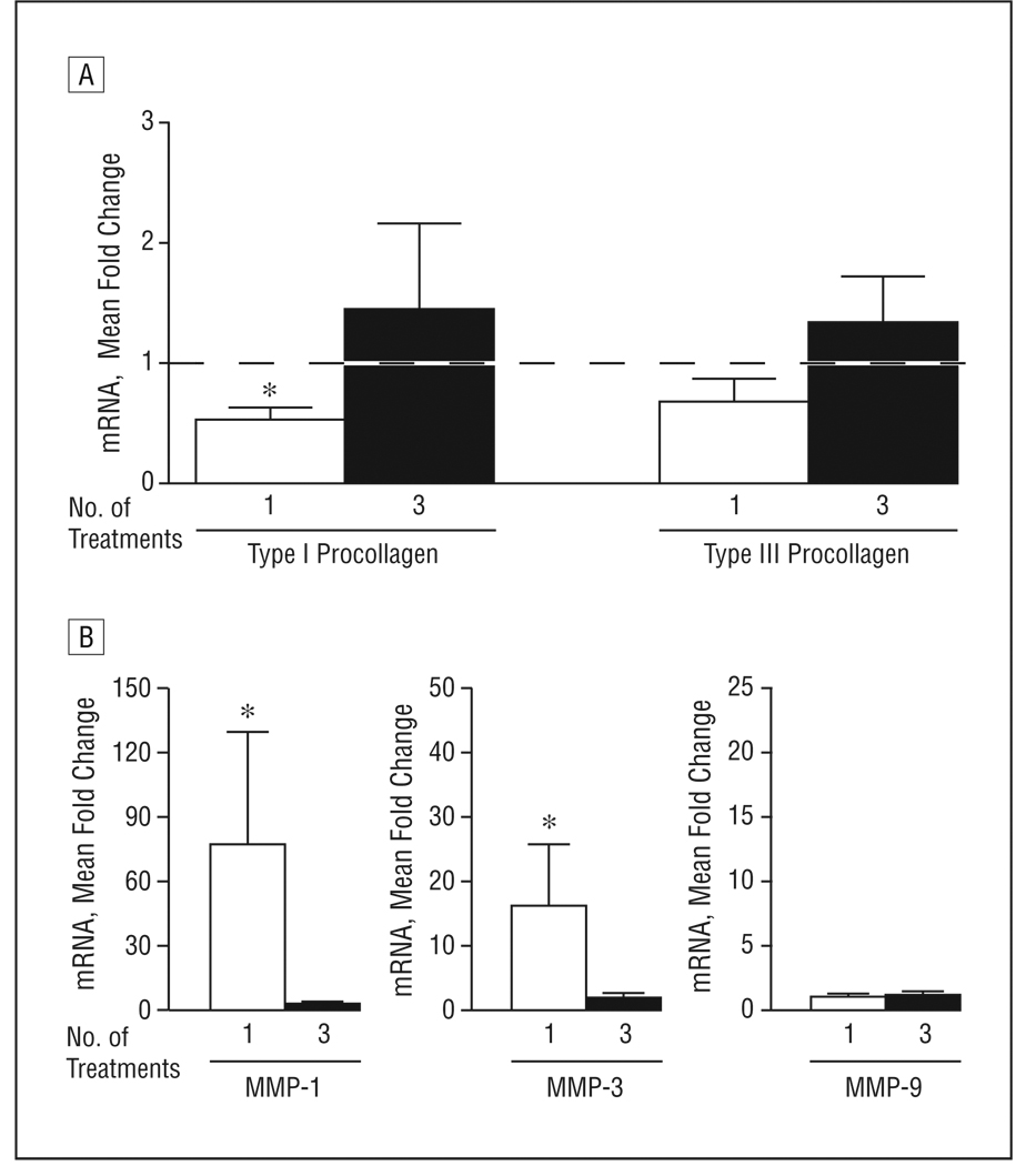
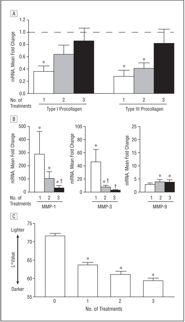
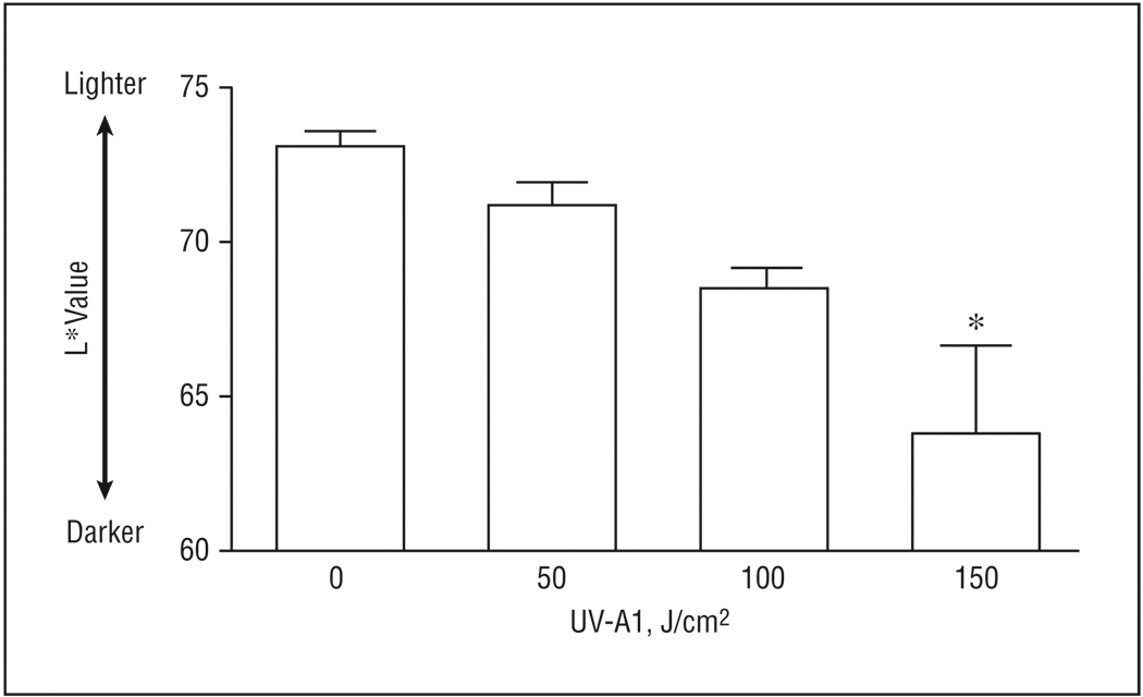
Results: In patients with sclerotic skin treated with high-dose UV-A1 irradiation, clinical scores for induration modestly decreased. To investigate what factors prevented further improvement (ie, complete clearance), normal skin with light pigmentation was exposed to UV-A1 irradiation (70-150 J/cm(2)) and was assessed for antifibrotic responses. A single high-dose exposure (110-150 J/cm(2)) elicited substantial antifibrotic responses and induced skin darkening. This skin darkening attenuated responses to subsequent UV-A1 exposures and was dose dependent. Thus, to minimize skin darkening, additional patients with sclerotic skin were treated with medium-dose UV-A1 phototherapy, which was no less effective than high-dose therapy.

Conclusion: Clinical responses of sclerotic skin to UV-A1 phototherapy were modest because of UV-A1-induced skin darkening, which is photoprotective and attenuates antifibrotic responses.

Trial registration: clinicaltrials.gov Identifier: NCT00129415.


Figures

Figure 1
Figure 1
Mean induration and pigmentation of sclerotic skin in 12 patients treated with high-dose UV-A1 irradiation (130 J/cm2) 3 times weekly. A, Induration was assessed on a scale from 1 (very mild) to 9 (very severe). B, Pigmentation of treated areas was assessed by using a chromameter (L* [luminescence] value). Error bars represent SEM. *P<.05 compared with before therapy (week 0).
Figure 2
Figure 2
Duration of antifibrotic responses after a single high-dose UV-A1 exposure. Eight healthy subjects with lightly pigmented skin (L* [luminescence] >65) were exposed once to high-dose UV-A1 irradiation (130 J/cm2). Biopsies were performed 3, 5, and 7 days later. Real-time polymerase chain reaction was used to assess expression for the indicated messenger RNA (mRNA) transcripts: type I and type III procollagen (A) and matrix metalloproteinases (MMPs) 1, 3, and 9 (B). Data are presented as mean fold change relative to untreated skin (normalized to 1). Error bars represent SEM. In A, the horizontal dashed line indicates fold change of untreated skin. *P<.05 compared with untreated skin.
Figure 3
Figure 3
Refractory antifibrotic responses with repetitive high-dose UV-A1 irradiation. Eight healthy subjects with lightly pigmented skin (L* [luminescence] >65) were treated with high-dose UV-A1 irradiation (110 J/cm2) either once (Friday) or 3 times (Monday, Wednesday, and Friday). Four days later (the following Tuesday), all biopsy samples were collected and assessed by real-time polymerase chain reaction for the indicated transcripts: type I and type III procollagen (A) and matrix metalloproteinases (MMPs) 1, 3, and 9 (B). Data are presented as mean fold change relative to untreated skin (normalized to 1). Error bars represent SEM. In A, the horizontal dashed line indicates fold change of untreated skin. *P<.05 compared with untreated skin. mRNA indicates messenger RNA.
Figure 4
Figure 4
Attenuation of antifibrotic responses with weekly high-dose UV-A1 phototherapy. Ten healthy subjects with lightly pigmented skin (L* [luminescence] >65) were treated with high-dose UV-A1 phototherapy (130 J/cm2) once per week. Biopsy samples and skin pigmentation readings were obtained 24 hours after 1, 2, and 3 weekly treatments. A and B, The indicated transcripts (type I and type III procollagen [A] and matrix metalloproteinases [MMPs] 1, 3, and 9 [B]) were assessed by real-time polymerase chain reaction. Data are presented as mean fold change relative to untreated skin (normalized to 1). In A, the horizontal dashed line indicates fold change of untreated skin. C, Skin pigmentation is shown as mean L* value. Error bars represent SEM. *P<.05 compared with untreated skin. †P<.05 compared with a single UV-A1 exposure. mRNA indicates messenger RNA.
Figure 5
Figure 5
Rapid darkening of skin with high-dose UV-A1 irradiation. Eight healthy subjects with lightly pigmented skin (L* [luminescence] >65) were treated once with the indicated UV-A1 doses. After 24 hours, chromameter readings (L* values) were obtained. Error bars represent SEM. *P<.05 compared with untreated skin.
Figure 6
Figure 6
Abrogation of UV-A1–induced antifibrotic responses in skin with darker pigmentation. Based on L* (luminescence) values, 28 healthy subjects were stratified according to skin pigmentation as light (n=8), medium (n=8), or dark (n=12). Skin was then exposed to the indicated UV-A1 doses. Biopsies were performed 24 hours later, and the indicated transcripts were assessed by real-time polymerase chain reaction: type I procollagen (A), type III procollagen (B), matrix metalloproteinase (MMP) 1 (C), MMP-3 (D), and MMP-9 (E). Data are presented as mean fold change relative to untreated skin (normalized to 1). Error bars represent SEM. *P<.05 for light skin vs untreated skin. †P<.05 for medium skin vs untreated skin.

Comment in

References

    1. Charles C, Clements P, Furst DE. Systemic sclerosis: hypothesis-driven treatment strategies. Lancet. 2006;367(9523):1683–1691. - PubMed
    1. Denton CP, Black CM. Targeted therapy comes of age in scleroderma. Trends Immunol. 2005;26(11):596–602. - PubMed
    1. Breuckmann F, Gambichler T, Altmeyer P, Kreuter A. UVA/UVA1 phototherapy and PUVA photochemotherapy in connective tissue diseases and related disorders: a research based review. BMC Dermatol. 2004;4(1):11. - PMC - PubMed
    1. Fisher GJ, Kang S. Phototherapy for scleroderma: biologic rationale, results, and promise. Curr Opin Rheumatol. 2002;14(6):723–726. - PubMed
    1. Denton CP, Black CM, Abraham DJ. Mechanisms and consequences of fibrosis in systemic sclerosis. Nat Clin Pract Rheumatol. 2006;2(3):134–144. - PubMed

Publication types

MeSH terms

Associated data